- Infectious Diseases of Livestock
- Part 3
- Borrelia suilla infection
- GENERAL INTRODUCTION: SPIROCHAETES
- Swine dysentery
- Borrelia theileri infection
- Borrelia suilla infection
- Lyme disease in livestock
- Leptospirosis
- GENERAL INTRODUCTION: AEROBIC ⁄ MICRO-AEROPHILIC, MOTILE, HELICAL ⁄ VIBROID GRAM-NEGATIVE BACTERIA
- Genital campylobacteriosis in cattle
- Proliferative enteropathies of pigs
- Campylobacter jejuni infection
- GENERAL INTRODUCTION: GRAM-NEGATIVE AEROBIC OR CAPNOPHILIC RODS AND COCCI
- Moraxella spp. infections
- Bordetella bronchiseptica infections
- Pseudomonas spp. infections
- Glanders
- Melioidosis
- Brucella spp. infections
- Bovine brucellosis
- Brucella ovis infection
- Brucella melitensis infection
- Brucella suis infection
- Brucella infections in terrestrial wildlife
- GENERAL INTRODUCTION: FACULTATIVELY ANAEROBIC GRAM NEGATIVE RODS
- Klebsiella spp. infections
- Escherichia coli infections
- Salmonella spp. infections
- Bovine salmonellosis
- Ovine and caprine salmonellosis
- Porcine salmonellosis
- Equine salmonellosis
- Yersinia spp. infections
- Haemophilus and Histophilus spp. infections
- Haemophilus parasuis infection
- Histophilus somni disease complex in cattle
- Actinobacillus spp. infections
- Actinobacillus equuli infections
- Gram-negative pleomorphic infections: Actinobacillus seminis, Histophilus ovis and Histophilus somni
- Porcine pleuropneumonia
- Actinobacillus suis infections
- Pasteurella and Mannheimia spp. infections
- Pneumonic mannheimiosis and pasteurellosis of cattle
- Haemorrhagic septicaemia
- Pasteurellosis in sheep and goats
- Porcine pasteurellosis
- Progressive atrophic rhinitis
- GENERAL INTRODUCTION: ANAEROBIC GRAM-NEGATIVE, IRREGULAR RODS
- Fusobacterium necrophorum, Dichelobacter (Bacteroides) nodosus and Bacteroides spp. infections
- GENERAL INTRODUCTION: GRAM-POSITIVE COCCI
- Staphylococcus spp. infections
- Staphylococcus aureus infections
- Exudative epidermitis
- Other Staphylococcus spp. infections
- Streptococcus spp. infections
- Strangles
- Streptococcus suis infections
- Streptococcus porcinus infections
- Other Streptococcus spp. infections
- GENERAL INTRODUCTION: ENDOSPORE-FORMING GRAM-POSITIVE RODS AND COCCI
- Anthrax
- Clostridium perfringens group infections
- Clostridium perfringens type A infections
- Clostridium perfringens type B infections
- Clostridium perfringens type C infections
- Clostridium perfringens type D infections
- Malignant oedema⁄gas gangrene group of Clostridium spp.
- Clostridium chauvoei infections
- Clostridium novyi infections
- Clostridium septicum infections
- Other clostridial infections
- Tetanus
- Botulism
- GENERAL INTRODUCTION: REGULAR, NON-SPORING, GRAM-POSITIVE RODS
- Listeriosis
- Erysipelothrix rhusiopathiae infections
- GENERAL INTRODUCTION: IRREGULAR, NON-SPORING, GRAM-POSITIVE RODS
- Corynebacterium pseudotuberculosis infections
- Corynebacterium renale group infections
- Bolo disease
- Actinomyces bovis infections
- Trueperella pyogenes infections
- Actinobaculum suis infections
- Actinomyces hyovaginalis infections
- GENERAL INTRODUCTION: MYCOBACTERIA
- Tuberculosis
- Paratuberculosis
- GENERAL INTRODUCTION: ACTINOMYCETES
- Nocardiosis
- Rhodococcus equi infections
- Dermatophilosis
- GENERAL INTRODUCTION: MOLLICUTES
- Contagious bovine pleuropneumonia
- Contagious caprine pleuropneumonia
- Mycoplasmal pneumonia of pigs
- Mycoplasmal polyserositis and arthritis of pigs
- Mycoplasmal arthritis of pigs
- Bovine genital mycoplasmosis
- Neurotoxin-producing group of Clostridium spp.
- Contagious equine metritis
- Tyzzer's disease
- MYCOTIC AND ALGAL DISEASES: Mycoses
- MYCOTIC AND ALGAL DISEASES: Pneumocystosis
- MYCOTIC AND ALGAL DISEASES: Protothecosis and other algal diseases
- DISEASE COMPLEXES / UNKNOWN AETIOLOGY: Epivag
- DISEASE COMPLEXES / UNKNOWN AETIOLOGY: Ulcerative balanoposthitis and vulvovaginitis of sheep
- DISEASE COMPLEXES / UNKNOWN AETIOLOGY: Ill thrift
- Eperythrozoonosis
- Bovine haemobartonellosis
Borrelia suilla infection
This content is distributed under the following licence: Attribution-NonCommercial CC BY-NC  View Creative Commons Licence details here
Borrelia suilla infection
G C BISHOP*
Introduction
According to Henning6 spirochaetosis in pigs was first described in South Africa by Dodd in 1906. The condition, which is frequently associated with conditions of poor hygiene, is also known as porcine ulcerative granuloma, and has been reported in New Zealand, Australia, the USA and the UK.1, 6 The disease is seen infrequently today and in South Africa it is not considered to be of any economic importance.
Aetiology and epidemiology
Borrelia suilla is 9 to 12 µm in length and has two to six spirals. According to Canham,4 Neitz believed that the spirochaete was definitely not identical to B. theileri. The bacteria stain readily with Giemsa, May-Grünwald, Gram’s stain and 1 per cent carbol fuchsin, and using dark-field microscopy and fresh specimens they may be observed as actively motile spirochaetes.
The primary role of B. suilla (synonym Spirochaeta suilla) in the ulcerative granuloma syndrome is uncertain. Borrelia suilla has never been cultured on artificial media1 and it is almost always to be found together with other bacteria in the lesions. The bacterium is not accorded species status in Bergey’s Manual of Determinative Bacteriology.3
Osborne8 reproduced typical lesions by subcutaneous injection of material from natural cases which also contained actively motile spirochaetes, into healthy pigs. Although it has not yet been proved,5 some workers believe that predisposing factors (such as trauma due to biting, or self-trauma due to the irritation caused by mange or flea infestations) are prerequisites for infection and that the spirochaete may not be the primary pathogen.
Spirochaetosis was commonly seen in pigs of six to ten weeks of age, especially following castration.6 Generally, less severe lesions were seen in older pigs.1, 5
Pathogenesis, clinical signs and pathology
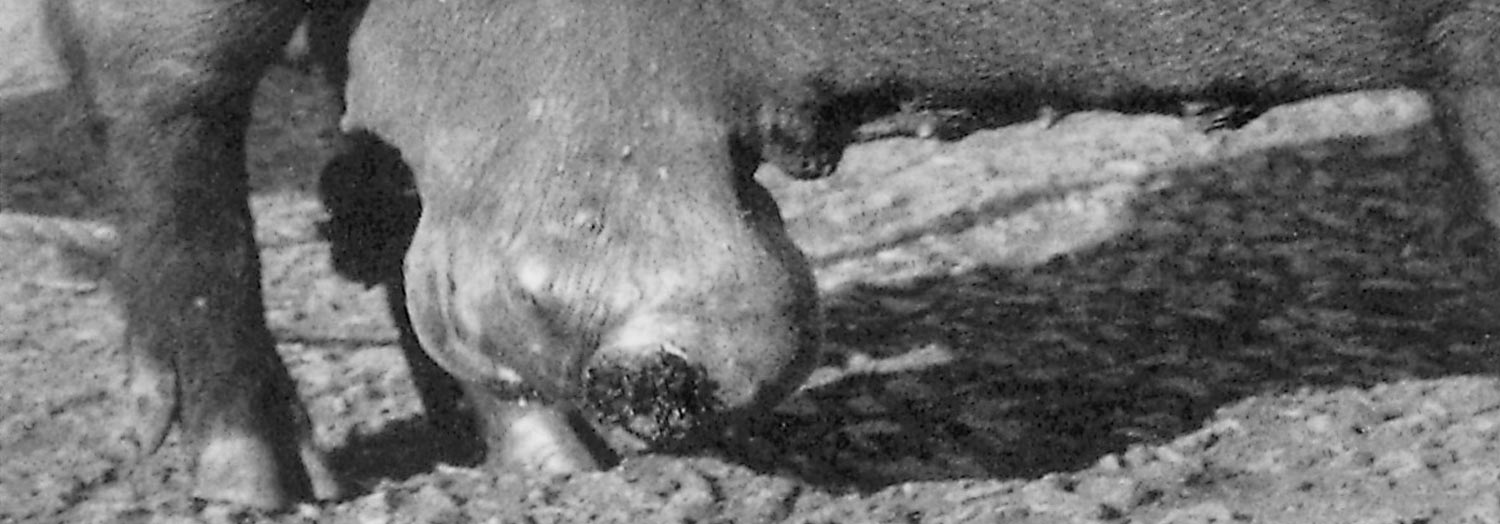
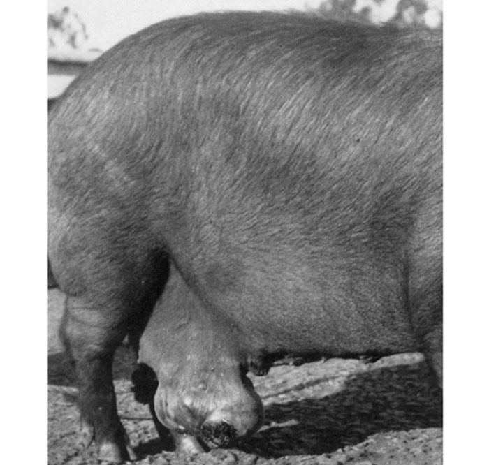
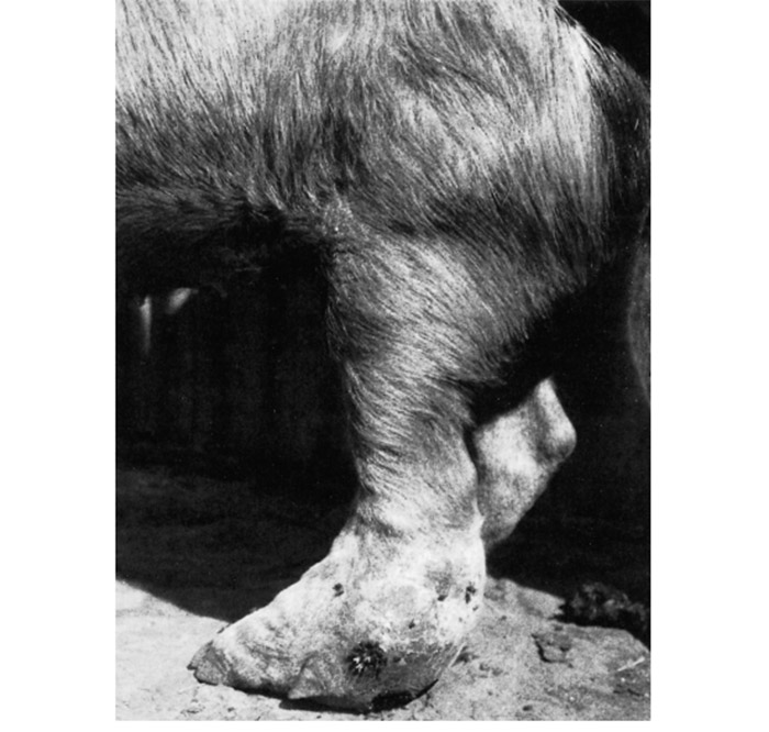
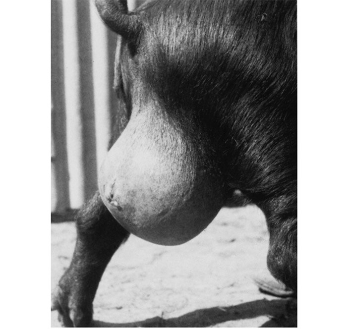
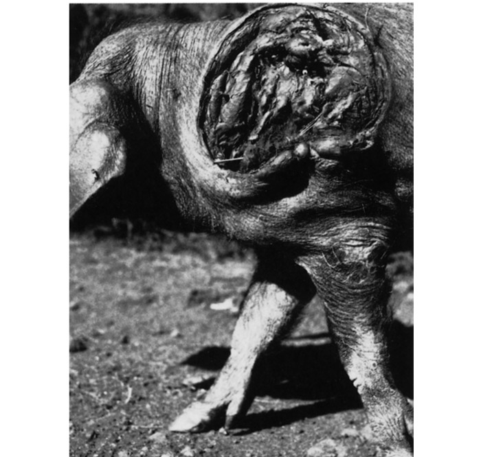
The spirochaetes usually enter the body through superficial wounds and provoke the formation of abscesses or ulcerative lesions of varying severity in the subcutaneous tissues.6 Lesions may occur on the face of suckling pigs, the ventral abdominal wall of sows,2 the legs, which may result in varying degrees of lameness, or in the testes (Figures 134.1 to Figures 134.4).1, 4
More recently, lesions confined mainly to the ears, lips, gums, tongue and larynx have been reported in pigs in Britain.1 Superficial abscesses may attain considerable dimensions and, on rupturing, may leave open wounds which persistently discharge necrotic material.6 The surrounding skin, and sometimes the underlying tissues, may slough (Figures 134.4).
Lesions vary widely in appearance.7 Chronic ulcerations, accompanied by considerable fibrosis of the surrounding tissue, may develop. Histologically, numerous spirochaetes may be found in the necrotic tissue and the active margins of the ulcers.
Diagnosis, differential diagnosis and control
The demonstration of spirochaetes in tissue specimens by dark-field microscopy or in tissue sections using the usual stains, support the diagnosis.6 In young pigs lesions should be differentiated from necrotic lesions due to snout-rubbing, ear-biting and self-trauma resulting from mange infestation.2
Potassium iodide administered orally, penicillin injections, or topical tetracycline treatment have been used successfully.2, 5 Prevention of the condition by improvement of hygiene, disinfection and treatment of skin wounds, should reduce its occurrence in affected piggeries.
References
- BLANDFORD, T.B., BYGRAVE, A.C., HARDING, D.J. & LITTLE, T.W.A., 1972. Suspected porcine ulcerative spirochaetosis in England. The Veterinary Record, 90, 15.
- BLOOD, D.C., RADOSTITS, O.M. & HENDERSON, J.A., 1983. Veterinary Medicine. 6th edn. London: Bailliere Tindall.
- BUCHANAN, R.E. & GIBBONS, N.E., 1974. Bergey’s Manual of Determinative Bacteriology. 8th edn. Baltimore: The Williams & Wilkins Co
- CANHAM, A.S., 1947. Spirochaetosis in pigs. Journal of the South African Veterinary Medical Association, 18, 32–38.
- HARCOURT, R.A., 1973. Porcine ulcerative spirochaetosis. The Veterinary Record, 92, 647–648.
- HENNING, M.W., 1956. Animal Diseases in South Africa. 3rd edn. South Africa: Central News Agency Ltd.
- JUBB, K.F.E. & KENNEDY, P.C., 1970. Pathology of Domestic Animals. 2nd edn. Vol. II, New York: Academic Press.
- OSBORNE, H.G., 1955. Some aspects of the pathology, aetiology and therapeutics of footrot in pigs. New Zealand Veterinary Journal, 3, 91–99.
To see the full item, register today:


