- Infectious Diseases of Livestock
- Part 2
- Rabies
- GENERAL INTRODUCTION: PARAMYXOVIRIDAE AND PNEUMOVIRIDAE
- Rinderpest
- Peste des petits ruminants
- Parainfluenza type 3 infection
- Bovine respiratory syncytial virus infection
- Hendra virus infection
- Paramyxovirus-induced reproductive failure and congenital defects in pigs
- Nipah virus disease
- GENERAL INTRODUCTION: CALICIVIRIDAE AND ASTROVIRIDAE
- Vesicular exanthema
- Enteric caliciviruses of pigs and cattle
- GENERAL INTRODUCTION: RETROVIRIDAE
- Enzootic bovine leukosis
- Jaagsiekte
- Visna-maedi
- Caprine arthritis-encephalitis
- Equine infectious anaemia
- GENERAL INTRODUCTION: PAPILLOMAVIRIDAE
- Papillomavirus infection of ruminants
- Papillomavirus infection of equids
- GENERAL INTRODUCTION: ORTHOMYXOVIRIDAE
- Equine influenza
- Swine influenza
- GENERAL INTRODUCTION: CORONAVIRIDAE
- Porcine transmissible gastroenteritis
- Porcine respiratory coronavirus infection
- Porcine epidemic diarrhoea
- Porcine haemagglutinating encephalomyelitis virus infection
- Porcine deltacoronavirus infection
- Bovine coronavirus infection
- Ovine coronavirus infection
- Equine coronavirus infection
- GENERAL INTRODUCTION: PARVOVIRIDAE
- Porcine parvovirus infection
- Bovine parvovirus infection
- GENERAL INTRODUCTION: ADENOVIRIDAE
- Adenovirus infections
- GENERAL INTRODUCTION: HERPESVIRIDAE
- Equid herpesvirus 1 and equid herpesvirus 4 infections
- Equid gammaherpesvirus 2 and equid gammaherpesvirus 5 infections
- Equine coital exanthema
- Infectious bovine rhinotracheitis/infectious pustular vulvovaginitis and infectious pustular balanoposthitis
- Bovine alphaherpesvirus 2 infections
- Malignant catarrhal fever
- Pseudorabies
- Suid herpesvirus 2 infection
- GENERAL INTRODUCTION: ARTERIVIRIDAE
- Equine viral arteritis
- Porcine reproductive and respiratory syndrome
- GENERAL INTRODUCTION: FLAVIVIRIDAE
- Bovine viral diarrhoea and mucosal disease
- Border disease
- Hog cholera
- Wesselsbron disease
- Louping ill
- West nile virus infection
- GENERAL INTRODUCTION: TOGAVIRIDAE
- Equine encephalitides caused by alphaviruses in the Western Hemisphere
- Old World alphavirus infections in animals
- Getah virus infection
- GENERAL INTRODUCTION: BUNYAVIRIDAE
- Diseases caused by Akabane and related Simbu-group viruses
- Rift Valley fever
- Nairobi sheep disease
- Crimean-Congo haemorrhagic fever
- GENERAL INTRODUCTION: ASFARVIRIDAE
- African swine fever
- GENERAL INTRODUCTION: RHABDOVIRIDAE
- Rabies
- Bovine ephemeral fever
- Vesicular stomatitis and other vesiculovirus infections
- GENERAL INTRODUCTION: REOVIRIDAE
- Bluetongue
- Ibaraki disease in cattle
- Epizootic haemorrhagic disease
- African horse sickness
- Equine encephalosis
- Palyam serogroup orbivirus infections
- Rotavirus infections
- GENERAL INTRODUCTION: POXVIRIDAE
- Lumpy skin disease
- Sheeppox and goatpox
- Orf
- Ulcerative dermatosis
- Bovine papular stomatitis
- Pseudocowpox
- Swinepox
- Cowpox
- Horsepox
- Camelpox
- Buffalopox
- GENERAL INTRODUCTION: PICORNAVIRIDAE
- Teschen, Talfan and reproductive diseases caused by porcine enteroviruses
- Encephalomyocarditis virus infection
- Swine vesicular disease
- Equine picornavirus infection
- Bovine rhinovirus infection
- Foot-and-mouth disease
- GENERAL INTRODUCTION: BORNAVIRIDAE
- Borna disease
- GENERAL INTRODUCTION: CIRCOVIRIDAE AND ANELLOVIRIDAE
- Post-weaning multi-systemic wasting syndrome in swine
- GENERAL INTRODUCTION: PRION DISEASES
- Scrapie
- Bovine spongiform encephalopathy
- Transmissible spongiform encephalopathies related to bovine spongiform encephalopathy in other domestic and captive wild species
Rabies
This content is distributed under the following licence: Attribution-NonCommercial CC BY-NC View Creative Commons Licence details here
NJ Maclachlan and M-L Penrith (Editors). W Markotter, L H Nel, A Fooks and R Swanepoel, Rabies, 2018.

Rabies
Previous authors: R SWANEPOEL
Current authors:
W MARKOTTER - Professor, PhD with specialisation in Virology, University of Pretoria, Room 2-68, Pathology Building, 5 Bophelo Road, Prinshof Campus, Corner of Steve Biko and Dr Savage St, Pretoria, Gauteng, 0001, South Africa
L H NEL - Professor, BSc, BSc HONS, MSc, PhD, Faculty of NAS, Department of Biochemistry, Genetics and Microbiology, University of Pretoria, Lunnon Road, Pretoria, 0001, South Africa
A FOOKS - Group Leader: ALPHA, BSc (Hons), MBA, PhD, CBiol: FRSB, Animal and Plant Health Agency, Weybridge, Surrey, KT15 3NB, United Kingdom
R SWANEPOEL - Extraordinary Lecturer, BVSc, DTVM, PhD, Department of Veterinary Tropical Diseases, Faculty of Veterinary Science, University of Pretoria, Private Bag X04, Onderstepoort, Pretoria, Gauteng, 0110, South Africa
Introduction
Rabies (rabidus, L. = mad) is a highly fatal disease of humans and all other warm-blooded vertebrates, caused by a lyssavirus which is present in the saliva during the late stage of the disease and which is generally transmitted by the bite of diseased animals, most commonly dogs. From the bite wound the virus enters peripheral nerves and, during an incubation period of weeks to months, it spreads to the spinal cord and brain to produce a range of severe neurological clinical signs.
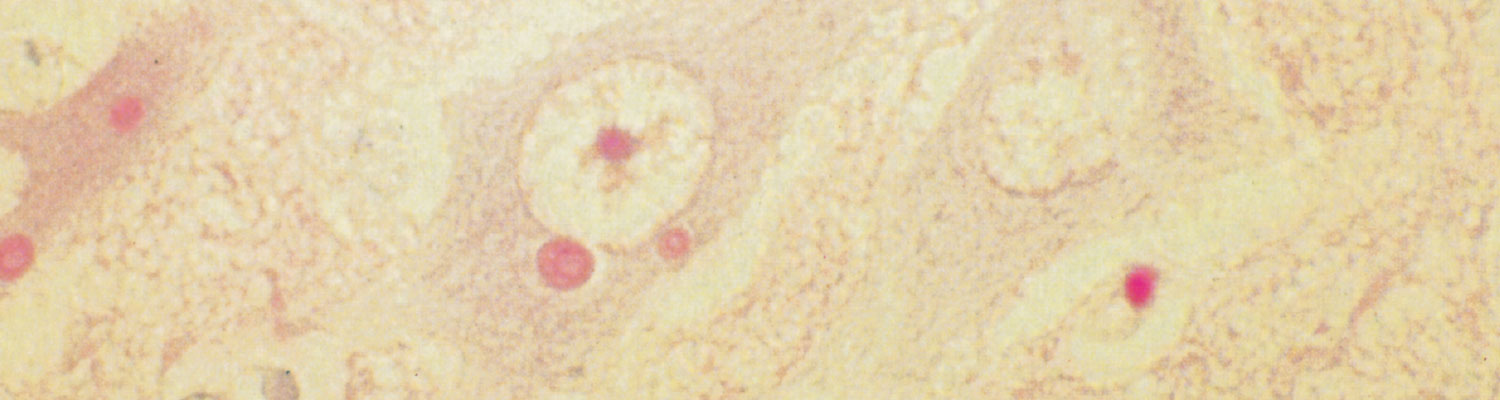
Rabies occurred widely in Europe, Asia and Africa throughout recorded history, but did not had a significant impact on human and livestock populations similar to diseases such as bubonic plague, smallpox, contagious bovine pleuropneumonia or rinderpest. Descriptions of the disease can be traced back further in early Chinese, Egyptian, Greek and Roman records than descriptions of any other infectious disease.252, 301 Controversy raged for centuries as to whether the disease arose spontaneously, or was caused by an agent transmitted by a bite, and it was not until 1804 that Zinke309 published a description of the experimental transmission of the disease to dogs and cats by brushing saliva from a rabid dog into wounds. Galtier102 described the transmission of the disease to a laboratory rabbit, and Pasteur soon thereafter established the association of the causative agent of rabies with nerve tissue. He demonstrated by serial intracerebral passage of infected nerve tissue in laboratory animals that wild or ‘street virus’ could be transformed into ‘fixed virus’ with a shortened and reproducible incubation period.205 In a logical extension of their work, Pasteur and his associates reasoned that vaccine could be administered to humans after exposure to rabies virus to induce immunity before the infection became established in the victim. They 'attenuated’ the virus by desiccating strips of infected rabbit spinal cord over potassium hydroxide and administered suspensions of increasing ‘virulence’ to patients, starting with material dried for 14 days and ending with material dried for two days. The technique was first applied in 1885 on a nine-year-old boy, who survived, and within a short period the technique found widespread application and had a lasting impact on rabies immunization practices.203 Despite the development of a vaccine, the true nature of the infectious agent remained obscure and it was not until 1903 that Remlinger213 demonstrated that it passed through filters that retained bacteria, and thus conformed to the newly defined group of agents known as viruses. In the same year, Negri195 described the occurrence of cytoplasmic inclusions in infected nerve cells.
Aetiology
Viruses in the order Mononegavirales, family Rhabdoviridae (rhabdos, Gr. = rod), genus Lyssavirus cause rabies. The genus was initially divided into serotypes according to antigenic cross-reactivity and later genotypes based on genetic characterization. It is now delineated according to viral species based on several criteria including genetic distance, antigenic patterns in reaction with anti-nucleocapsid monoclonal antibodies and, where available, information on ecological properties such as host and geographic range. Currently, 16 lyssavirus species are recognized by the International Committee on Taxonomy of Viruses (ICTV); comprising rabies lyssavirus, the type species and the other 15 species are referred to as rabies- related lyssaviruses3, 261 (Table 1). Two additional bat- associated viruses, Taiwan bat lyssavirus from Taiwan118 and Kotalahti bat lyssavirus from Finland201 are not yet officially classified. Lyssavirus species are also divided into phylogroups I, II and III (Table 1) based on immunological and pathological characteristics.
Table 1 Lyssavirus species occurring worldwide
Phylogroup | Lyssavirus species | Geographical distribution | Reservoir/Host species | Spill-over infections reported in other animals | Human deaths |
I | Rabies lyssavirus | Worldwide (except for designated rabies-free countries) | Terrestrial carnivores and bats (bats only in the Americas) | Yes (can infect all warm blooded mammals including domestic, livestock and wildlife species) | Yes (> 59 000) p.a |
I | Aravan lyssavirus | Kyrgyzstan | Lesser mouse-eared bat (Myotis blythii) | None | None |
I | Australian bat lyssavirus | Australia | Flying fox species (Pteropus spp.) |
To see the full item, register today: