- Infectious Diseases of Livestock
- Part 2
- Suid herpesvirus 2 infection
- GENERAL INTRODUCTION: PARAMYXOVIRIDAE AND PNEUMOVIRIDAE
- Rinderpest
- Peste des petits ruminants
- Parainfluenza type 3 infection
- Bovine respiratory syncytial virus infection
- Hendra virus infection
- Paramyxovirus-induced reproductive failure and congenital defects in pigs
- Nipah virus disease
- GENERAL INTRODUCTION: CALICIVIRIDAE AND ASTROVIRIDAE
- Vesicular exanthema
- Enteric caliciviruses of pigs and cattle
- GENERAL INTRODUCTION: RETROVIRIDAE
- Enzootic bovine leukosis
- Jaagsiekte
- Visna-maedi
- Caprine arthritis-encephalitis
- Equine infectious anaemia
- GENERAL INTRODUCTION: PAPILLOMAVIRIDAE
- Papillomavirus infection of ruminants
- Papillomavirus infection of equids
- GENERAL INTRODUCTION: ORTHOMYXOVIRIDAE
- Equine influenza
- Swine influenza
- GENERAL INTRODUCTION: CORONAVIRIDAE
- Porcine transmissible gastroenteritis
- Porcine respiratory coronavirus infection
- Porcine epidemic diarrhoea
- Porcine haemagglutinating encephalomyelitis virus infection
- Porcine deltacoronavirus infection
- Bovine coronavirus infection
- Ovine coronavirus infection
- Equine coronavirus infection
- GENERAL INTRODUCTION: PARVOVIRIDAE
- Porcine parvovirus infection
- Bovine parvovirus infection
- GENERAL INTRODUCTION: ADENOVIRIDAE
- Adenovirus infections
- GENERAL INTRODUCTION: HERPESVIRIDAE
- Equid herpesvirus 1 and equid herpesvirus 4 infections
- Equid gammaherpesvirus 2 and equid gammaherpesvirus 5 infections
- Equine coital exanthema
- Infectious bovine rhinotracheitis/infectious pustular vulvovaginitis and infectious pustular balanoposthitis
- Bovine alphaherpesvirus 2 infections
- Malignant catarrhal fever
- Pseudorabies
- Suid herpesvirus 2 infection
- GENERAL INTRODUCTION: ARTERIVIRIDAE
- Equine viral arteritis
- Porcine reproductive and respiratory syndrome
- GENERAL INTRODUCTION: FLAVIVIRIDAE
- Bovine viral diarrhoea and mucosal disease
- Border disease
- Hog cholera
- Wesselsbron disease
- Louping ill
- West nile virus infection
- GENERAL INTRODUCTION: TOGAVIRIDAE
- Equine encephalitides caused by alphaviruses in the Western Hemisphere
- Old World alphavirus infections in animals
- Getah virus infection
- GENERAL INTRODUCTION: BUNYAVIRIDAE
- Diseases caused by Akabane and related Simbu-group viruses
- Rift Valley fever
- Nairobi sheep disease
- Crimean-Congo haemorrhagic fever
- GENERAL INTRODUCTION: ASFARVIRIDAE
- African swine fever
- GENERAL INTRODUCTION: RHABDOVIRIDAE
- Rabies
- Bovine ephemeral fever
- Vesicular stomatitis and other vesiculovirus infections
- GENERAL INTRODUCTION: REOVIRIDAE
- Bluetongue
- Ibaraki disease in cattle
- Epizootic haemorrhagic disease
- African horse sickness
- Equine encephalosis
- Palyam serogroup orbivirus infections
- Rotavirus infections
- GENERAL INTRODUCTION: POXVIRIDAE
- Lumpy skin disease
- Sheeppox and goatpox
- Orf
- Ulcerative dermatosis
- Bovine papular stomatitis
- Pseudocowpox
- Swinepox
- Cowpox
- Horsepox
- Camelpox
- Buffalopox
- GENERAL INTRODUCTION: PICORNAVIRIDAE
- Teschen, Talfan and reproductive diseases caused by porcine enteroviruses
- Encephalomyocarditis virus infection
- Swine vesicular disease
- Equine picornavirus infection
- Bovine rhinovirus infection
- Foot-and-mouth disease
- GENERAL INTRODUCTION: BORNAVIRIDAE
- Borna disease
- GENERAL INTRODUCTION: CIRCOVIRIDAE AND ANELLOVIRIDAE
- Post-weaning multi-systemic wasting syndrome in swine
- GENERAL INTRODUCTION: PRION DISEASES
- Scrapie
- Bovine spongiform encephalopathy
- Transmissible spongiform encephalopathies related to bovine spongiform encephalopathy in other domestic and captive wild species
Suid herpesvirus 2 infection
This content is distributed under the following licence: Attribution-NonCommercial CC BY-NC  View Creative Commons Licence details here
Suid herpesvirus 2 infection
N EDINGTON
Introduction
Suid herpesvirus 2 (SHV-2), also called porcine cytomegalovirus (PCMV), is an ubiquitous virus that may cause foetal or neonatal deaths and has been associated with runting, rhinitis and pneumonia in piglets less than three weeks of age, or rhinitis in growing pigs. With the advent of xenotransplantation there is renewed interest in this virus.
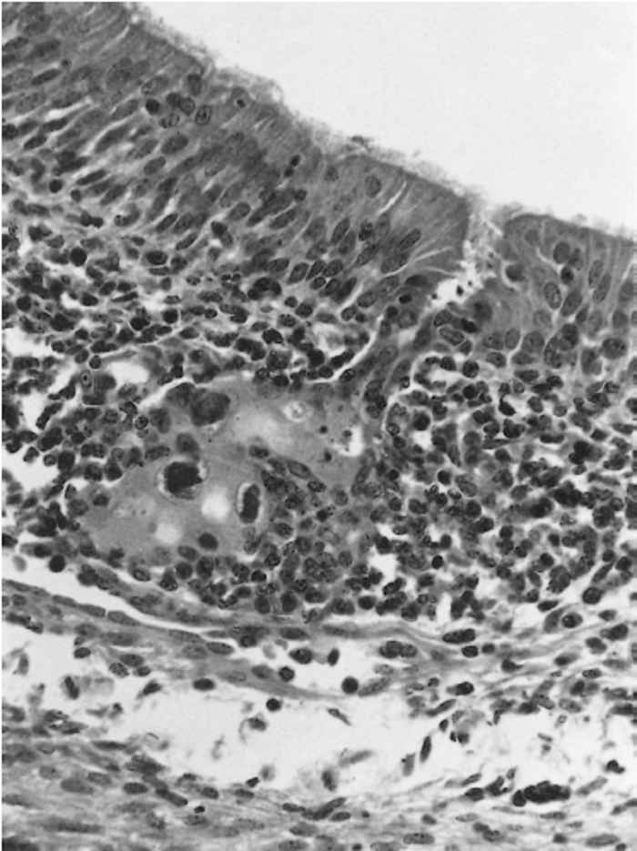
Suid herpesvirus 2 was first identified by Done6 as a cause of cytomegalic cells with characteristic basophilic intranuclear inclusion bodies in the mucosal glands of turbinates of pigs. Restriction of viral replication to pigs, localization of the virus in the nasal mucosa, lachrymal and salivary glands, and occasionally in renal tubular epithelium, as well as ultrastructural studies,8, 28 indicated that this was a cytomegalovirus of the pig similar to those described for some other animal species.20, 22, 30 These viruses usually cause clinically silent infections in healthy adults but often induce severe, generalized infections in young animals. Virus may also cross the placenta. Like other herpesviruses, SHV-2 induces latent infection. A cell-associated viraemia can occur in the presence of circulating antibody.
In herds where good management is applied, and in which the virus is endemic, there may be no clinical signs or economic loss.
Suid herpesvirus 2 is likely to be present in countries whose domestic pig populations are of European or Asian origin.
Aetiology
Suid herpesvirus 2 is a beta herpesvirus, with an 80 to 100 nm icosahedral capsid surrounded by an envelope, making the complete virion 120 to 150 nm in diameter. The 45 to 70 nm core is usually elongated and may be oval, rectangular or dumb-bell shaped.8, 28 So far only the sequences of the DNA polymerase gene have been published,31 supporting its classification as a beta virus. The virus is sensitive to chloroform and ether.2
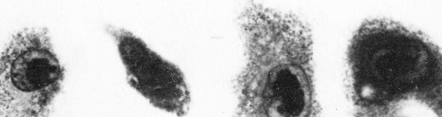
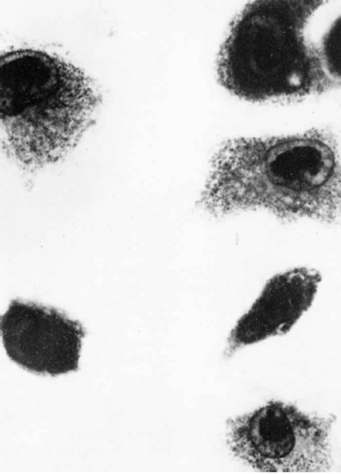
Watt29 found pulmonary macrophage cultures derived from three- to five-week-old piglets to be the most sensitive system for both primary isolation and propagation of virus. Cytomegaly and intranuclear inclusions develop 11 to 14 days after inoculation (Figure 83.1) and infectivity titres of up to 105,5 TCID50 may occur. This culture system has limitations because of the fastidiousness and variability of the cells, and because of residual adventitious viruses. Primary pig lung cells16 and a cell line derived from porcine Fallopian tube 3 also support viral replication, while it has been reported that 12-O-tetradecanoylphorbol 13-acetate accelerates viral replication in this system.15 Suid herpesvirus 2 appears to be host-specific both in vivo and in vitro.
In cultures, infected cells enlarge by up to six times and their mitochondria, endoplasmic reticulum, and Golgi apparatus swell.8 The large, intranuclear inclusions contain the nuclear capsids, often in crystalline array.27 The capsids acquire an electron-dense coat in the nucleus, the envelope derives from the inner nuclear membrane, and the virions come to lie free inside intracytoplasmic cisternae.
Epidemiology
Suid herpesvirus 2 has been detected wherever it has been sought. In the UK a serological survey indicated that over 95 per cent of herds had been exposed to the virus.19 The ubiquity of the infection and the possibility of transplacental infection indicate that even hysterotomy-derived littersmaybe infected.
In infected neonatal piglets the virus is shed predominantly in nasal secretions, but also in urine and ocular secretions. 11, 19 Levels of passive antibody fall initially during the period of virus excretion, but rise when the piglets are between 8 and 11 weeks old as a result of active production. They continue to increase until at least 23 weeks after infection. Susceptible sows show a similar pattern of excretion but, in addition, shedding of virus from the cervix occurs in some pregnant sows.
Recovery of virus from lung macrophages of apparently healthy pigs suggests that the virus either persists at this site or that it may establish a true latent infection. Infection may be reactivated after administration of corticosteroids.11
Pathogenesis
The mucous glands of the nasal mucosa are the primary site of viral replication. Thereafter a cell-associated viraemia develops which is detectable 14 to 16 days after infection in pigs older than three weeks, and from 5 to 19 days after infection in neonatal piglets.11
In animals older than three weeks, the virus occurs most commonly in the glands of the nasal mucosa but it may also be present in the Harderian and lachrymal glands and in the renal tubules. On rare occasions it is found in the mucous glands of the oesophagus and duodenal epithelium.
In foetuses or neonates, the virus has a predilection for reticuloendothelial cells. Inclusions occur in capillary endothelium, the sinus and sinusoidal cells of lymphoid...
To see the full item, register today:


