Helminths of poultry and ostriches
This content is distributed under the following licence: Attribution-NoDerivs CC BY-ND  View Creative Commons Licence details here

Helminths of poultry and ostriches
Author: C NKUNA
Introduction
Worm infections are a common problem in indigenous chickens, free-range commercial layers, breeders, and other types of poultry that have direct contact with their faeces and the intermediate hosts of helminths. Worms are usually not a problem in broilers due to their very short lifespan which is too short for the worms to complete their life cycles. In addition, end-of-cycle cleanouts of broiler houses also ensure that any worms in the litter are cleaned out.
In a study conducted by Mukaratirwa et al. (2001) on free-range chickens in KwaZulu-Natal, 16 helminth species were found, 12 nematodes and 4 cestodes. This supports previous studies that demonstrated that nematodes were dominant in the free-range birds in Zimbabwe and Ghana. Molla et al. (2012) found that in local backyard chickens on Ethiopia ’s North Gondar –although there were more species of cestodes isolated, the infection burden of the nematodes was higher than that of cestodes (60.38% compared with 54.62%).
Commercially farmed ostriches are generally kept on pasture for very long periods since they are grazers. They are therefore exposed to both nematodes and cestodes. Worm infestation can severely affect health and viability –especially of young ostriches.
Nematodes
Nematodes are the commonest and most important helminth group in poultry. More than 50 species have been described in poultry. Of these, most cause pathological damage to the host. The nematodes of poultry –unlike those of mammalian livestock –often have intermediate hosts, which is probably an adaptation to their foraging lifestyle and omnivorous feeding habits.
Crop and oesophagus
Capillary or Threadworms
Capillaria species
The Capillaria spp. are small roundworms and are found in many mammals –but are most important in poultry. Various Capillaria species are found in different organs in the birds. Species of importance in poultry are: C. annulata, C. anatis, C. obsignata, C. caudinflata, C. aerophilia and C. contorta. C. annulata and C. contorta are found in the crop and oesophagus.
Morphology
Capillaria species are very small and hairlike worms that are very difficult to see in the stomach contents. Size ranges from as small as 6 mm through to 80 mm. C. annulata females are the largest at 37- 80 mm long, with the males being 15-25 mm long. The eggs have bipolar plugs and measure 60 x 25 μm. C. contorta males are the same length as C. annulata males, while the females are shorter –measuring only 27-38 mm. The C. contorta eggs are equal in size to the C. annulata eggs –at 60 x 25 μm.
Life cycle
C. annulata and C. contorta have indirect life cycles –with earthworms being the intermediate hosts. Unembryonated eggs are shed in the faeces and are ingested by the earthworms, where they develop into the first larval stage in 9 to 14 days. The host then ingests the earthworm and becomes infected. C. contorta can also have a direct life cycle, with the L1 embryonated eggs as the infective stage. This means that poultry kept in houses –away from the intermediate host –can still be infected.
Clinical signs and pathogenicity
Capillaria spp. can be highly pathogenic for birds kept in deep-litter and freerange systems –both commercial and indigenous –where a build-up of infective eggs in litter or soil can occur. Young birds are more susceptible than older birds. Adult worms burrow into the anterior end of the oesophageal mucosa –causing inflammation. Heavy burdens can result in inflammation and thickening of the oesophageal and crop mucosal walls, which can cause death. C. contorta infections can result in severe anaemia, which can be fatal. Less severe burdens can cause loss of weight and condition, which in turn leads to a loss of production.
Zigzag worm
Gongylonema ingluvicola
G. ingluvicola is a roundworm with a predilection for the crop, oesophagus, and rarely the proventriculus, of chickens, turkeys, pheasants and quails.
Morphology
The female worm is 32-55 mm long and the males are 17-20 mm long. The eggs are approximately 35 x 58 μm. The anterior part of the body has numerous characteristic round or oval thickenings –called cuticular plaques –on the cuticle.
Life cycle
The life cycle is indirect and includes the beetle Copris minutus and the cockroach Blatella germanica as intermediate hosts. The eggs are shed in the faeces and are eaten by beetles and cockroaches. The worm develops in the intermediate host into infective larvae over 30 days. Birds become infected after eating the intermediate hosts that contain the infective larvae L3.
Clinical signs and pathogenicity
Adult parasites may cause inflammation and hypertrophy –with cornification of the epithelium in chronic infections. The pathogenicity is dependent on the burden of infection.
Proventriculus and gizzard
Tetrameres species
T. americana and T. fissispina are found in various bird species in Africa. The adult worms are found in the proventriculus –detriculus and the walls of the gizzard.
info missing??
Clinical signs and pathogenicity
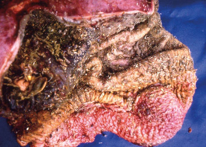
Infected birds may be emaciated and sluggish or weak. The worm infection weakens the immunity of the birds –making then susceptible to other infections. Adult D. nasuta attach to the wall of the proventriculus, causing ulcerations at the attachment sites. When the worm burdens are high, a proliferative proventriculitis with necrosis and sloughing of the mucosa is evident. The inside of the proventriculus is then filled with thick, white, slimy mucous and sloughed gastric tissue. There is also thickening of the tissues below the lining of the proventriculus. The lumen of the proventriculus can become completely blocked –thereby preventing the passage of food. Some birds may die from starvation in very severe cases. Adult worms may be found beneath and in the proliferating tissue. The proventriculus may become enlarged and flaccid due to the destruction of the glandular tissue and muscle layer. In some severe cases, it may be perforated –resulting in peritonitis.
Wireworm or stomach worm of ostriches
Libyostrongylus douglassi
L. douglassi is a parasite of the digestive system causing libyostrongylosis (rotten stomach/vrotmaag ). It is the most economically significant gastro-intestinal parasite in ostriches. The wireworm is a nematode in the family Trichostrongylidae (Nemejc et al ., 2012). In South Africa, it has been reported to cause as much as 50% mortality in juvenile ostriches (Reinecke, 1983).
Morphology
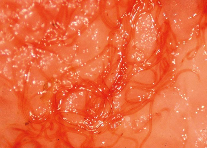
Wireworms are very small, round, wirelike, yellowish-red worms. They are about 3 mm long (males 4-6 mm and females 5-6 mm). The mature worms and late larval stages live in the crypts of the glandular portion of the proventriculus and gizzard wall ( Figure 83 ).
Life cycle
Eggs shed in the faeces of the infected host hatch and develop from first to second and finally to the third larval stage. This development takes place at temperatures ranging from 7-10 °C to a maximum of 37 °C (McKenna, 2005). Infective larvae tend to climb to the tips of blades of grass in films of moisture (Anderson, 1992; McKenna, 2005). Infection of susceptible hosts is by ingestion of third-stage larvae. The fourth-stage larvae develop in the proventriculus some 4 to 5 days later. The development of fifth-stage larvae and the production of eggs occur at about 20 days and 33 days post infection, respectively. The eggs will be passed from the proventriculus and appear in the faeces after 3 to 4 days (McKenna, 2005).
Clinical signs and pathogenicity
The different stages have predilection for different sites within the gut. The immatures penetrate deep into the glands of the proventriculus and adults are found on the surface of the proventriculus, where they cause damage by sucking blood –resulting in severe inflammation (McKenna, 2005) ( Figure 84 ). This causes proventriculitis which can lead to impaction of the organ (McKenna, 2005). Clinical signs in young birds are wasting, anorexia, anaemia and death. Mature birds can sustain infections with no visible clinical signs (McKenna, 2005).
Control
Young ostriches raised in commercial systems should be raised in pens which have the floors regularly cleaned. Regular faecal monitoring should be done to check for worms. Infections can be treated with injections of ivermectin or dosed with levamisole or fenbendazole. Young ostriches should be fed lucerne from pastures which have been ostrich free for three years.
Acuaria hamulosa
A. hamulosa is found in chickens and turkeys worldwide. The adults have an affinity for the area below the keratinised layer of the gizzard –where they embed themselves.
Morphology
The males are 10-14 mm long –while the females are longer at 16-29 mm. The worms have four cuticular cordons, which are irregular and wavy –extending twothirds the length down the body. The males have long and slender spicules on the left side measuring 1.63-1.8 mm, while on the right side they are only 0.23-0.25 mm long.
Life cycle
The worms have an indirect life cycle –with grasshoppers, beetles, sandhoppers and weevils as possible intermediate hosts. The eggs are found in the faeces and are ingested by the birds when they eat the intermediate hosts. The eggs develop into the infective stage inside the intermediate hosts over 21 days.
Clinical signs and pathogenicity
The clinical signs seen in birds with heavy infections include droopiness, weakness, anaemia and emaciation. In severe cases the gizzard may rupture. The worms are found in soft, yellow-red nodules. The keratinised layer of the gizzard may be destroyed or can even die off in severe infections.
Small Intestine
Ascarid worms
Ascaridia galli, A. dissimilis ,
A. columbae
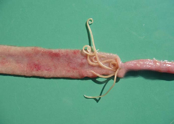
Ascaridia spp. are large, round white worms which infect various avian species. Of the many species, those important in domestic birds are: A. galli in fowl, A. dissimilis in turkeys, and A. columbae in pigeons. They are found mainly in the intestines (jejunum and duodenum) where they can cause obstruction, when in large numbers. The adult worms can migrate through the large intestine and cloaca to the oviduct –where they can be found in the eggs.
Morphology
These are large worms; the females can be as long as 72-116 mm, while the males are 51-76 mm long ( Figure 85 ). The mouthpart has three prominent lips. The male has a pre-anal sucker and two equal spicules which are 1.0-2.4 mm long. The females have an opening in the middle of the body. The eggs are oval, with smooth shells, and are 73-92 x 45-57 μm in size.
Life cycle
The ascarids have a direct life cycle. Sexually mature worms live in the lumen of the small intestine, and infective-stage larvae (L3) develop in the environment (Permin, 1997; Tarbiat, 2012). The route of infection is oral –usually by direct ingestion of the embryonated egg –and there is a 5-10 week prepatent period, which is shorter in young birds. The larvae moult in the eggs until L3. The development process from fertile eggs to fully mature infective larvae (L3) takes 7 to 20 days –depending on environmental conditions (Permin et al ., 1998; Tarbiat, 2012).
Clinical signs and pathogenicity
Penetration of the parasite into the duodenal or jejunal mucosa may cause haemorrhagic enteritis. In some cases the larvae can cause destruction of the glandular epithelium and adhesions of the mucosal villi (Permin, 1997; Tarbiat 2012). Ascarids can cause a variety of symptoms, including anorexia, diarrhoea, dehydration, stunted growth, unthriftiness, drooping wings, ruffled feathers, weight loss, and misshapen eggs with soft and thin shells. Clinical signs are more pronounced in chickens up to 3 months of age, after which the worm burden normally decreases, but can still be very high.
Harteria gallinarum
H. gallinarum closely resembles Ascaridia galli . It is found in the small intestine of chickens in West and South Africa –as well as Asia.
Morphology
The females are 60-100 mm long, while the males are only 28-40 mm long. Embryonated eggs are shed in the faeces. They have thick shells and measure 45- 53 by 27-33 μm.
Life cycle
The parasite has an indirect life cycle –with termites as intermediate hosts.
Clinical signs and pathogenicity
Severe infections cause diarrhoea, weight loss, and decreased egg production. The birds become emaciated and very weak.
Caeca and large intestine
Heterakis species
Heterakis are among the most important roundworms in poultry. They are seen in chickens, turkeys, ducks, geese, guinea fowls and pheasants. There are three species that are believed to be important: H. gallinarum, H. isolonche and H. dispar.
All three species are found in the lumen of the caeca.
Morphology
Heterakis spp. have typical roundworm morphology –with features such as a cuticle, an oesophagus ending in a valved bulb, and three papillae-lined lips and alae. The alae, which run almost the entire length of the body, are ridges formed by the thickening of the cuticle –that may act as receptors for molecules which stimulate reproduction. Adult female and male caecal worms differ in length, with the female (10-15 mm) generally being larger than the male (7-13 mm). Both sexes have a pointed tail, and males having a precloacal sucker at the posterior end. The eggs of H. gallinarum are approximately 65-77 x 35-48 μm –with visibly thick, smooth shells (Carron, 2012). The eggs are thick, smooth-shelled and very similar to those of A. galli .
Life cycle
The eggs develop to the infective stage in 12 to 14 days at 22 °C and can remain infective for 4 years in the soil. Infection occurs when eggs are eaten by the host. Earthworms and houseflies can act as transport or paratenic hosts –in which the worm can survive for a number of years. The second stage juveniles hatch in the gizzard or duodenum and pass down to the caeca. Most complete their development in the lumen, but some penetrate the mucosa where they remain for 2 to 5 days without further development. Returning to the lumen, they mature about 14 days after infection. If eaten by an earthworm, the juvenile may hatch and become dormant in the earthworm ’s tissues, remaining infective to chickens for at least a year.
Clinical signs and pathogenicity
The damage is evident on the cloacal mucosa which becomes inflamed and thickened, with petechial haemorrhages. The clinical signs are not readily visible. However, infections with H. isolonche can produce nodular typhilitis, diarrhoea, emaciation and death. The most important feature of H. gallinurum is that it can transmit the protozoon Histomonas meleagridis to fowls. The H. meleagridis parasite can remain viable in H. gallinarum eggs for many years.
Codiostomum struthionis
C. struthionis is a roundworm found in the large intestine and distal third of the ostrich caecum.
Morphology
The parasite is about 1.0-1.5 cm long and white in colour. The infective larva of C. struthionis has a tapered tail-end –otherwise its morphology is very similar to that of Libyostrongylus dentatus (Fagundes et al ., 2012).
Life cycle
The life cycle of C. struthionis has not yet been determined –but it is believed to be simple and direct (Fagundes, 2012).
Clinical signs and pathogenicity
Heavy infestations can be pathogenic. C. struthionis causes lesions in the caecum of ostriches; the severity of the lesions depends on the worm burden (Fagundes et al ., 2012).
Eyes and nose
Oxyspirura mansoni (Cobbold, 1879; Permin, 1998)
O. mansoni infections occur in chickens, turkeys, guinea fowl and peafowl in tropical and subtropical areas. The parasite is located under the nictitating membrane and in the naso-lacrimal ducts or conjunctival sacs.
Morphology
The female worm is 12-19 mm long and the male reaches 10-16 mm in length. The worm is slender and the cuticle smooth. The pharynx is shaped like an hourglass. The male tail is curved ventrally and has no alae. The two spicules are uneven in size; the left is slender, 3-3.5 mm long and the right one is stout and 0.2-0.22 mm long. The vulva is to the posterior end of the female worm.
Life cycle
The life cycle is indirect. After the eggs have passed through the lacrimal duct, are swallowed and passed out with the faeces, the intermediate stages develop in cockroaches (Pycnoscelus surinamensis) . After ingestion of the intermediate host, the larvae migrate via the oesophagus, pharynx and lacrimal duct to the eye.
Clinical signs and pathogenicity
The eyes become irritated and the birds start to scratch them. After a while, the affected birds develop ophthalmitis with inflamed and watery eyes.
Respiratory system
Gapeworm
Syngamus trachea
(syn. S. parvis, S. gracilis)
S. trachea is found in chickens, turkeys, pheasants, guinea fowl, geese and various wild birds throughout the world. The adult worms are found in the trachea or in the lungs.
Morphology
The worms are red in colour and the two sexes are found in permanent copulation. The female is bigger than the male –measuring 5-20 mm –while the male is 2-6 mm long. S. trachea has a wide mouth opening, without leaf-crowns. The buccal capsule is cup-shaped with 6 to 10 teeth at the base. The males have two spicules which measure 53- 82 μm. The eggs have a thick operculum in both poles and they measure 70-100 x 43-46 μm.
Life cycle
The life cycle may be direct or indirect: some intermediate hosts are earthworms, snails, flies or other arthropods. When these hosts are swallowed by poultry the larvae migrate through the intestinal wall and are carried by the blood to the lungs. Here they develop into the adult stage. The prepatent period is three weeks. Eggs are coughed up and swallowed and passed with the faeces. Depending on the temperature and humidity, the eggs become infective in 2 to 7 days. Infections with S. trachea mainly affect young birds –except for turkeys which are affected at any age. Pheasants and other reared game birds are highly susceptible.
Clinical signs and pathogenicity
The characteristic signs of S. trachea infections are dyspnoea due to mucous accumulating in the trachea (gaping). Asphyxia and death follow when the mucus blocks the trachea. Emaciation, anaemia and weakness are also seen as clinical signs. On post-mortem examination the carcass is emaciated and anaemic and the adult worm is seen macroscopically when opening the trachea.
Diagnosis and control of nematodes of poultry
In commercial systems –for example free-range layers or breeders –faecal monitoring for nematode eggs may be necessary, especially if clinical signs are present. Careful post-mortem examinations of any birds dying should be done as a routine.
Birds with clinical signs can be treated with nematocides registered for use in poultry –such as ivermectin (injectable), levamisole, piperazine or one of the benzimidazoles –by dosing, or in feed or water.
Regular rotation of camps will help reduce the level of infection.
Cestodes
Poultry reared under free-range conditions are likely to be infected with tapeworms. All tapeworms of poultry have indirect life cycles, with intermediate hosts like earthworms, beetles, flies, ants or grasshoppers. The intermediate hosts are essential to perpetuate the life cycle –and infections are therefore rare in indoor systems. More than 1 400 tapeworm species have been described in domesticated poultry and wild birds. The pathogenicity of most of these species is unknown. A great number are harmless or have a mild pathogenicity, but a few species cause severe reactions in the host. Poultry tapeworms are mainly intestinal and most are small –but some may reach 30-50 cm in length.
Small intestine
Broad-Headed and nodular Tapeworms
Raillietina spp.
There are three species of importance in poultry. These are: R. cesticillus (Skrijabinia cesticillus) (broad-headed tape worm), R. echinobothrida (nodular tapeworm), and R. tetragona . These tapeworms have chickens, turkey, geese and other wild and domestic birds as their final hosts (Junquera, 2013).
Morphology
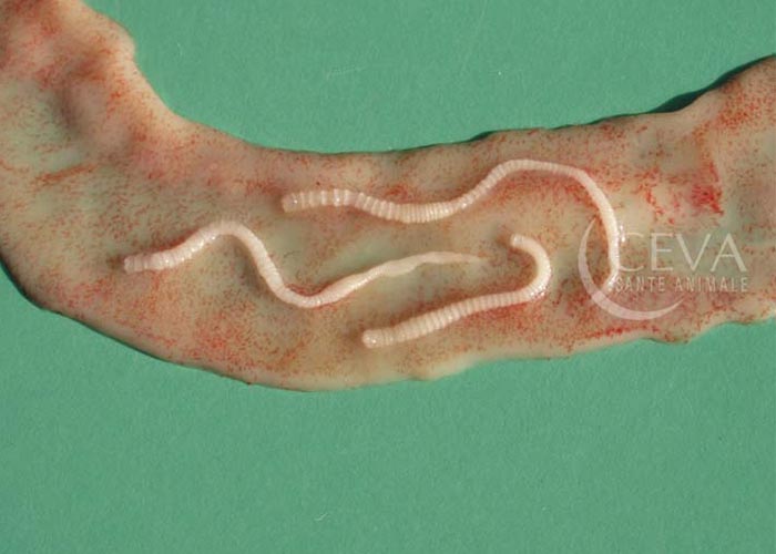
Raillietina adults are whitish, mediumsized tapeworms measuring 5-30 cm long and 1-4 mm wide. The head –termed the scolex –has hooks, spines and four suckers to attach to the wall of the host. The segments of the worm have both male and female organs. The segments also have excretory cells known as flame cells. The eggs are 74 x 93 μm and the highest number is found in the gravid proglottid of R. tetragona .
Life cycle
The gravid proglottids are shed with the faeces, and the eggs can survive for years outside the host. The intermediate hosts are beetles for R. cesticillus , ants for R. echinobothrida , and ants and houseflies for R. tetragona (Junquera, 2013). The intermediate hosts get infected when they ingest individual eggs, and the larvae hatch in the intestine of the intermediate host. The larvae then develop further into cysticercoids in the intestine of the intermediate host –until ingested by the final host. The cysticercoids are activated by the bile and release the young tapeworms that attach to the mucosa in the small intestine ( Figure 86 ). The prepatent period lasts 2 to 3 weeks.
Clinical signs and pathogenicity
The worms affect the weight gain and egg production of various poultry species. R. echinobothrida can cause the appearance of large nodules in the gut –a phenomenon called ‘nodular tapeworm disease ’.
Heavy, chronic infections may cause diarrhoea, anaemia, weight loss and intestinal inflammation and haemorrhage. Young birds and free-range birds are commonly affected (Junquera, 2013).
Choanotaenia infundibulum
C. infundibulum is a tapeworm that affects domestic fowls and turkeys. The worms have a predilection for the mucosa of the upper end of the small intestine.
Morphology
The worms are 56 mm long and 1.8 mm wide in the region of mature proglottids. The scolex is 0.456 mm in diameter, and 0.373 mm to 0.581 mm in length. The neck is 0.47-0.52 mm long (Sawada, 1970). The segments are wider at the posterior end of the parasite. The eggs have a distinctly long filament and measure 47 x 54 μm.
Life cycle
The eggs are excreted with the faeces and are swallowed by the intermediate hosts. Houseflies and beetles are natural hosts. Experimental hosts include beetles, grasshoppers and termites. The gravid proglottids are released into the intestines of the fowls after swallowing the intermediate host.
Clinical signs and pathogenicity
Adult worms cause weight loss.
Minute tapeworm
Davainea proglottina
This very small tapeworm is found in chickens, turkey, guinea fowl, and other domestic and wild birds. They have a predilection for the mucosa of the duodenum. The worms are found worldwide –and most commonly in free-range poultry (Junquera, 2013).
Morphology
The adult worms are 0.5-3 mm long and have 4 to 9 proglottids. The eggs are 28- 40 nm in size. The scolex has numerous hammer-shaped hooks, and also suckers armed with spines and numerous hooks. Only the last segment is gravid. Each segment has both male and female reproductive organs (Junquera, 2013).
Life cycle
The gravid proglottids are shed with the faeces. The eggs hatch after being swallowed by various species of gastropod molluscs –such as species of Limax, Cepaea, Agriolimax and Arion . Cystecercoids develop after 3 weeks and develop into adult tapeworms 2 weeks after ingestion by the final hosts.
Clinical signs and pathogenicity
D. proglottina is one of the most pathogenic tapeworms. Sudden massive infections can cause haemorrhagic enteritis and intestinal necrosis that can be fatal for young birds. Clinical signs include dull plumage, slow movements, reduced weight gain, emaciation, difficulty in breathing, leg paralysis, and death. Histology shows mucosal thickening with haemorrhages, and necrosis.
Amoebataenia sphenoides
(A. cuneata)
This is a tapeworm of chickens and other domesticated and wild birds. It has predilection for the small intestine.
Morphology
A. spheniodes are very small –no longer that 4 cm in length. The head has suckers and 12 to 14 hooks and there are no more than 25 segments. The segments have both male and female reproductive organs. The embryonated eggs are almost spherical, and about 35 μm in diameter (Junquera, 2013).
Life cycle
A spheniodes has an indirect life cycle –with earthworms as the intermediate hosts. The gravid segments are passed with the faeces and earthworms ingest them. The eggs develop to cysticercoids in the earthworm ’s body cavity. The birds become infected by ingesting the infected earthworms. After ingestion, the earthworms release young tapeworms that attach in the gut of the bird. The time between infection and shedding of the first eggs is 4 weeks.
Clinical signs and pathogenicity
High worm burdens can cause haemorrhagic enteritis. Severely affected birds can be apathetic and tend to isolate themselves (Junquera, 2013).
Ostrich tapeworm
Houttuynia struthionis
H. struthionis is a cestode occurring in the intestines of ostriches in Africa.
Morphology
The tapeworms are long, large, flat, segmented, and about 50-100 cm long. The scolex has a retractile rostellum bearing two rows of large hammer-shaped hooks, and is equipped with four unarmed suckers. The proglottids are wider than long.
Life Cycle
Unknown.
Clinical signs and pathogenicity
Ostrich chicks are most susceptible and show gradual loss of condition, lethargy, anaemia, loss of appetite, and sometimes mild diarrhoea (Nemejc, 2012).
Hymenolepis species
There are three pathogenic and economically important Hymenolepis species that affect poultry. These are H. carioca and H. contaniana which are seen in fowls in most parts of the world, and Drepanidotaenia lanceolata which is seen in geese and ducks.
Morphology
H. carioca is 20-100 mm long. The neck is 75-150 μm wide and the posterior end is 0.4-0.8 mm wide (Guberlet, 1919). The suckers are armed with hooks. The segments have both male and female reproductive organs. H. contaniana is smaller and may reach a length of 20 mm. The adults of D. lanceolata may reach 130 mm in length and 18 mm in wide –with segments wider than long.
Life cycle
Hymenolepids have an indirect life cycle –with beetles as intermediate hosts for H. carioca and H. contaniana . Crustaceans are intermediate hosts for D. lanceolata. The prepatent time is 3 to 4 weeks.
Clinical signs and pathogenicity
Heavy infections with thousands of worms may result in catarrhal enteritis, diarrhoea and death. Severe signs have been seen with D. lanceolata in ducks and geese.
Diagnosis and control of cestodes in poultry
- Post-mortem examination of the intestinal tract when mortality is seen.
- Treatment –if necessary –with cestodicides (like niclosamide, resorantel and praziquantel) which are registered for use in birds.
Trematodes
All poultry trematodes belong to the subclass Digenea which utilise an intermediate host. Some species use a second or even a third intermediate host. More than 500 species are known from birds, but only a few are known to be pathogenic. Digenean life cycles vary in complexity and can involve up to four hosts –but two or three is more common. After the hatching of the egg in water (usually) or in the gut of the host after ingestion of the egg (more rare) the miracidium is released and penetrates the tissues of a mollusc –and develops into a mother sporocyst.
Germinal cells in the mother sporocyst give rise to daughter sporocysts or rediae. Germinal cells in the daughter sporocysts or rediae develop into cercariae. The cercariae then leave the snail, may encyst in the open or after penetrating another host –or may not encyst at all. Each cercaria gives rise to one metacercaria which in turn gives rise to one adult after it enters the gut or other appropriate site in the final host (Permin, 1998).
Echinostoma revolutum
The primary hosts for E. revolutum are ducks and geese, but the species may also be found in other water birds, pigeons, fowls, and humans. The worms are located in the rectum and caeca. The parasite can be found in the snail species Lymnaea elodes and in other lymnaeid species.
Morphology
E. revolutum is 10-22 mm long and 2.25 mm wide. Echinostomes have a headcollar armed with spines. The eggs are 90-126 μm x 59-71 μm.
Life cycle
Eggs are shed in the faeces and mature in three weeks –under conditions of high humidity and temperature. The miracidium penetrates the snail, and in the snail the cercariae develop in 2 to 3 weeks. These either encyst or enter into another snail. The birds become infected when they eat infected snails. The prepatent period is 15 to 19 days.
Clinical signs and pathogenicity
Heavy infections result in emaciation and catarrhal diarrhoea. Young animals may die from heavy infections.
Prosthogonimus species
There are three Prosthogonimus species of interest: P. pellucidus, P. macrorchis, and P. ovatus. They are trematode parasites of chickens, ducks, turkey and other domestic and wild birds as final hosts (Junquera, 2013). The adult worms are located in the Bursa of Fabricius, the oviduct, and in the cloaca/rectum (Permin, 1998).
Morphology
Prosthogonimus has a complex, indirect life cycle with two intermediate hosts: a freshwater snail (e.g. Bithynia) as the first intermediate host, and dragonflies as second intermediate hosts. The fluke eggs shed in the faeces of a final host continue development only after getting into water. They are eaten by aquatic snails. Inside the snails ’intestine they develop to miracidia that penetrate the intestine ’s wall and develop into sporocysts. Sporocysts multiply asexually to daughter sporocysts –and to the next larval stage, the cercariae. Mature cercariae leave the snail with its faeces and can swim. In the water, cercariae find a dragonfly larva (= naiad) and penetrate its body through the anus. They encyst in the muscles of the abdominal wall and form metacercariae. About two months later these metacercariae become infective for birds.
Chickens, ducks, geese and other birds become infected by eating contaminated dragonflies –either naiads or adult. Inside the bird ’s gut the metacercariae release the young flukes, which migrate to the Bursa of Fabricius and later on to the oviduct through the cloaca, where they complete development to mature flukes and start producing eggs again. The prepatent period in the birds takes one to several weeks –depending on the fluke and the bird species (Junquera, 2013).
Clinical signs and pathogenicity
Prosthogonimus are the most pathogenic of trematodes that affect fowls and ducks. Clinical signs include a milky discharge from the cloaca. The birds may lay softshelled or shell-less eggs. In chronic cases, peritonitis may occur (Permin, 1998). Heavily affected birds may refuse to feed, become listless and thirsty, walk abnormally, show difficulty in breathing and have a tense and hot abdomen. In some cases, mortalities may occur (Junquera, 2013).


