- Infectious Diseases of Livestock
- Part 1
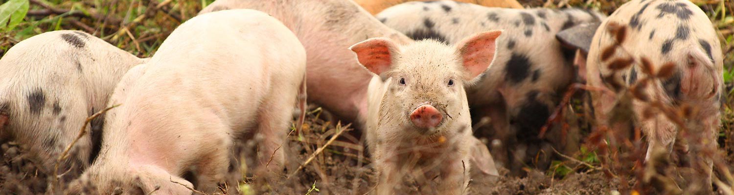
- Porcine babesiosis
- Vectors: Ticks
- Vectors: Tsetse flies
- Vectors: Muscidae
- Vectors: Tabanidae
- Vectors: Culicoides spp.
- Vectors: Mosquitoes
- Classification, epidemiology and control of arthropod-borne viruses
- Special factors affecting the control of livestock diseases in sub-Saharan Africa
- The control of infectious diseases of livestock: Making appropriate decisions in different epidemiological and socioeconomic conditions
- Infectious diseases of animals in sub-Saharan Africa: The wildlife⁄livestock interface
- Vaccination: An approach to the control of infectious diseases
- African animal trypanosomoses
- Dourine
- Trichomonosis
- Amoebic infections
- GENERAL INTRODUCTION: COCCIDIA
- Coccidiosis
- Cryptosporidiosis
- Toxoplasmosis
- Besnoitiosis
- Sarcocystosis
- Balantidiosis
- Leishmaniosis
- Neosporosis
- Equine protozoal myeloencephalitis
- GENERAL INTRODUCTION: BABESIOSES
- Bovine babesiosis
- Equine piroplasmosis
- Porcine babesiosis
- Ovine babesiosis
- GENERAL INTRODUCTION: THEILERIOSES OF CATTLE
- East Coast fever
- Corridor disease
- Zimbabwe theileriosis
- Turning sickness
- Theileria taurotragi infection
- Theileria mutans infection
- Theileria annulata theileriosis
- Theileriosis of sheep and goats
- Theileria buffeli⁄orientalis infection
- Non-pathogenic Theileria species in cattle
- GENERAL INTRODUCTION: RICKETTSIAL, CHLAMYDIAL AND HAEMOTROPIC MYCOPLASMAL DISEASES
- Heartwater
- Lesser known rickettsial infections in animals and humans
- Chlamydiosis
- Q fever
- Eperythrozoonosis
- Bovine Haemobartonellosis
- Potomac horse fever
- GENERAL INTRODUCTION: ANAPLASMOSES
- Bovine anaplasmosis
- Ovine and caprine anaplasmosis
Porcine babesiosis
This content is distributed under the following licence: Attribution-NonCommercial CC BY-NC  View Creative Commons Licence details here
Porcine babesiosis
Previous authors: D T DE WAAL
Current authors:
D T DE WAAL - Associate Professor, BVSc, PhD, DipDatMet, HDipUTL, DipEVPC, MRCVS, School of Veterinary Medicine, University College Dublin, Room 034, UCD Veterinary Sciences Centre, Belfield, Dublin, D04 W6F6, Ireland
Introduction
Porcine babesiosis is a tick-transmitted, protozoal disease caused by either Babesia trautmanni or Babesia perroncitoi and characterized by fever, anaemia, icterus and haemoglobinuria. Babesia perroncitoi does not occur in southern Africa.
The disease was first reported in Russia in 1911 by Dementjew.8 According to Knuth and Du Toit the first reliable description of the disease was given by Trautmann, who studied the condition in Tanzania in 1914.8 The protozoan concerned was later named Babesia trautmanni.8 In 1939, Cerruti identified another protozoan parasite in the red blood cells of pigs in Italy that were exhibiting clinical signs of babesiosis, and he named this parasite Babesia perroncitoi.1 Babesia trautmanni also occurs in pigs in Ghana,17 Sardinia, Zaire10 and Nigeria.3, 15 More recently, B. perroncitoi has been identified in pigs in China,6 Egypt,5 Ghana,17 and Senegal.23
In southern Africa, porcine babesiosis was described in 1948 in pigs near the Pongola River in the south-eastern region of the Limpopo Province, South Africa,7 and in Zimbabwe.9 It was also reported in 1958 in the Soutpansberg district in the Limpopo Province.13 In all three outbreaks the causative organism was B. trautmanni.
Although the disease is probably endemic in certain parts of the world, it is seldom reported and is not considered economically important.
Aetiology and life cycle
Babesia trautmanni is a large Babesia found in the red blood cells of infected pigs. The merozoites (which are usually paired) measure 2,5–4 × 1,5–2μm and are oval or pyriform in shape. Single forms, probably trophozoites, are amoeboid or round. The host cell usually contains between one and four parasites.
Babesia perroncitoi is smaller than B. trautmanni, the majority of parasites being pleomorphic, annular and measuring 0,7 to 2 μm in diameter.
A variety of other forms, such as oval, quadrangular, and pyriform, also occur. These vary in size from 1,2 to 2,6 μm in length and 0,7 to 1,9 μm in width.1
The life cycles of the organisms in the tick vector are as yet unknown.
Epidemiology
Porcine babesiosis has been reported in the former Soviet Union, Southern Europe, Africa,8, 16, 22 and more recently from China6 but has not been studied extensively and remains a neglected disease.
Clinical cases of babesiosis caused by B. trautmanni is seldom reported and have only been observed in domestic pigs in southern Africa and Italy. The bushpig (Potamochoerus porcus) has been shown to be capable of harbouring the parasite for at least 24 days after artificial infection, but without apparent disease occurring.20 However, wild boar sampled in Sardinia (Italy) all tested negative in an area that overlapped with that of domestic pigs that were found positive for B. trautmanni.24 Clinical cases of B. perroncitoi are rarely reported – the only recent case was from a farm in Inner Mongolia where 20 per cent of pigs died.6
The disease could potentially occur in those parts of the world where the tick vector(s) and in southern Africa where both bushpigs and tick vector(s) are found. Bushpigs are particularly associated with forests, thickets, riparian undercover, reed beds or heavy cover of tall grass where there is water.21 Agricultural developments have favoured them. In South Africa, bushpigs occur in the Limpopo Province, eastern parts of Mpumalanga, central and eastern KwaZulu-Natal, and along the coast in the Eastern Cape Province. They also occur in Zimbabwe, Botswana and Mozambique south of the Zambezi River. Bushpigs are widespread but do not occur in the more arid west of Zimbabwe, while in Botswana they are mainly confined to the Okavango Swamps and adjacent river systems.21 They have not been recorded in Namibia.21
Infection is probably endemic in domestic pigs kept under free-ranging systems of management in endemic areas, but clinical disease is rare. Although intensely farmed pigs are usually not exposed to the parasites, clinical cases of babesiosis may occur when they are turned out to pasture, or when infected ticks are introduced into pigsties via grass used for bedding.
Circumstantial evidence suggests that the ticks Rhipicephalus (Boophilus) decoloratus8 and R. turanicus14 are the vectors of B. trautmanni in Africa, and it has been successfully transmitted by R. turanicus11 as well as R. simus.2
The nymph and adult progeny of experimentally infected female R. simus and R. turanicus ticks transmitted the infection to pigs. The prepatent period varied from 6 to 15 days after tick infestation. Attempts at transmitting the disease with Hyalomma marginatum, H. truncatum, H. marginatum rufipes, R. sanguineus, R. maculatus, R. rossicus, Amblyomma hebraeum and Dermacentor silvarum have failed.11, 14 The possibility of mechanical transmission by haematophagous flies, such as tabanids and Stomoxys spp., has been suggested.3
Although not proven, R. sanguineus, D. reticulatus and H. aegyptium have been suggested as vectors of B. perroncitoi in Europe.1
Clinical signs and pathology
Babesiosis caused by B. trautmanni is a mild disease4, 12, 13 which, in endemic situations, usually only results in severe clinical signs when the animals are under stress, such as that caused by malnutrition or helminth infestations. Healthy adult pigs exposed to infection for the first time, however, may contract a severe or even fatal disease.
The disease is less severe in younger pigs than in adults. Clinical signs include listlessness, fever, anorexia and anaemia. Icterus and haemoglobinuria may appear during the later stages of the disease and abortions associated with the fever may result.
Necropsy findings include anaemia, icterus, a slight increase in volume of the fluids in the body cavities, pulmonary congestion and oedema, petechiae on the serous membranes, epi- and endocardial haemorrhages, splenomegaly, and hepatomegaly with pigmentation.
Diagnosis
The history and clinical signs may raise suspicion of porcine babesiosis, but a specific diagnosis can only be made when Babesia parasites are demonstrated in red blood cells. Blood smears, preferably stained with Romanovsky-type stains, are therefore essential. The percentage of parasitized erythrocytes may be as high as 60 per cent.
Blood smears, preferably stained with Romanovsky-type stains, are therefore essential. The percentage of parasitized erythrocytes may be as high as 60 per cent.
Recently a molecular assay has been described using primers targeting the entire Babesia 18S rRNA gene24 in a study of Babesia spp. from pigs (morphologically similar to B. trautmanni) in Italy.
Differential diagnosis
Porcine babesiosis should be differentiated from mycoplasmosis (eperythrozoonosis or yellow belly) and leptospirosis.
Of the two haemoparasitic species of Mycoplasma that occur in only Mycoplasma suis (syn. Eperythrozoon suis) is pathogenic. These rickettsia- like parasites can easily be distinguished from Babesia spp. by blood smear examination. Mycoplasmosis (eperythrozoonosis) is an acute disease of fattening pigs characterized by anorexia and pyrexia in the early stage of the disease, and by anaemia and icterus in the later stages. It has low morbidity and mortality. The distribution of porcine babesiosis in southern Africa is restricted to areas where bushpigs occur.
Acute leptospirosis (see Leptospirosis) in pigs is manifested by septicaemia, fever, anorexia, haemolytic anaemia and icterus. Appropriate laboratory procedures must be taken in order to exclude it.
Control
Parenteral administration of diminazene leads to a rapid regression of clinical signs for both Babesia spp.6, 18 Treatment with phenamidine isothionate is effective against both Babesia spp., while trypan blue is only effective against B. trautmanni.
It seems that animals that have recovered from the disease will have life-long immunity, but there is a chance that relapses may occur.10 It has been claimed that B. trautmanni can persist in pigs for up to eight months after infection.19
Control of the disease should be aimed at tick control as well as the prevention of contact between domestic pigs and bushpigs and potentially wild boar.
No vaccine is available.
References
- CERRUTI, C. G., 1939. Recherches sur les piroplasmoses du porc. Annales de Parasitologie Humaine et Comparée, 17, 114–136.
- DE WAAL, D. T., LOPEZ-REBOLLAR, L. M. & POTGIETER, F. T., 1992. The transovarial transmission of Babesia trautmanni by Rhipicephalus simus to domestic pigs. Onderstepoort Journal of Veterinary Research, 59, 219–221.
- DIPEOLU, O. O., MAJARO, O. M. & AKINBOADE, O. A., 1982. Studies on the blood parasites of pigs in Ibadan, Nigeria. Veterinary Parasitology, 10, 87–90.
- DIPEOLU, O. O., OTESILE, E. B., ADETUNJI, A. & FAGBEMI, B. O., 1983. Studies on blood parasites of pigs in Nigeria: Pathogenicity of Babesia trautmanni in experimentally infected pigs. Zentralblatt für Veterinärmedizin, Reie B, 30, 97–102.
- EL-SHABRAWY, M. N., 1978. On Babesia perroncitoi (Cerruti, 1939) from pigs in Egypt. Journal of the Egyptian Veterinary Medical Association, 38, 29–33.
- GUO, Y., YANG, X., HUHEBATER BAI, Y. & LIU, Z., 1997. The cure of acute Babesiasis perroncitoi in swine. Tropical Animal Health and Production, 29, 64S-65S.
- HENNING, M. W., 1956. Animal Diseases in South Africa. 3rd edn, South Africa: Central News Agency, Ltd.
- KNUTH, P. & DU TOIT, P. J., 1921. Die Piroplasmose des Schweines. In: Tropen-Krankheiten der Haustiere. Leipzig: Barth.
- LAWRENCE, D. A., 1948. Annual Report of the Director of Veterinary Services for the Year 1948, Southern Rhodesia, 10.
- LAWRENCE, D. A. & SHONE, D. K., 1955. Porcine piroplasmosis. Babesia trautmanni infection in Southern Rhodesia. Journal of the South African Veterinary Medical Association, 26, 89–93.
- LOPEZ-REBOLLAR, L. M. & DE WAAL, D. T., 1994. Tick vectors of Babesia trautmanni in domestic pigs in South Africa. Proceedings of the 8th International Congress of Parasitology, 10–14 October 1994, Izmir, Turkey, 231.
- NARDI, E., 1954. Ulteriori osservazioni sulla piroplasmosi suina. Veterinaria Italiana, 5, 803–811.
- NAUDÉ, T. W., 1962. An outbreak of porcine babesiosis in the northern Transvaal. Journal of the South African Veterinary Medical Association, 33, 209–211.
- NEITZ, W. O., 1956. Classification, transmission and biology of piroplasms of domestic animals. Annals of the New York Academy of Science, 64, 56–111.
- OKON, E. D., 1976. Blood parasites of local pigs in Ibadan. Tropical Animal Health and Production, 8, 96.
- PENZHORN, B. L., 2006. Babesiosis of wild carnivores and ungulates. Veterinary Parasitology, 138, 11-21.
- PERMIN, A., YELIFARI, L., BLOCH, P., STEENHARD, N., HANSEN, N. P. & NANSEN, P., 1999. Parasites in cross-bred pigs in the Upper East region of Ghana. Veterinary Parasitology, 87, 63-71.
- PUCCINI, V., LO MUZIO, F. & GIANNUBILO, G., 1958. Efficacia del ‘Berenil’ nella cura della piroplasmosi suina da Piroplasma trautmanni e da Babesiella perroncitoi. Veterinaria Italiana, 9, 611–616.
- RIEK, R. F., 1968. Babesiosis. In: WEINMANN, D. & RISTIC, M., (eds). Infectious Blood Diseases of Man and Animals. Vol. II. New York, London: Academic Press.
- SHONE, D. K. & PHILIP, J. R., 1960. The susceptibility of the African bush pig, Potamochoerus porcus maschona, Lannberg, to infection with Babesia trautmanni. Journal of the South African Veterinary Medical Association, 31, 451–453.
- SMITHERS, R. H. N., 1983. The Mammals of the Southern African Subregion. Pretoria. Published by the University of Pretoria.
- UILENBERG, G., 2006. Babesia — A historical overview. Veterinary Parasitology, 138, 3-10.
- VERCRUYSSE, J. & PARENT, R., 1981. Observation d’une epizootie de babesiose porcine a Babesia perroncitoi, Cerruti, 1939 au Senegal. Annales de la Société Belge de Médecine Tropicale, 61, 125–131.
- ZOBBA, R., NUVOLI, A. M., SOTGIU, F., LECIS, R., SPEZZIGU, A., DORE, G. M., MASIA, M. A., CACCIOTTO, C., PARPAGLIA, M. L., DESSI, D., PITTAU, M. & ALBERTI, A., 2014. Molecular epizootiology and diagnosis of porcine babesiosis in Sardinia, Italy. Vector Borne and Zoonotic Diseases, 14, 716-723.


