- Infectious Diseases of Livestock
- Part 1
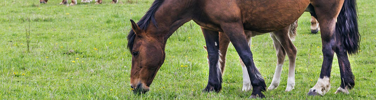
- Potomac horse fever
- Vectors: Ticks
- Vectors: Tsetse flies
- Vectors: Muscidae
- Vectors: Tabanidae
- Vectors: Culicoides spp.
- Vectors: Mosquitoes
- Classification, epidemiology and control of arthropod-borne viruses
- Special factors affecting the control of livestock diseases in sub-Saharan Africa
- The control of infectious diseases of livestock: Making appropriate decisions in different epidemiological and socioeconomic conditions
- Infectious diseases of animals in sub-Saharan Africa: The wildlife⁄livestock interface
- Vaccination: An approach to the control of infectious diseases
- African animal trypanosomoses
- Dourine
- Trichomonosis
- Amoebic infections
- GENERAL INTRODUCTION: COCCIDIA
- Coccidiosis
- Cryptosporidiosis
- Toxoplasmosis
- Besnoitiosis
- Sarcocystosis
- Balantidiosis
- Leishmaniosis
- Neosporosis
- Equine protozoal myeloencephalitis
- GENERAL INTRODUCTION: BABESIOSES
- Bovine babesiosis
- Equine piroplasmosis
- Porcine babesiosis
- Ovine babesiosis
- GENERAL INTRODUCTION: THEILERIOSES OF CATTLE
- East Coast fever
- Corridor disease
- Zimbabwe theileriosis
- Turning sickness
- Theileria taurotragi infection
- Theileria mutans infection
- Theileria annulata theileriosis
- Theileriosis of sheep and goats
- Theileria buffeli⁄orientalis infection
- Non-pathogenic Theileria species in cattle
- GENERAL INTRODUCTION: RICKETTSIAL, CHLAMYDIAL AND HAEMOTROPIC MYCOPLASMAL DISEASES
- Heartwater
- Lesser known rickettsial infections in animals and humans
- Chlamydiosis
- Q fever
- Eperythrozoonosis
- Bovine Haemobartonellosis
- Potomac horse fever
- GENERAL INTRODUCTION: ANAPLASMOSES
- Bovine anaplasmosis
- Ovine and caprine anaplasmosis
Potomac horse fever
This content is distributed under the following licence: Attribution-NonCommercial CC BY-NC  View Creative Commons Licence details here
Potomac horse fever
J E PALMER
Introduction
Potomac horse fever is an acute enterocolitis of equids caused by a group of closely related intracellular-occurring ehrlichias collectively known as Ehrlichia risticii, first recognized in 1979 along the Potomac River in Maryland, USA.29 Ehrlichia risticii infection in horses has been referred to as Potomac horse fever, equine monocytic ehrlichiosis, and equine ehrlichial colitis. It has been recognized to occur throughout North America. The original term, ‘Potomac horse fever’, was coined by a television reporter covering the original epidemic along the Potomac River. Although it is the least descriptive term, its wide acceptance and its high level of recognition ensures that it will remain the most common term for the disease. Equine ehrlichial colitis is a more descriptive term for the originally described syndrome of fever, anorexia, depression and diarrhoea. Even mild cases not manifesting diarrhoea have evidence of colitis. Equine ehrlichial abortion is an appropriate term for the abortion syndrome caused by E. risticii.
Aetiology
The causative organism of Potomac horse fever is a member of the tribe Ehrlichieae.23 Based on nucleotide sequence, the genus Ehrlichia is phylogenetically incoherent. Ehrlichia risticii is most closely related to Ehrlichia sennetsu and Neorickettsia helminthoeca (levels of sequence similarity, > 95 per cent).55 Although only definitively proven for N. helminthoeca, it is likely that these three organisms also share the unique property of being the only known obligate intracellular bacteria that are transmitted via a helminth vector.
Although E. risticii grows readily in a number of tissue culture lines as long as antimicrobials are not used in the culture media, the continuous murine macrophage cell line P388D1 is frequently used for isolation and propagation. The original Maryland isolate grows readily in this cell line, initially appearing as a cluster of singly occurring elementary bodies followed by the development of initial bodies and later forming morulae (mature inclusions). Heavily infected cells eventually disintegrate, releasing loosely packed groups of organisms held together by cytoplasmic stroma. 69
There is considerable biological diversity of E. risticii isolates from clinical cases of Potomac horse fever. Morphologically, some form large cytoplasmic morulae (inclusions), whereas others form small morulae or are individually dispersed in the cytoplasm in murine P388D1 cells.11 Similarly, patterns of antigenic proteins may differ considerably between isolates.11 The sequences of the 16S rRNA genes of isolates may differ between each other more than from the next most closely related Ehrlichia sp., E. sennetsu. It is evident that Potomac horse fever is not caused by one Ehrlichia sp. but rather by several closely related but distinct Ehrlichia spp.83 This antigenic variation of isolates causing the same clinical disease has resulted in incomplete protection from the first generation of vaccines which all originated from the same type strain of E. risticii.
Epidemiology
Potomac horse fever is confined to North America where it has been reported in a wide variety of geographic regions in most USA states and Canada. Surveys in the USA have shown that 16 to 33 per cent of horses with no history of illness have antibodies to E. risticii18, 39, 65 and that these are seasonal fluctuations with the highest number of seropositive horses and highest clinical occurrence in July, August and September.18, 39 Over a five-year period 70 per cent of 900 clinical cases recorded in Maryland occurred during the same months.53
Clinical disease shows an unusual sporadic pattern with a low prevalence rate (< 5 per cent) on any one farm despite its frequent occurrence in an endemic area. In fact, when multiple cases occur on a large farm, the pattern is also temporally (within the season of the disease) and geographically (among the pastures and barns on the farm) sporadic. Occasionally an epidemic form occurs which is characterized by a high attack rate (20 to 50 per cent) on an individual farm. The disease may be concentrated on a particular farm or race track, resulting in an outbreak simultaneously involving a large number of horses. The reason for this epidemiological variation is unknown.45, 48
The mode of transmission has been extensively studied but significant questions continue to exist. Direct contact transmission does not occur.44 The seasonal occurrence leads to early investigation of a possible arthropod vector. Common vectors of other ehrlichial agents are ticks.67 The only adult tick found feeding on horses along the Potomac River during the original epidemic in the early 1980s was Dermacentor variabilis.9, 72 The larval and nymphal stages of this tick feed on small ground mammals such as the white-footed mouse (Peromyscus leucopus).9 Mice are highly susceptible to experimental E. risticii infection.24 However, attempts to transmit E. risticii to horses using field-captured adult ticks (D. variabilis) from endemic farms failed.72 Furthermore, larval and nymphal stages of D. variabilis fed on ehrlichaemic mice did not transmit the organism to other mice or to ponies,20, 30 and white-footed mice infested with immature D. variabilis and living in endemic areas of the disease have consistently been found to be seronegative.9 Dogs, the primary host of adult D. variabilis, are uniformly seronegative on endemic farms53 despite being susceptible to infection.68 Dermacentor variabilis is not the vector of E. risticii and attempts to transmit the pathogen with other ticks including Rhipicephalus sanguineus, Ixodes scapularis, and Amblyomma americanum have also failed.20
Despite the wide experimental host susceptibility of E. risticii, 13, 24, 68, 77 non-equine mammals in endemic areas show a surprisingly low frequency of seroconversion.9, 19, 53, 71 One investigation revealed that a high percentage of cats on some endemic farms were seropositive,53 but transmission studies using the cat flea (Ctenocephalides felis) and the chigger mite (Neotrombicula whartoni), both common ectoparasites of cats on endemic farms, failed when E. risticii-infected horses or mice were used as donors and mice were used as recipients.71
The ehrlichaemia that occurs during the acute stage of the disease makes transmission by haematophagous flies possible. The role of adult stable flies (Stomoxys calcitrans) has been investigated:8 despite demonstrating ehrlichaemia in donor horses, recipient horses did not seroconvert or develop signs of Potomac horse fever and recovery of E. risticii from the flies was not successful.
All the evidence indicates that blood-sucking arthropods are not responsible for transmitting E. risticii. Oral infection produces clinical disease which is identical to that seen when the organism is given intravenously.44 If horses are exposed orally to a sufficient number of viable ehrlichias, infection will occur. Large numbers of E. risticii are shed into the lumen of the colon in exfoliating epithelial cells.64 Thus large numbers of ehrlichias are present in the faeces of infected horses.6, 38 However, direct contact does not result in transmission,44 so it seems unlikely that contaminated faeces play a direct role in the transmission of the disease. Indirect oral transmission may occur through concentration of the ehrlichias in a vector. Neorickettsia helminthoeca and Neorickettsia elokominica, two other members of the tribe Ehrlichieae which share common antigens59 and DNA homology55 with E. risticii, are transmitted through ingestion of infected helminths.22, 23 The metacercaria of the salmon fluke (Nanophyetus salmincola) carries these pathogens. Dogs are infected by ingestion of fluke-infested salmon or trout. Similarly, E. risticii may be concentrated in infected helminths carried by arthropods which are inadvertently ingested. Ehrlichia sennetsu, a closely related pathogen of humans, has been suspected of being transmitted by ingestion of a fish nematode. 28 The involvement of an arthropod vector or helminth would be consistent with the seasonality of the disease.
Recently a reservoir of E. risticii has been identified in trematode cercariae (virgulate cercariae) parasitizing freshwater stream snails and aquatic insects in two geographically distinct areas of the USA.2, 10, 25, 56, 58 The trematodes which can become infected with E. risticii appear to have a broad intermediate host range, having been found in abundance in adult and immature forms of the following aquatic insects: caddisflies (Trichoptera), mayflies (Ephemeroptera), damselflies (Odonata, Zygoptera), dragonflies (Odonata, Anisoptera), and stoneflies (Plecoptera).10 In one study, the prevalence of E. risticii was 32 per cent in 13 of 17 aquatic species of insects acting as hosts for the trematodes.10
The discovery of an aquatic reservoir is not surprising considering the early observations that the disease outbreaks centred along major rivers.48 This finding could easily explain the observation that the disease appears to be associated with areas and not horses in the areas. One of the original index farms along the Potomac River was abandoned and left without horses for several years because of the high prevalence of the disease. It was then repopulated with a new herd. Potomac horse fever re-emerged on the farm during the first spring thereafter, a high attack rate being recorded in the newly introduced horses.
The connection between the aquatic reservoir and transmission to horses has not been clearly characterized. Transmission attempts using naturally infected trematodes through skin contact with water harbouring E. risticii infected cercariae, through ingestion of water harbouring E. risticii-infected cercariae or through ingestion of different aquatic insects — caddisfly larvae (Limnephilidae), adult caddisflies (Leptoceridae, Limnephilidae), nymph and adult mayflies (Heptageniidea) and stonefly nymphs (Perlodidae) — harbouring E. risticii-infected metacercariae were only successful in one horse which received large numbers of adult caddisflies.
Two other horses receiving fewer adult caddisflies did not develop disease, seroconvert, develop ehrlichaemia or shed ehrlichias in their faeces.33 Considering the susceptibility of horses to oral infections, it is surprising that it was not easier to transmit the organism through ingestion of the trematodes.44 The failure to do so is probably associated with lack of sufficient viable ehrlichias.
How horses would ingest sufficient numbers of these aquatic insects to result in infection has not been explored. Caddisflies spend their life primarily on the water or in vegetation near water. They may be attracted by lights at night. When the ecosystem of horses and caddisflies, or other aquatic insects, would allow for transmission is unclear. Another issue that needs resolution is the observation that cases of Potomac horse fever can occur in horses at significant distances from major waterways as well as in horses that have no apparent access to streams or other aquatic environments. 42, 45, 48
The faecal shedding of E. risticii, which appears to be a consistent finding in infected horses,6 is an ideal way to seed aquatic environments with the organism, and introduction of recently infected horses could conceivably convert a previously disease-free area into one potentially endemic for the disease, if aquatic environments do prove to harbour the source of endemic disease in horses. It is apparent that, although a freshwater reservoir is probably important in the epidemiology of E. risticii, many questions have been left unanswered.
Pathogenesis
Although the mode of transmission is unknown, it is clear that within hours of infection E. risticii can be found in a subpopulation of blood monocytes. Although the pathogen is readily phagocytized by monocytes,84 it appears to elude the host’s defence mechanisms by inhibiting lysosomal fusion with phagosomes.82 The ehrlichaemia persists throughout and beyond the clinical period. The pathogen has a predilection for the mucosa of the caecum and large colon but is occasionally found in the mucosa of the jejunum and small colon.64 Intestinal epithelial cells, mast cells and macrophages are the targets of infection. Lesions are confined to the intestinal tract.12 The inflammatory response to E. risticii infection, which appears to vary in intensity, is mediated through cytokines. Preliminary studies in mice suggest that increased macrophage production of interleukin-1 alpha, but not tumour necrosis factor-alpha, interleukin-6 or prostaglandin E2, may be primarily involved in the pathogenesis of the disease.79
Ehrlichia risticii causes significant immune depression in mice and measurable alterations of the equine immune system, probably through modification by inflammatory cytokines. Whether or not the immune depression is clinically important in horses is unclear. Infected mice show evidence of significant depression of both humoral and cellular immunity62 and develop lymphoid depletion in lymphoid tissues.63 Although infected horses have a transient decrease in antibody production16 and lymphocyte function, 62 no histologic lesions develop in the lymphoid system.12, 63 In rickettsial infections antibodies can be protective when they block the pathogen’s attachment to or penetration of host cells.21 Anti-E. risticii equine IgG inhibits E. risticii internalization and interferes with the metabolic activity of E. risticii, rendering them incapable of proliferation in host cells and resulting in the eventual destruction of the organisms.36 Opsonization of E. risticii with anti-E. risticii serum renders E. risticii more susceptible to macrophage destruction.84 Mice can be passively protected from E. risticii infection by antibody transfer.26 However, ehrlichias can be isolated from animals in the presence of a rising antibody titre,14 and presence of antibodies does not always correlate with clearance of ehrlichias or with presence of protective immunity.60 Effective neutralizing antibodies are the important subset of the general antibody response to ehrlichial infections. In horses, clinical disease occurs in the face of rising antibody titres. Modulation of the immune response by the pathogen so that the initial antibodies are not protective may be an important aspect of the pathogen’s virulence.
Clinical signs
There is considerable variation in clinical manifestations of Potomac horse fever. The clinical signs include fever, depression, anorexia, ileus, colic, diarrhoea and laminitis. 40, 45, 48, 51, 52, 69 It is a common misconception that typical cases show most of these signs. On the contrary, any combination of these signsmaybe present. Common to all cases are the clinical manifestations of colitis which do not always result in colic or diarrhoea. Mild colitis can result in depression and anorexia, with or without fever. Normally formed faeces may be passed without signs of colic being present. Cases with equivocal signs of gastrointestinal tract involvement typically have, on careful auscultation of the abdomen, a remarkable decrease in borborygmal sounds, confirming gastrointestinal disease and long periods of silence broken by short bursts of loud, high-pitched sounds produced by gas passing through a tense intestine with a gas-fluid interface will be revealed. Simultaneous auscultation and finger percussion often result in a high-pitched resonance in the dorsal abdomen. Simultaneous auscultation and ballottement of the abdomen reveal splashing sounds confirming a gas-fluid interface. These signs of ileus are the most consistent clinical findings in all cases, and take on added significance when they occur in the face of an absence of clinical signs of colic, and the passage of normally formed faeces.
The fever is frequently biphasic, although the first phase is often not detected. The initial increased rectal temperature ranges from 39,4 to 41,1 °C but, since it is not accompanied by other signs, the presence of this initial fever will not be realized unless the horse’s temperature is measured frequently. It typically resolves within hours and is followed in three to seven days by a more persistent fever accompanied by other clinical signs.
Although diarrhoea is often thought of as a cardinal sign, it develops in less than 60 per cent of cases.40 When present, faecal consistency can vary from cow-like to watery, pipestream, but the diarrhoea is not as profuse as that associated with enteric salmonellosis in horses. The duration of diarrhoea may be as short as one day and rarely lasts as long as 10 days. Chronic diarrhoea is not a manifestation of E. risticii infection.
Decreased feed intake may be a prominent sign. Some horses may have complete anorexia for up to 10 days in the absence of other signs. The major manifestation in other cases may be a profound ileus evident by severe colic, severe toxaemia and dehydration. In some outbreaks severe laminitis in all four hooves may occur in up to 40 per cent of affected horses, but in most areas it is a rare sign.40, 45, 48 The laminitis, when present, may be the major manifestation of disease and result in humane euthanasia of the horse within days of onset. More often, however, it occurs together with other signs, is mild and completely resolves within three to five days. A few cases develop chronic laminitis.
The course of disease without therapeutic intervention is usually 5 to 10 days. Chronic manifestations, with the exception of laminitis, do not occur. The case fatality rate of untreated clinical cases varies from 5 to 30 per cent. Some fatalities are related to an apparent uncontrolled sepsis, the horse suddenly dying despite not being severely dehydrated or having extreme acid/base or electrolyte imbalances. This may be an extreme inflammatory response orchestrated by a modified systemic inflammatory response syndrome response. Other horses may have to be euthanased because of severe laminitis.40
Ehrlichia risticii can cause abortion. Experimentally, naive pregnant mares infected between 100 and 160 days of gestation aborted between 190 and 250 days of gestation. The mares developed typical signs of Potomac horse fever and appeared to have recovered fully before aborting. Abortion is accompanied by placentitis, detectable histologically, and a retained placenta in most cases. Foals which are carried to term may or may not have a presuck titre of serum antibodies to E. risticii. In either case full-term foals are healthy and are not ehrlichaemic.15, 31, 32
Pathology
The leukocyte count during the early phases of clinical signs may be variable. Leukopenia with a neutropenia and lymphopenia producing a haemogram similar to that typically seen in equine salmonellosis may be present, but many horses have normal leukocyte counts despite other signs of enteritis.
A marked leukocytosis can occur within a few days of onset of clinical signs in some cases. A total white blood cell count of 20 000 to 30 000 cells/μl may be found after an initial leukopenia. Although this profound leukocytosis does not occur consistently with E. risticii infection, it is a rare finding in mature horses suffering from most other diseases. If it is detected in a horse with consistent clinical signs, suspicion of Potomac horse fever should be high.
Gross necropsy, even in fatal cases of Potomac horse fever, is generally unimpressive. The most consistent postmortem findings are increased amounts of fluid contents in the caecum and large colon, and focal areas of mild hyperaemia primarily in the mucosa of the caecum and large colon but occasionally also in that of the small intestine. The intestinal wall is not oedematous and the mucosa is intact.12 By light microscopy a colitis is usually evident and ehrlichias can be identified in tissue sections stained with a silver stain.76 Ehrlichias may also be detected by electron microscopy, and by a tissue polymerase chain reaction (PCR) assay.41 The histologic appearance is somewhat variable but generally includes multifocal necrosis limited to the mucosa with a mixed inflammatory infiltrate. The histologic lesion often appears mild in relation to the severe clinical disease which has led to a fatal outcome.
In addition to the placentitis, histologic examination of foetal tissues reveals a remarkable colitis, periportal hepatitis and lymphoid hyperplasia of the mesenteric lymph nodes and spleen.
Diagnosis
Although the epidemiology, clinical presentation, haematology findings and response to treatment may all be suggestive of Potomac horse fever, its diagnosis is based on serology, PCR and/or isolation of E. risticii in cultures. Although the organism does parasitize circulating monocytes and can be seen by light microscopy of blood smears stained with common haematology stains, only a small percentage of circulating monocytes are infected; this renders the examination of peripheral smears a not very rewarding diagnostic procedure.
Ehrlichia risticii can be isolated from monocyte cultures of blood within days of experimental infection and consistently until 30 days after infection.44 Ehrlichia risticii may also be cultured from affected foetal tissues, especially when the period between infection and abortion is long. The placenta is usually retained and bacterial contamination of it interferes with the isolation of E. risticii from it. However, isolation and growth of E. risticii in antibiotic-free tissue culture (co-cultivation of tissue or blood monocytes with murine P388D1 cells) are research techniques not easily adapted to diagnostic laboratories. Although a definitive diagnosis of Potomac horse fever requires that E. risticii be isolated, in practice reliance is placed on serology or PCR for its diagnosis.
Serology is not a straightforward diagnostic technique because of the somewhat unique kinetics of antibody production stimulated by E. risticii. Infected horses have a rapid rise in fluorescent antibody titre, which level out within a few days. In the absence of a blood sample for serology taken in the early stages of the disease, detecting a significant rise in antibody titre may be impossible. Even samples taken within hours of the onset of clinical signs can show fluorescent antibody titres of 1:2 560 or higher. As a result, a decrease in titre is as common as a rise in titre and must be taken into account when antibody titres in acute and convalescent serum samples are compared. Thus a four-fold or greater increase or decrease in fluorescent antibody titre can be considered diagnostic. Approximately half of acutely affected clinical cases will have no significant change in titre because the antibody level has reached a plateau before the first sample is drawn.35
The determination of antibody titres in single serum samples serves no purpose. Although clinical cases will have titres greater than 1:80, there is no correlation between height of titre and likelihood of acute disease. Antibody titres in horses that have been exposed to Potomac horse fever for longer than a year before antibody determinations are done may be as high as 1:5 120. On the other hand, if a horse is found to have a negative titre after clinical signs develop, it is probable that it is not suffering from the disease andother diagnoses should be pursued. Treatment with oxytetracycline early in the clinical course of the disease does not affect the development of high antibody levels. Horses which have been vaccinated with a Potomac horse fever vaccine may have titres as high as 1:640. In general, titres consistently decrease after vaccination and after six to nine months have disappeared. When vaccinated animals develop the clinical disease (e.g. when a vaccine ‘break’ occurs) antibody titres may rise dramatically. In such cases antibody titres are often over 1:100 000 despite the presence of clinical signs of the disease.35, 40, 70
The first blood sample for diagnostic serum antibody determinations should be taken early in the course of the clinical disease and the second five to seven days after the first to maximize the chance of seeing a significant change. This short time interval between the first and second antibody determinations is contrary to what is generally the case for most other infectious diseases in which, for diagnostic purposes, reliance is also placed on the differences in antibody levels in acute and convalescent phase sera, the latter being determined two to three weeks or longer after the former. In Potomac horse fever this longer time interval has no advantage. It is of utmost importance to have the antibody levels in both the acute and chronic phase sera determined simultaneously in the same laboratory.
Great care must be taken in choosing a laboratory to assay serum samples. The most common technique used is an indirect fluorescent antibody test. Laboratories adept at running other indirect fluorescent antibody assays may still have difficulty with the procedure for Potomac horse fever.
Inconsistent antigen preparations, improper use of positive and negative controls, confusing nonspecific fluorescence, and inexperience with identifying specific fluorescence can all lead to erroneous results. The most consistent results will be obtained from laboratories which frequently run the assay and take great care in running both negative and positive controls.
A recent study has shown that even when performed by experienced research laboratories, the serologic test results may not be accurate. Serum samples from suspect cases and a sample from an experimentally infected horse were sent blindly to several university and private laboratories. Most laboratories reported high titres in many of the samples. Close scrutiny of the samples by Western blot analysis showed that the only true positive sample came from the experimentally infected horse. The rest were false positives.34 This suggests that caution should be used in interpreting the results of serological tests when used for diagnostic purposes. Serology should be abandoned in favour of more accurate diagnostic methods, as they become available.
A PCR technique has been adapted to detect E. risticii. 3, 5, 6, 38, 42, 57 The organism can be identified in blood samples and tissues41 without the need for culturing, and it takes less than a day. Although false negative and false positive results54 can occur, with proper controls the PCR assay should be a rapid, definitive diagnostic test and should replace serology as the preferred clinical diagnostic method. This testing procedure should not be viewed as a ‘gold’ standard since doing so may lead to unnecessary morbidity and mortality from unnecessary antimicrobial therapy resulting in antimicrobial-induced complications, such as antimicrobial-associated enteritis and renal disease, as has recently been reported in human medicine associated with the diagnosis of Lyme borreliosis.37, 50
Ehrlichial abortion can be tentatively diagnosed by histologic examination of the foetal colon. In fact, the colonic lesions are as helpful in establishing a diagnosis of E. risticii abortion as the foetal liver lesions are in equid herpesvirus abortion. Confirmation is possible through a PCR assay of foetal tissues.41
Differential diagnosis
The differential diagnosis of Potomac horse fever includes salmonellosis, intestinal clostridiosis, nonsteroidal antiinflammatory drug toxicity, antimicrobial-associated enteritis, colitis x, fungal colitis, and toxic enteritis.
Control
Ehrlichia risticii is sensitive in vitro to demeclocycline, doxycycline, oxytetracycline and minocycline.61 Oxytetracycline is the drug of choice for treating E. risticii infection in horses. The organism is an obligate intracellular parasite that survives within phagosomes in macrophages by inhibiting phagosome-lysosome fusion.82
This inhibition is reversed in vitro by oxytetracycline, resulting in destruction of the ehrlichias by the host cell.82 Oxytetracycline may interfere with the production of a cell wall protein that is responsible for the phagosome-lysosome fusion inhibition. Oxytetracycline administered intravenously at the dosage rate of 6,6 mg/kg once a day for five days is a very effective treatment for Potomac horse fever when given early in the course of the disease. A response to treatment is seen within 12 hours as noted by a decrease in rectal temperature, followed by an improvement in appetite, habitus and borborygmal sounds.47 Diarrhoea, if present, does not respond to oxytetracycline therapy.47 Since destroying the pathogen would be expected to take longer than 12 hours, the rapid clinical response to treatment implies that metabolic by-products acting through the production of cytokines result in the systemic inflammatory response syndrome which may be responsible for many of the clinical signs. Once treatment is begun, progression of the disease stops. The response to oxytetracycline therapy is so dramatic that it can be used to strengthen a clinical diagnosis. If the diagnosis is correct, a response to therapy should be clearly seen within 12 hours (with the exception of a change in diarrhoea). If no improvement occurs within 24 hours, the diagnosis should be reconsidered. If therapy is begun early in the course of the clinical disease, clinical signs resolve by the third day of treatment and no more than five days of therapy are needed.47 If a relapse occurs after discontinuing therapy, a second course of oxytetracycline is usually as successful as the first.
When treatment is begun early in the course of some rickettsial diseases, relapses may occur.27, 75, 78 Relapses are thought to be due to lack of sufficient time for adequate immunestimulation of the host resulting in a lack of protection.75 In experimental E. risticii infection, relapsesdonot occurwhen therapy is initiated early in the clinical course.47 However, treatment during the incubation period merely prolongs the incubation period but does not prevent clinical disease from developing.49 Early treatment does not interfere with the serologic response or the development of protection.47
A combination of erythromycin (25 mg/kg) and rifampin (10 mg/kg) administered orally twice a day is also effective in treating Potomac horse fever when given early in the course of clinical disease.43 The response to this treatment is similar to that to oxytetracycline, with the exception that there is a slightly longer delay before the animal becomes afebrile (24 hours vs. 12 hours).43, 47 Such a delay in the induction of apyrexia has also been observed when rifampin has been used to treat rickettsial infections in humans. The time period between initiation of therapy and return to normal of other physical findings is similar for both treatment regimes.43, 47 Although no experimental evidence is available in horses, it is considered that doxycycline administered orally should also be effective in the therapy of the disease.62
As in any horse suffering from colitis, the administration of fluids is an important consideration. Fluid and electrolyte replacement therapy should be tailored to suit the individual case. Many of the clinical signs in acute colitis cases can be attributed to inflammatory cytokine responses. These cases may benefit from therapy with anti-inflammatory mediators. When choosing such therapy, it should be borne in mind that experimental evidence suggests interleukin- 1 alpha may be much more important than tumour necrosis factor alpha, interleukin-6 or prostaglandins in the inflammatory response caused by E. risticii.79 Secondary salmonellosis is not uncommon in horses suffering from Potomac horse fever and this possibility should not be overlooked. For this reason, and because a definitive diagnosis is often difficult, it is felt that horses suspected of having Potomac horse fever should be isolated despite the lack of direct contact transmission.
Until the mode of transmission is clarified and the vector unequivocally identified, attempts to prevent exposure are unlikely to succeed. Although aquatic environments appear to be a location of one reservoir for the organism, there is no current evidence that the avoidance of such areas will prevent infection since the disease also occurs in horses which have had no direct contact with them.
After infection with E. risticii, protective immunity develops to the infecting strain as evidenced by clinical protection to reinfection for as long as 20 months.46 Protective immunity can be passively transferred,26 suggesting that it is primarily humoral. Neutralizing antibodies to specific protective antigens appears to be an important aspect in the induction of an effective immunity.17, 66, 74, 81 These antibodies appear both to block internalization of E. risticii and to interfere with its metabolic activity in host cells, rendering it incapable of intracellular proliferation, and result in its eventual destruction.36 Thus it should be possible to induce immunity by the administration of a vaccine.
A number of inactivated, partially purified, whole cell vaccines are commercially available in the USA. Vaccination has been reported to protect 78 per cent of experimentally infected horses from all clinical manifestations of Potomac horse fever except, in most cases, the fever (22 per cent of them had no clinical signs, 56 per cent developed fever only, 22 per cent developed fever and other signs of disease).70 Vaccination-induced protection is incomplete at best, which may be associated with poor stimulation of neutralizing antibodies by the vaccine.17 The protection induced by vaccination wanes much more rapidly than that induced by experimental infection. Only 50 per cent of vaccinates are fully protected at six months after vaccination, 73 and protection decreases to 33 per cent after nine months.4 Based on this evidence, revaccination at fourmonth intervals appears to be necessary to maintain a reasonable likelihood of protection. In most endemic areas the disease occurs seasonally. In these areas, if any of the currently available inactivated vaccines are used, horses should be vaccinated in the spring one month before the first cases are expected and again four months later, if cases are still occurring in the area.
During the past few years, protection by vaccination seems to have become less efficacious. Initially, after the introduction of the vaccines, vaccinated animals which did develop disease appeared to have a mild form of it. More recently, however, some vaccinated horses have developed the severe natural disease from which some died despite the fact that they were apparently adequately vaccinated. In some areas, the prevalence rate of the disease and treatment expenses are not decreased by vaccination.1 The reason for these vaccine failures may be associated with inconsistent antigen preparation or, more likely, it is associated with the appearance of several genetically distinct strains of E. risticii which vary in their ability to induce the formation of protective antibodies.7, 17, 80 All vaccines currently available contain only the strain that was originally isolated. Polyvalent recombinant vaccines containing antigens which elicit protective antibody formation may, in future, become available. These should increase the efficacy of protection afforded by vaccination.
References
- ATWILL, E.R.& MOHAMMED, H.O., 1996. Evaluation of vaccination of horses as a strategy to control equine monocytic ehrlichiosis. Journal of the American Veterinary Medical Association, 208, 1290–1294.
- BARLOUGH, J.E., REUBEL, G.H., MADIGAN, J.E., VREDEVOE, L.K., MILLER, P.E.& RIKIHISA, Y., 1998. Detection of Ehrlichia risticii, the agent of Potomac horse fever, in freshwater stream snails (Pleuroceridae: Juga spp.) from northern California. Applied & Environmental Microbiology, 64, 2888–2893.
- BARLOUGH, J.E., RIKIHISA, Y. & MADIGAN, J.E., 1997. Nested polymerase chain reaction for detection of Ehrlichia risticii genomic DNA in infected horses. Veterinary Parasitology, 68, 367–373.
- BIGBEE, H.G., 1989. Schering-Plough Animal Health Corporation, P.O. Box 3182, Union, N.J. 07083–1982. Personal communication.
- BISWAS, B., MUKHERJEE, D., MATTINGLY-NAPIER, B.L. & DUTTA, S.K., 1991. Diagnostic application of polymerase chain reaction for detection of Ehrlichia risticii in equine monocytic ehrlichiosis (Potomac horse fever). Journal of Clinical Microbiology, 29, 2228–2233.
- BISWAS, B., VEMULAPALLI, R. & DUTTA, S.K., 1994. Detection of Ehrlichia risticii from feces of infected horses by immunomagnetic separation and PCR. Journal of Clinical Microbiology, 32, 2147–2151.
- BISWAS, B., VEMULAPALLI, R. & DUTTA, S.K., 1998. Molecular basis for antigenic variation of a protective strain-specific antigen of Ehrlichia risticii. Infection & Immunity, 66, 3682–3688.
- BURG, J.G., ROBERTS, A.W., WILLIAMS, N.M., POWELL, D.G., & KNAPP F.W., 1990. Attempted transmission of Ehrlichia risticii (Rickettsiaceae) with Stomoxys calcitrans (Diptera: Muscidae). Journal of Medical Entomolology, 27, 874–877.
- CARROLL, J.F., SCHMIDTMANN, E.T. & RICE, R.M., 1989. White-footed mice: Tick burdens and role in the epizootiology of Potomac horse fever in Maryland. Journal of Wildlife Diseases, 25, 397–400.
- CHAE, J.S., PUSTERLA, N., JOHNSON, E., DEROCK, E., LAWLER, S.P. & MADIGAN, J.E., 2000. Infection of aquatic insects with trematode metacercariae carrying Ehrlichia risticii, the cause of Potomac horse fever. Journal of Medical Entomology, 37, 619–625.
- CHAICHANASIRIWITHAYA, W., RIKIHISA, Y., YAMAMOTO, S., REED, S., CRAWFORD, T.B., PERRYMAN, L.E. & PALMER, G.H., 1994. Antigenic, morphologic, and molecular characterization of new Ehrlichia risticii isolates. Journal of Clinical Microbiology, 32, 3026–3033.
- CORDES, D.O., PERRY, B.D., RIKIHISA, Y. & CHICKERING, W.R., 1986. Enterocolitis caused by Ehrlichia sp. in the horse (Potomac horse fever). Veterinary Pathology, 23, 471–477.
- DAWSON, J.E., ABEYGUNAWARDENA, I., HOLLAND, C.J., BUESE, M.M. & RISTIC, M., 1988. Susceptibility of cats to infection with Ehrlichia risticii, causative agent of equine monocytic ehrlichiosis. American Journal of Veterinary Research, 49, 2096–2100.
- DAWSON, J.E. & EWING, S.A., 1992. Susceptibility of dogs to infection with Ehrlichia chaffeensis, causative agent of human ehrlichiosis. American Journal of Veterinary Research, 53, 1322–1327.
- DAWSON, J.E., RISTIC, M., HOLLAND, C.J., WHITLOCK, R.H. & SESSIONS, J., 1988. Isolation of Ehrlichia risticii from a fetus of a mare with Potomac horse fever. In Proceedings from a Symposium on Potomac Horse Fever, Louisville, pp. 107–111.
- DUTTA, S.K., MATTINGLY, B.L. & SHANKARAPPA, B., 1989. Antibody response to Ehrlichia risticii and antibody reactivity to the component antigens in horses with induced Potomac horse fever. Infection & Immunity, 57, 2959–2962.
- DUTTA, S.K., VEMULAPALLI, R. & BISWAS, B., 1998. Association of deficiency in antibody response to vaccine and heterogeneity of Ehrlichia risticii strains with Potomac horse fever vaccine failure in horses. Journal of Clinical Microbiology, 36, 506–512.
- GOETZ, T.E., HOLLAND, C.J., DAWSON, J.E., RISTIC, M., SKIBBE, K., KEEGAN, K.G., JOHNSON, P.J., SCHAEFFER, D.J. & BAKER G.J., 1989. Monthly prevalence (in 1986) of antibody titres against equine monocytic ehrlichiosis in apparently healthy horses in Illinois. American Journal of Veterinary Research, 50, 1936–1939.
- GORDON, J.C., BECH-NIELSEN, J.C., FARRAR, W., FOSTER, W., PARSONS, M. & KOHN, K., 1988. Epidemiologic investigation of affected farms. Proceedings of a Symposium on Potomac Horse Fever, Louisville, KT, USA. pp. 21–25.
- HAHN, N.E., FLETCHER, M., RICE, R.M., KOCAN, K.M., HANSEN, J.W., HAIR, J.A., BARKER, R.W., & PERRY, B.D., 1990. Attempted transmission of Ehrlichia risticii, causative agent of Potomac horse fever, by the ticks, Dermacentor variabilis, Rhipicephalus sanguineus, Ixodes scapularis and Amblyomma americanum. Experimental & Applied Acarology, 8, 41–50.
- HANSON, B.A., 1983. Effect of immune serum on infectivity of Rickettsia tsutsugamushi. Infection & Immunity, 42, 341–349.
- HIBLER, S.C., HOSKINS, J.D. & GREENE, C.E., 1986. Rickettsial infections in dogs part III. Salmon disease complex and haemobartonellosis. Compendium of Continuing Education in Veterinary Practice, 8, 251– 256.
- HOLT, J.G. & KRIEG, N.R., 1984. Tribe Ehrlichieae. In: Bergey’s Manual of Systematic Bacteriology. Baltimore: Williams and Wilkins, pp. 704–711.
- JENKINS, S.J., JONES, N.K. & JENNY, A.L., 1985. Potomac horse fever agent in mice. The Veterinary Record, 117, 556–557.
- KANTER, M., MOTT, J., OHASHI, N., FRIED, B., REED, S., LIN, Y.C. & RIKIHISA, Y., 2000. Analysis of 16S rRNA and 51-kilodalton antigen gene and transmission in mice of Ehrlichia risticii in virgulate trematodes from Elimia livescens snails in Ohio. Journal of Clinical Microbiology, 38, 3349–3358.
- KAYLOR, P.S., CRAWFORD, T.B., MCELWAIN, T.F. & PALMER, G.H., 1991. Passive transfer of antibody to Ehrlichia risticii protects mice from ehrlichiosis. Infection and Immunity, 59, 2058–2062.
- KENYON, R.H. & PEDERSEN JR., C.E., 1980. Immune responses to Rickettsia akari infection in congenitally athymic nude mice. Infection and Immunity, 21, 310–313.
- KITAO, T., FARRELL, R.K. & FUKUDA, T., 1978. Differentiation of Salmon poisoning disease and Elokomin fluke fever: Fluorescent antibody studies with Rickettsia sennetsu. American Journal of Veterinary Research, 34, 927–928.
- KNOWLES, R.C., ANDERSON, C.W., SHIPLEY, W. D., WHITLOCK, R. H., PERRY, B.D. & DAVIDSON, J.P., 1983. Acute equine diarrheal syndrome (AEDS): a preliminary report. Proceedings of the 29th American Association of Equine Practitioners, Las Vegas, NV, USA 29, 353–357.
- LEVINE, J.F., LEVY, M.G., NICHOLSON, W.L. & GAGER, R.B., 1990. Attempted Ehrlichia risticii transmission with Dermacentor variabilis (Acari: Ixodidae). Journal of Medical Entomology, 27, 931–933.
- LONG, M.T., GOETZ, T.E., KAKOMA, I., WHITELEY, H.E., LOCK, T.E., HOLLAND, C.J., FOREMAN, J.H. & BAKER, G.J., 1995. Evaluation of fetal infection and abortion in pregnant ponies experimentally infected with Ehrlichia risticii. American Journal of Veterinary Research, 56, 1307–1316.
- LONG, M.T., GOETZ, T.E., WHITELEY, H.E., KAKOMA, I. & LOCK, T.E., 1995. Identification of Ehrlichia risticii as the causative agent of two equine abortions following natural maternal infection. Journal of Veterinary Diagnostic Investigation, 7, 201–205.
- MADIGAN, J.E., PUSTERLA, N., JOHNSON, E., CHAE, J.S., PUSTERLA, J.B., DEROCK, E. & LAWLER, S.P., 2000. Transmission of Ehrlichia risticii, the agent of Potomac horse fever, using naturally infected aquatic insects and helminth vectors: preliminary report. Equine Veterinary Journal, 32, 275–279.
- MADIGAN, J.E., RIKIHISA, Y., PALMER, J.E., DEROCK, E. & MOTT, J., 1995. Evidence for a high rate of false-positive results with the indirect fluorescent antibody test for Ehrlichia risticii antibody in horses. Journal of the American Veterinary Medical Association, 207, 1448–1453.
- MEINERSMANN, R.J., PALMER, J.E., BULLS, J.A., GANSER, R.D., BENSON, C.E., & WHITLOCK, R.H., 1988. Serology for the diagnosis of equine ehrlichial colitis. Proceedings of a Symposium on Potomac Horse Fever, Louisville, KT, USA. 29 May, 1987, pp. 33–35.
- MESSICK, J.B. & RIKIHISA, Y., 1994. Inhibition of binding, entry, or intracellular proliferation of Ehrlichia risticii in P388D1 cells by anti-E. risticii serum, immunoglobulin G, or Fab fragment. Infection and Immunity, 62, 3156–3161.
- MOLLOY, P.J., PERSING, D.H. & BERARDI, V.P., 2001. False-positive results of PCR testing for Lyme disease. Clinical Infectious Diseases, 33, 412–413.
- MOTT, J., RIKIHISA, Y., ZHANG, Y., REED, S.M. & YU, C.Y., 1997. Comparison of PCR and culture to the indirect fluorescent-antibody test for diagnosis of Potomac horse fever. Journal of Clinical Microbiology, 35, 2215–2219.
- OLCHOWY, T.W., AMES, T.R. & MOLITOR, T.W., 1990. Serodiagnosis of equine monocytic ehrlichiosis in selected groups of horses in Minnesota. Journal of the American Veterinary Medical Association, 196, 1967–1970.
- PALMER, J.E., 1987. Potomac horse fever. In: ROBINSON, N.E., (ed.). Current Equine Therapy 2, Philadelphia: WB. Saunders Company, pp. 92–93.
- PALMER, J.E., 1992. New Bolton Center, University of Pennsylvania, Kennett Square, PA 19348, unpublished data.
- PALMER, J.E., 1993. Potomac horse fever. Veterinary Clinics of North America—Equine Practice, 9, 399–410.
- PALMER, J.E. & BENSON, C.E., 1992. Effect of treatment with erythromycin and rifampin during the acute stages of experimentally induced equine ehrlichial colitis in ponies. American Journal of Veterinary Research, 53, 2071–2076.
- PALMER, J.E. & BENSON, C.E., 1994. Studies on oral transmission of Potomac horse fever. Journal of Veterinary Internal Medicine, 8, 87–92.
- PALMER, J.E., BENSON, C.E. & WHITLOCK, R.H., 1986. Potomac horse fever: Diagnostic criteria and recognition of endemic areas. Proceedings of the Second Symposium on Equine Colic Research, Athens, GA, 157–160.
- PALMER, J.E., BENSON, C.B. & WHITLOCK, R.H., 1990. Resistance to development of equine ehrlichial colitis in experimentally inoculated horses and ponies. American Journal of Veterinary Research, 51, 763– 765.
- PALMER, J.E., BENSON, C.E. & WHITLOCK, R.H., 1992. Effect of treatment with oxytetracycline during the acute stages of experimentally induced equine ehrlichial colitis in ponies. American Journal of Veterinary Research, 53, 2300–2304.
- PALMER, J.E., WHITLOCK, R.H. & BENSON, C.E., 1986. Equine ehrlichial colitis (Potomac horse fever): Recognition of the disease in Pennsylvania, New Jersey, New York, Ohio, Idaho, and Connecticut. Journal of the American Veterinary Medical Association, 189, 197–199.
- PALMER, J.E., WHITLOCK, R.H. & BENSON, C.E., 1988. Equine ehrlichial colitis: effect of oxytetracycline treatment during the incubation period of Ehrlichia risticii infection in ponies. Journal of the American Veterinary Medical Association, 192, 343–345.
- PATEL, R., GROGG, K.L., EDWARDS, W.D., WRIGHT, A.J. & SCHWENK, N.M., 2000. Death from inappropriate therapy for Lyme disease. Clinical Infectious Diseases, 31, 1107–1109.
- PERRY, B.D., PALMER, J.E., BIRCH, J.B., MAGNUSSEN, R.A., MORRIS, D.D. & TROUT, H.F., 1984. Epidemiological characterization of an acute diarrhoea syndrome: The case-control approach. Proceedings of the Society of Veterinary Epidemiology and Preventative Medicine, Edinburgh, pp. 148–153.
- PERRY, B.D., PALMER, J.E., TROUT, H.F., BIRCH, J., MORRIS, D., EHRICH, M. & RIKIHISA, Y. , 1986. A case-control study of Potomac horse fever. Preventative Veterinary Medicine, 4, 69–82.
- PERRY, B.D., SCHMIDTMANN, E.T., RICE, R.M., HANSEN, J.W., FLETCHER, M., TURNER, E.C., ROBL, M.G. & HAHN, N.E., 1989. Epidemiology of Potomac horse fever: An investigation into the possible role of non-equine mammals. The Veterinary Record, 125, 83–86.
- PERSING, D.H., 1993. Amplification product contamination control. In: PERSING, D.H., TENOVER, F., SMITH, T.F. & WHITE, T., (eds). Diagnostic Molecular Microbiology: Principles and Applications, pp. 105–121.
- PRETZMAN, C., RALPH, D., STOTHARD, D.R., FUERST, P.A. & RIKIHISA, Y., 1995. 16S rRNA gene sequence of Neorickettsia helminthoeca and its phylogenetic alignment with members of the genus Ehrlichia. International Journal of Systematic Bacteriology, 45, 207–211.
- PUSTERLA, N., JOHNSON, E., CHAE, J., PUSTERLA, J.B., DEROCK, E. & MADIGAN, J.E., 2000. Infection rate of Ehrlichia risticii, the agent of Potomac horse fever, in freshwater stream snails (Juga yrekaensis) from northern California. Veterinary Parasitology, 92, 151–156.
- PUSTERLA, N., LEUTENEGGER, C.M., SIGRIST, B., CHAE, J.S., LUTZ, H. & MADIGAN, J.E., 2000. Detection and quantitation of Ehrlichia risticii genomic DNA in infected horses and snails by real-time PCR. Veterinary Parasitology, 90, 129–135.
- REUBEL, G.H., BARLOUGH, J.E. & MADIGAN, J.E., 1998. Production and characterization of Ehrlichia risticii, the agent of Potomac horse fever, from snails (Pleuroceridae: Juga spp.) in aquarium culture and genetic comparison to equine strains. Journal of Clinical Microbiology, 36, 1501–1511.
- RIKIHISA, Y., 1991. Cross-reacting antigens between Neorickettsia helminthoeca and Ehrlichia species, shown by immunofluorescence and Western immunoblotting. Journal of Clinical Microbiology, 29, 2024–2029.
- RIKIHISA, Y., 1991. The tribe Ehrlichieae and ehrlichial diseases. Clinical Microbiology Reviews, 4, 286–308.
- RIKIHISA, Y. & JIANG, B.M., 1988. In vitro susceptibilities of Ehrlichia risticii to eight antibiotics. Antimicrobial Agents and Chemotherapy, 32, 986–991.
- RIKIHISA, Y. & JIANG, B.M., 1989. Effect of antibiotics on clinical, pathologic and immunologic responses in murine Potomac horse fever: protective effects of doxycycline. Veterinary Microbiology, 19, 253–262.
- RIKIHISA, Y., JOHNSON, G.C. & BURGER, C.J., 1987. Reduced immune responsiveness and lymphoid depletion in mice infected with Ehrlichia risticii. Infection and Immunity, 55, 2215–2222.
- RIKIHISA, Y., PERRY, B.D. & CORDES, D.O., 1985. Ultrastructural study of ehrlichial organisms in the large colons of ponies infected with Potomac horse fever. Infection and Immunity, 49, 505–512.
- RIKIHISA, Y., REED, S.M., SAMS, R.A., GORDON, J.C., & PRETZMAN, C.I., 1990. Serosurvey of horses with evidence of equine monocytic ehrlichiosis. Journal of the American Veterinary Medical Association, 197, 1327–1332.
- RIKIHISA, Y., WADA, R., REED, S.M. & YAMAMOTO, S., 1993. Development of neutralizing antibody in horses infected with Ehrlichia risticii. Veterinary Microbiology, 36, 139–147.
- RISTIC, M., 1986. Pertinent characteristics of leukocytic Rickettsiae of humans and animals. In: Microbiology, pp. 182–187.
- RISTIC, M., DAWSON, J., HOLLAND, C.J. & JENNY, A., 1988. Susceptibility of dogs to infection with Ehrlichia risticii, causative agent of equine monocytic ehrlichiosis (Potomac horse fever). American Journal of Veterinary Research, 49, 1497–1500.
- RISTIC, M., HOLLAND, C.J., DAWSON, J.E., SESSIONS, J. & PALMER, J.E., 1986. Diagnosis of equine monocytic ehrlichiosis (Potomac horse fever) by indirect immunofluorescence. Journal of the American Veterinary Medical Association, 189, 39–46.
- RISTIC, M., HOLLAND, C.J. & GOETZ, T.E., 1988. Evaluation of a vaccine for equine monocytic ehrlichiosis. Proceedings of a Symposium on Potomac Horse Fever, Louisville, KT, USA, pp. 89–100.
- SCHMIDTMANN, E.T., RICE, R.M., ROBLE, M.G. & AZAD, A.F., 1988. Search for an arthropod vector of Ehrlichia risticii. Proceedings of a Symposium on Potomac Horse Fever, Louisville, KT, USA, pp. 9–16.
- SCHMIDTMANN, E.T., ROBLE, M.G. & CARROLL, J.F., 1986. Attempted transmission of Ehrlichia risticii by field-captured Dermacentor variabilis (Acari: Ixodidae). American Journal of Veterinary Research, 47, 2393–2395.
- SESSIONS, J.E. & DAWSON, J., 1988. Product evaluation: Maryland field evaluation of the Potomac horse fever vaccine. Equine Practice, 10, 7–12.
- SHANKARAPPA, B., DUTTA, S.K. & MATTINGLY-NAPIER, B., 1992. Identification of the protective 44-kilodalton recombinant antigen of Ehrlichia risticii. Infection and Immunity, 60, 612–617.
- SMADEL, J.E., TRAUB, R., LEY, H.L., SAUNDERS, J.P., HUXSULL, D.L., & GROVES, M.G., 1950. Chloramphenicol (chloromycetin) in the chemoprophylaxis of scrub typhus (Tsutsugamushi disease) II. Results with volunteers exposed in hyperendemic areas of scrub typhus. American Journal of Hygiene, 50, 75–91.
- STEELE, K.E., RIKIHISA, Y. & WALTON, A.M., 1986. Ehrlichia of Potomac horse fever identified with a silver stain. Veterinary Pathology, 23, 531–533.
- STEPHENSON, E.H., 1990. Experimental ehrlichiosis in nonhuman primates. In: Ehrlichiosis. A vector-borne disease of animals and humans. Current Topics in Veterinary Medicine and Animal Science, 54, 93–99.
- TWARTZ, J.C., SHIRAI, A., SELVARAJU, G., SAUNDERS, J.P., HUXSULL, D.L., & GROVES, M.G., 1982. Doxycycline prophylaxis for human scrub typhus. Journal of Infectious Diseases, 146, 811–818.
- VAN HEECKEREN, A.M., RIKIHISA, Y., PARK, J. & FERTEL, R., 1993. Tumor necrosis factor alpha, interleukin-1 alpha, interleukin-6, and prostaglandin E2 production in murine peritoneal macrophages infected with Ehrlichia risticii. Infection and Immunity, 61, 4333–4337.
- VEMULAPALLI, R., BISWAS, B. & DUTTA, S.K., 1995. Pathogenic, immunologic, and molecular differences between two Ehrlichia risticii strains. Journal of Clinical Microbiology, 33, 2987–2993.
- VEMULAPALLI, R., BISWAS, B. & DUTTA, S.K., 1998. Studies with recombinant proteins of Ehrlichia risticii: identification of strain-specific antigen as a protective antigen. Veterinary Parasitology, 76, 189–202.
- WELLS, M.Y. & RIKIHISA, Y., 1988. Lack of lysosomal fusion with phagosomes containing Ehrlichia risticii in P388D1 cells: Abrogation of inhibition with oxytetracycline. Infection and Immunity, 56, 3209–3215.
- WEN, B., RIKIHISA, Y., FUERST, P.A. & CHAICHANASIRIWITHAYA, W., 1995. Diversity of 16S rRNA genes of new Ehrlichia strains isolated from horses with clinical signs of Potomac horse fever. International Journal of Systematic Bacteriology, 45, 315–318.
- WILLIAMS, N.M. & TIMONEY, P.J., 1993. In vitro killing of Ehrlichia risticii by activated and immune mouse peritoneal macrophages. Infection and Immunity, 61, 861–867.


