- Infectious Diseases of Livestock
- Part 1
- Classification, epidemiology and control of arthropod-borne viruses
- Vectors: Ticks
- Vectors: Tsetse flies
- Vectors: Muscidae
- Vectors: Tabanidae
- Vectors: Culicoides spp.
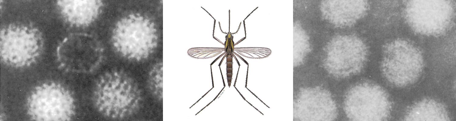
- Vectors: Mosquitoes
- Classification, epidemiology and control of arthropod-borne viruses
- Special factors affecting the control of livestock diseases in sub-Saharan Africa
- The control of infectious diseases of livestock: Making appropriate decisions in different epidemiological and socioeconomic conditions
- Infectious diseases of animals in sub-Saharan Africa: The wildlife⁄livestock interface
- Vaccination: An approach to the control of infectious diseases
- African animal trypanosomoses
- Dourine
- Trichomonosis
- Amoebic infections
- GENERAL INTRODUCTION: COCCIDIA
- Coccidiosis
- Cryptosporidiosis
- Toxoplasmosis
- Besnoitiosis
- Sarcocystosis
- Balantidiosis
- Leishmaniosis
- Neosporosis
- Equine protozoal myeloencephalitis
- GENERAL INTRODUCTION: BABESIOSES
- Bovine babesiosis
- Equine piroplasmosis
- Porcine babesiosis
- Ovine babesiosis
- GENERAL INTRODUCTION: THEILERIOSES OF CATTLE
- East Coast fever
- Corridor disease
- Zimbabwe theileriosis
- Turning sickness
- Theileria taurotragi infection
- Theileria mutans infection
- Theileria annulata theileriosis
- Theileriosis of sheep and goats
- Theileria buffeli⁄orientalis infection
- Non-pathogenic Theileria species in cattle
- GENERAL INTRODUCTION: RICKETTSIAL, CHLAMYDIAL AND HAEMOTROPIC MYCOPLASMAL DISEASES
- Heartwater
- Lesser known rickettsial infections in animals and humans
- Chlamydiosis
- Q fever
- Eperythrozoonosis
- Bovine Haemobartonellosis
- Potomac horse fever
- GENERAL INTRODUCTION: ANAPLASMOSES
- Bovine anaplasmosis
- Ovine and caprine anaplasmosis
Classification, epidemiology and control of arthropod-borne viruses
This content is distributed under the following licence: Attribution-NonCommercial CC BY-NC  View Creative Commons Licence details here
Classification, epidemiology and control of arthropod-borne viruses
R SWANEPOEL
Introduction
As recently as the late 1960s the term arboviruses, a partial acronym derived from arthropod-borne viruses, was used in a taxonomic sense to denote the existence of an inferred underlying relationship between viruses with similar modes of transmission. After morphological and physicochemical characterization, it became evident that the viruses actually belonged to a range of widely disparate families, and since then there has been a tendency to stress the diversity of the viruses, at the cost of overlooking shared epidemiological features which dictate a common approach to the investigation of arbovirus diseases. Indeed, the study of the arthropod-borne viruses has always been a specialized branch of virology in which there is, nevertheless, a need for co-operation between such varied investigators as virologists, medical and veterinary clinicians, entomologists, zoologists, ecologists and climatologists,amongothers. Except where otherwise indicated, the following outline of the subject is drawn from a number of general accounts of arboviruses.22, 109, 228, 240
For centuries it was suspected that certain diseases were transmitted by arthropods, but it was not until 1878 that Patrick Manson demonstrated in China that mosquitoes serve as vectors for the filarial worm Wuchereria bancrofti. In 1893 Smith and Kilborne established that ticks transmit the babesia, which causes Texas redwater fever of cattle, and between 1895 and 1898 Ronald Ross performed investigations in India that proved that mosquitoes transmit the parasite of malaria. Shortly thereafter, in 1900, Walter Reed and his associates in Cuba demonstrated human to human transmission of the virus of yellow fever by mosquitoes, as had been postulated earlier by Carlos Finlay. Not only did this constitute the first proof of transmission of a virus by arthropods, but it was also the first occasion on which evidence was produced to indicate that an agent causing disease in humans, as opposed to lower animals, was caused by a virus. However, it was not until 1927 that Stokes and his co-workers in Nigeria succeeded in maintaining yellow fever virus by serial passage in monkeys. By then, several arboviruses of veterinary significance had been isolated, the first two being African horse sickness and bluetongue viruses in South Africa. Although the exact dates and authorship of isolation of these two viruses are debatable, they had been maintained by subinoculation in horses and sheep respectively, long before they were shown to be filterable agents or were adapted to laboratory hosts.4, 65, 164, 176, 216, 217, 239
By 1939, 16 arboviruses had been isolated (Table 7.1), although this figure is somewhat misleading as it does not take into account that African horse sickness and bluetongue viruses are each made up of multiple serotypes. Of the 19 arboviruses isolated between 1940 to 1949 (Table 7.2) eight were human pathogens from various parts of the world, but 11 were viruses of unknown significance that had been isolated from mosquitoes and a sentinel monkey in the course of yellow fever investigations in which the Rockefeller Foundation Virus Laboratories (RFVL) of New York played a leading role. During the Second World War, RFVL concentrated on the investigation of yellow fever, but during the 1950s it seconded staff to laboratories in many parts of the world, including what is now known as the National Institute for Communicable Diseases in Johannesburg, to establish and foster arbovirus units which were intended to continue operating as regional centres with locally trained staff.
The newly established arbovirus unit in South Africa, initially under the leadership of Smithburn of RFVL, conducted surveys for antibodies to a range of arboviruses on sera collected from humans and domestic animals in South Africa, 62, 124, 127, 148, 155, 213 Mozambique,123 Angola,116 Namibia and Botswana.129 The highest prevalence of antibodies was found in sera from Tongaland, a subtropical area occupying the coastal plain of the northern part of KwaZulu-Natal Province in South Africa, and consequent efforts to isolate new viruses were concentrated mainly on this area. In the decade from 1950 to 1959, members of the Onderstepoort Veterinary Institute isolated Wesselsbron virus from a lamb in the Wesselsbron district of the Free State Province, while personnel of the arbovirus unit in Johannesburg isolated 10 new viruses from Tongaland and four from elsewhere in southern African (Table 7.3).
Table 7.1 Arthropod-borne viruses isolated throughout the world prior to 1940. Information adapted from Karabatsos (1985)109
| VIRUS | ORIGINAL SOURCE | YEAR |
|---|---|---|
| Bluetongue | Sheep blood | 1904* |
| African horse sickness | Horse blood | 1905* |
| African swine fever | Pig blood | 1910 |
| Nairobi sheep disease | Sheep blood | 1910 |
| Vesicular stomatitis – Indiana | Cow tongue epithelium | 1925 |
| Yellow fever | Human blood | 1927 |
| Louping ill | Sheep brain | 1929 |
| Rift Valley fever | Sheep blood | 1930 |
| Western encephalitis | Horse brain | 1930 |
| Eastern encephalitis | Horse brain | 1933 |
| St Louis encephalitis | Human brain | 1933 |
| Japanese encephalitis | Human brain | 1935 |
| Sofjin ** | Human brain | 1937 |
| Bwamba | Human blood | 1937 |
| West Nile | Human blood | 1937 |
| Venezuelan encephalitis | Horse brain | 1938 |
* See text for derivation of the dates given here for isolation of bluetongue and African horse sickness viruses
** Sofjin = prototype strain of Russian spring-summer encephalitis virus
Table 7.2 New arthropod-borne viruses isolated throughout the world from 1940 to 1949 Information adapted from Karabatsos (1985).109
| VIRUS | ORIGINAL SOURCE | YEAR | COUNTRY |
|---|---|---|---|
| Wyeomyia | Mosquitoes | 1940 | Colombia |
| Anopheles A | Mosquitoes | 1940 | Colombia |
| Anopheles B | Mosquitoes | 1940 | Colombia |
| Semliki Forest | Mosquitoes | 1942 | Uganda |
| Ntaya | Mosquitoes | 1943 | Uganda |
| California encephalitis | Mosquitoes | 1943 | USA |
| Bunyamwere | Mosquitoes | 1943 | Uganda |
| Sandfly fever – Sicily | Human blood | 1943 | Italy |
| Colorado tick fever | Human blood | 1943 | USA |
| Dengue 1 | Human blood | 1944 | Hawaii |
| Ilheus | Mosquitoes | 1944 | Brazil |
| Dengue 2 | Human blood | 1944 | New Guinea |
| Sandfly fever – Naples | Human blood | 1944 | Italy |
| Zika | Sentinel monkey blood | 1947 | Uganda |
| Uganda S | Mosquitoes | 1947 | Uganda |
| Omsk haemorrhagic fever | Human blood | 1947 | Soviet Union |
| Hanzalova* | Human brain | 1948 | Czechoslovakia |
| Trivittatus | Mosquitoes | 1948 | USA |
| Negishi | Human cerebrospinal fluid | 1948 | Japan |
* Hanzalova = first strain of the virus of Central European encephalitis to have been isolated; indistinguishable by cross-neutralization from the virus of Russian spring-summer encephalitis
Most of these viruses were originally isolated from mosquitoes or birds collected in surveys and have not been found to be of great medical or veterinary significance, although a few of the same viruses were later isolated from the blood of persons with febrile disease. Since 1959, there has been less emphasis on surveys and only three new viruses have been isolated from arthropods, although several potential veterinary pathogens have been isolated in southern Africa (Table 7.4). A number of arboviruses that had originally been isolated elsewhere, were also encountered in southern Africa. These included Rift Valley fever and Akabane viruses and human pathogens such as chikungunya, West Nile and Sindbis viruses (Table 7.5).
There was a need to collate information on the wealth of new arboviruses isolated worldwide from 1950 onwards and in 1959 the RFVL organized meetings that led to the formation of the American Committee on Arthropod-borne Viruses (ACAV). In 1960 a Subcommittee on Information Exchange (SIE) produced the ‘Catalogue of Arthropod-borne Viruses of the World’, which is continually updated by the mailing of registration cards for new viruses. However, many of the more than 500 registered viruses have been proven not to be arthropod-borne but are nevertheless retained in the catalogue as they are likely to be encountered by arbovirologists in the course of testing materials such as organs and sera from small mammals and birds. The title of the catalogue has been altered to ‘International Catalogue of Arboviruses Including Certain Other Viruses of Vertebrates’.
Classification
The system of classifying arboviruses on the basis of immunological relationships, which was largely developed by Jordi Casals of RFVL and its successor, the Yale Arbovirus Research Unit (YARU), has been borne out by modern morphological and physicochemical characterization of the viruses. Serologically related viruses were first placed into groups A and B,35 and then later a group C and a Bunyamwera group were added.33, 34, 36, 37 Weak serological crossreactions which were observed to exist between group C, the Bunyamwera group and certain smaller groups led to the inclusion of these groups in a Bunyamwera supergroup.7 Many other unclassified arboviruses and rabies virus were found to be rod- or bullet-shaped and to share certain physicochemical properties. These were then placed together in a rhabdovirus group (rhabdos Gr. = rod).42, 100, 103, 115, 145, 163 As African horse sickness and bluetongue viruses were found to be resistant to lipid solvents and had a similar morphology to reoviruses,28, 173 it was suggested that they should be placed in an orbivirus group,28 named for the ring-shaped capsomeres seen on the surface of virus particles (orbis L. = ring).
The overwhelming majority of arthropod-borne viruses were thus placed into A, B, rhabdovirus and orbivirus groups plus the Bunyamwera supergroup. The ‘groups’ were initially raised to the status of genera,5, 269 and some were later designated as families as the general classification system of viruses evolved.
Table 7.3 New arthropod-borne viruses isolated in southern Africa from 1950 to 1959
| VIRUS | ORIGINAL SOURCE | LOCATION | YEAR | REFERENCE |
|---|---|---|---|---|
| Wesselsbron | Lamb liver | Wesselsbron, RSA* | 1955 | 264 |
| Pongola | Mosquitoes | Lake Simbu, RSA | 1955 | 128 |
| Simbu | Mosquitoes | Lake Simbu, RSA | 1955 | 262 |
| Spondweni | Mosquitoes | Lake Simbu, RSA | 1955 | 125 |
| Lebombo | Mosquitoes | Ndumu, RSA | 1956 | 22 |
| Banzi | Human blood | Ndumu, RSA | 1956 | 214 |
| Middelburg | Mosquitoes | Middelburg, Cape, RSA | 1957 | 117 |
| Nyamanini | Bird blood | Nyamanini Pan,RSA | 1957 | 228 |
| Germiston | Mosquitoes | Germiston, RSA | 1958 | 126 |
| Witwatersrand | Mosquitoes | Germiston, RSA | 1958 | 152 |
| Usutu | Mosquitoes | Ndumu, RSA | 1959 | 228 |
| Tete | Bird viscera | Ndumu, RSA | 1959 | SIE, 1970** |
| Mossuril | Mosquitoes | Lumbo, MOZ* | 1959 | 123 |
| Ndumu | Mosquitoes | Ndumu, RSA | 1959 | 120 |
| Ingwavuma | Bird viscera | Ndumu, RSA | 1959 | 153 |
* RSA = Republic of South Africa; MOZ = Mozambique
** SIE, 1970 = Subcommittee on Information Exchange of the American Committee on Arthropod-borne Viruses, 1970
Table 7.4 New arthropod-borne viruses isolated in southern Africa subsequent to 1959
| VIRUS | ORIGINAL SOURCE | LOCATION | YEAR | REFERENCE |
|---|---|---|---|---|
| Shokwe | Mosquitoes | Ndumu, RSA* | 1962 | 147 |
| Olifantsvlei | Mosquitoes | Johannesburg, RSA | 1963 | 109 |
| Cascara | Horse brain, viscera | Kimberley, RSA | 1967 | 70 |
| Apies River | Cattle blood | Onderstepoort, RSA | 1967 | 266 |
| Bovine ephemeral fever** | Cattle blood | Onderstepoort, RSA | 1967 | 253 |
| Gamil | Horse blood | Naboomspruit, RSA | 1971 | 71 |
| Pretoria | Argasid ticks | Pretoria, RSA, | 1973 | 50 |
| Nyabira | Cattle foetus | Nyabira, ZIM* | 1973 | 223 |
| Kaalplaas | Horse blood | Onderstepoort, RSA | 1974 | 71 |
| Kyalami | Horse viscera | Onderstepoort, RSA | 1974 | 69 |
| Gweru*** | Cattle foetus | Gweru, ZIM | 1976 | 265 |
| Bryanston | Horse viscera | Johannesburg, RSA | 1976 | 71 |
| Langeberg | Horse foetus | Parys, RSA | 1977 | 69 |
| Marondera | Cow viscera | Marondera, ZIM | 1978 | 265 |
* RSA = Republic of South Africa; ZIM = Zimbabwe
** Virus isolated in 1967 from cattle blood stored in 1958
*** Results of recent tests on unidentified viruses in storage at the National Institute for Communicable Diseases, indicate that Gweru virus was isolated from Culicoides midges in South Africa in 1969 and 1970, prior to its recognition as a new virus in Zimbabwe in 1976265
On the grounds that they possessed similar morphology with lipoprotein envelopes, groups A and B were initially included in the new Togavirus genus (toga L. = cloak),5 but were later designated as the Alphavirus and Flavivirus genera of a new family Togaviridae (‘flavi’ referring to the yellow of yellow fever virus, the type species for the genus Flavivirus).74 Subsequently, the elucidation of fundamental differences, including differences in replication strategy, led to the genus Flavivirus being placed in a new family Flaviviridae.30 However, the genus Alphavirus remained in the Togaviridae along with a few other genera containing non-arthropod-borne viruses such as rubella and those of hog cholera, bovine virus diarrhoea and equine viral arteritis.
Bunyamwera supergroup viruses, and serologically ungrouped viruses with similar morphology, were placed in the new family Bunyaviridae,73, 174, 181 which initially contained the single genus Bunyavirus, but to which was later added Nairovirus, Phlebovirus and Uukuvirus genera (named for Nairobi sheep disease, phlebotomus fever and Uukuniemi viruses respectively).24, 146 More recently, the genus Uukuvirus has been dropped and its members incorporated in the genus Phlebovirus on the basis of biochemical similarities, while a new genus Hantavirus (named for Hantaan virus), which includes a group of non-arthropod-borne, rodent-associated viruses producing haemorrhagic fever with an associated renal syndrome in humans has been added to the Bunyaviridae.96
Table 7.5 Arthropod-borne viruses originally isolated elsewhere and subsequently isolated in southern Africa
| VIRUS | SOURCE | LOCATION | YEAR | REFERENCE |
|---|---|---|---|---|
| Rift Valley fever | Cattle liver | Johannesburg, RSA* | 1951 | 3 |
| Sindbis | Mosquitoes | Springs, RSA | 1954 | 262 |
| Bunyamwera | Mosquitoes | Ndumu, RSA | 1955 | 118 |
| Chikungunya | Human blood | Letaba, RSA | 1956 | 84 |
| West Nile | Human blood | Ndumu, RSA | 1958 | 119 |
| Tahyna | Mosquitoes | Lumbo, MOZ* | 1959 | 122 |
| Semliki Forest | Mosquitoes | Namacurra, MOZ | 1959 | 156 |
| Quaranfil | Bird viscera | Naboomspruit, RSA | 1960 | 22 |
| Chenuda | Argasid ticks | Hartebeespoort, RSA | 1960 | 147** |
| Shaamonda | Midges | Onderstepoort, RSA | 1967 | 147** |
| Sabo | Cattle blood | Onderstepoort, RSA | 1967 | 56 |
| Shuni | Cattle blood | Onderstepoort, RSA | 1967 | 56 |
| Gomoka*** | Mosquitoes | Harare, ZIM* | 1969 | 25 |
| Thimiri | Midges | Lake Chrissie, RSA | 1971 | 25 |
| Arumowot | Mosquitoes | Port Shepstone, RSA | 1972 | 150 |
| Soldado | Argasid ticks | Lambert’s Bay, RSA | 1973 | 147** |
| Abadina | Cattle foetus | Nyabira, ZIM | 1976 | 265 |
| Israel turkey meningoencephalitis | Turkey ovaries | Krugersdorp, RSA | 1978 | 15 |
| Bagaza | Cattle foetus | Ofavi, NAM* | 1978 | 16 |
| Uganda S | Mosquitoes | Port Shepstone, RSA | 1979 | 147 |
| Crimean-Congo haemorrhagic fever | Human blood | Bloemhof, RSA | 1981 | 224 |
| Akabane | Midges | Mazowe, ZIM | 1982 | 26 |
* RSA = Republic of South Africa; MOZ = Mozambique; ZIM = Zimbabwe; NAM = Namibia
** Additional information from unpublished laboratory records, National Institute for Communicable Diseases
*** Gomoka and Thimiri viruses were recently identified in tests on unidentified isolates in storage at the National Institute for Communicable Diseases
The family Reoviridae, as originally established, included arthropod-borne members in a single genus Orbivirus, but later it was found that Colorado tick fever and related viruses differed from orbiviruses in the number of RNA segments in the genome, and they were moved to a new genus Coltivirus. Arthropod-borne members of the family Rhabdoviridae were placed in a genus Vesiculovirus (named for vesicular stomatitis virus).74
Altogether 497 of the 534 viruses (93 per cent) registered in the working catalogue at the end of December 2000 belonged to five families (Togaviridae, Flaviviridae, Bunyaviridae, Reoviridae and Rhabdoviridae) (Table 7.6) and, although not all of these viruses are known for certain to be arthropod-borne, very few of the remaining 37 viruses in the catalogue are considered to be definitely or probably arthropod- borne. New viruses are continually being registered in the catalogue and it is believed that only a small proportion of the arboviruses existing in nature have yet been discovered. Although it can be assumed that virtually all viruses that cause major epidemic diseases in livestock and humans are known, it is possible that many arboviruses that cause less obvious ‘erosive’ diseases such as sporadic abortion and teratology, remain to be discovered. Furthermore, it occasionally happens that pathogenic roles are discovered for viruses registered in the catalogue and which had no previously known medical or veterinary significance.
Names of arboviruses with a long history of general usage were accepted unaltered for registration in the catalogue. New viruses are named by the authors reporting the isolation, it being common usage to name arboviruses after the geographic location in which they are first discovered. Geographic names are spelt with capital initial letters, but arboviruses named for diseases are not capitalized — for example: Rift Valley fever, African horse sickness, and bluetongue.
Viruses registered in the catalogue are accorded abbreviations derived in a specified manner from their names.8
Epidemiology
Arboviruses are defined as viruses that are maintained in nature principally, or to a significant extent, through biological transmission between susceptible vertebrate hosts by haematophagous arthropods. They replicate and produce viraemia in at least one species of vertebrate in order to facilitate infection of haematophagous vectors. Following an extrinsic incubation period lasting several days and involving replication of the virus in the tissues of the arthropod, arboviruses are transmitted to new vertebrates by the bites of the arthropod vector. By contrast, mechanical transmission of arboviruses involves the direct subinoculation into a second vertebrate host of infected blood, contaminating the mouth parts of a biting arthropod during the feeding process. Robust tabanid flies, which are painful biters and are consequently frequently disturbed during feeding, constitute good mechanical vectors (see Vectors: Tabanidae).
Table 7.6 Taxonomic status of 534 viruses registered in the ‘International Catalogue of Arboviruses Including Certain Other Viruses of Vertebrates’, at the end of December 2000
| FAMILY | GENUS | NUMBER OF SEROGROUPS | NUMBER OF VIRUSES |
|---|---|---|---|
| Togaviridae | Alphavirus | 1 (formerly=group A arboviruses) | 27 |
| Unassigned | Ungrouped | 1 | |
| Flaviviridae | Flavivirus | 1 (formerly=group A arboviruses) | 68 |
| Bunyaviridae | Bunyavirus | 18 | 138 |
| Nairovirus | 6 | 24 | |
| Phlebovirus | 2 | 43 | |
| Hantavirus | 1 | 7 | |
| Unassigned | 8 | 41 | |
| Reoviridae | Orbivirus | 15 | 69 |
| Coltivirus | 1 | 2 | |
| Orthoreovirus | Ungrouped | 1 | |
| Unassigned | Ungrouped | 5 | |
| Rhabdoviridae | Lyssavirus | 3 | 16 |
| Vesiculovirus | 1 | 18 | |
| Unassigned | 6 | 37 | |
| Arenaviridae | Arenavirus | 1 | 13 |
| Arteriviridae | Arterivirus | 1 | 1 |
| Asfarviridae | Asfivirus | 1 | 1 |
| Filoviridae | Filovirus | 2 | 2 |
| Nodaviridae | Alphanodavirus | Ungrouped | 1 |
| Orthomyxoviridae | Thogotovirus | 1 | 1 |
| Ungrouped | Ungrouped | 2 | |
| Paramyxoviridae | Paramyxovirus | Ungrouped | 1 |
| Poxviridae | Unassigned | 1 | 1 |
| Unassigned | Ungrouped | 2 | |
| Unclassified | 3 | 12 | |
| 534 |
Arthropods that have been incriminated as biological vectors of viruses in animals and humans include dipterous insects of three families: mosquitoes, ceratopogonid midges (Culicoides) and phlebotomine flies (sandflies), as well as ticks (see Vectors: Ticks, and Vectors: Culicoides spp.). Of the 534 viruses registered in the arbovirus catalogue at the end of December 2000, 263 have been isolated from wild-caught mosquitoes (predominantly culicine rather than anopheline mosquitoes), 117 have been isolated from argasid and ixodid ticks, 45 from sandflies, 36 from midges and 17 from miscellaneous other arthropods. The figures provide an indication as to the relative importance of the various groups of arthropods as vectors, but definite proof of biological transmission in nature is lacking in most instances. However, much can be gleaned from analysis of supplementary evidence on virus transmission, and for this purpose ACAV established a Subcommittee on the Evaluation of Arthropod-borne Status (SEAS) in 1970.
Criteria used by SEAS when determining whether or not a virus should be regarded as arthropod-borne include:
- the frequency of isolation of the virus from naturally infected arthropods;
- demonstration of natural transmission of infection to suitable sentinel animals exposed under controlled conditions;
- demonstration of transmission by naturally infected, wild-caught arthropods after an appropriate interval in captivity to exclude mechanical transmission;
- demonstration of biological transmission following infection of putative arthropod vectors, either by feeding on an experimentally produced viraemic blood meal, or by direct inoculation of virus into the arthropods;
- demonstration of virus replication in experimentally infected arthropods, including transstadial persistence of infection during the life cycle of hemimetabolous arthropods such as ticks; and
- demonstration of the ability of the virus to produce viraemia in laboratory animals, suitably exposed sentinel animals or in naturally infected, free-ranging animals.
Supporting evidence includes taxonomic relationship to known arboviruses and strong epidemiological associations with arbovirus diseases, such as seasonal occurrence of infection that coincides with the biting activities of potential vectors.
Negative evidence which renders a virus unlikely to be arthropod-borne, includes:
- possession of physicochemical characters that are not typical of known arboviruses;
- failure to replicate in a battery of suspected arthropod vector species, including failure of infection to persist transstadially in hemimetabolous vectors; and
- isolation of the virus from salivary gland, urine or faeces of a vertebrate, since this implies the existence of an alternative mode of transmission.
On the basis of available evidence, SEAS classifies each registered virus as either arbovirus, probable arbovirus, possible arbovirus, probably not arbovirus, or not arbovirus.Of the 534 viruses registered in the catalogue at the end of December 2000, 214 (40 per cent) were rated as definitely or probably arboviruses and 33 (6 per cent) were rated as definitely or probably not arboviruses. In essence, this means that the information available on the remaining 287 viruses (54 per cent) rated as possible arboviruses is too meagre to allow meaningful conclusions to be drawn.
The concept of threshold intensity of viraemia for the infection of haematophagous arthropods was established with instantaneous feeders, namely mosquitoes. Threshold has been arbitrarily defined as the level of viraemia at which 1 to 5 per cent of arthropods taking a blood meal become infected with the virus concerned,39 and the concept has important epidemiological implications. For instance, the detection of antibodies in a survey would indicate which vertebrate species become infected with a virus in nature, but the performance of experimental infections would allow distinctions to be made between vertebrate species in which the intensity of viraemia exceeds the threshold for infection of arthropod vectors and vertebrates which may become infected yet fail to develop sufficient intensity of viraemia to infect arthropods.113, 114, 178, 193
It is a general property of arboviruses that they can replicate in a wide range of arthropods after being directly inoculated into the haemocoele, but that they tend to produce natural infection in only a limited range of specific vectors. The proportion of arthropods that becomes infected with a virus generally increases as the intensity of a viraemia rises above threshold value, but for each virus–vector relationship there is a maximum proportion of arthropods that become infected. Intense viraemia with a particular virus may produce infection in a maximum of 30 per cent of one species of mosquito, 80 per cent in another, and yet fail to produce infection in a third species of mosquito, however intense the viraemia. The exact determinants of specificity in vector–virus relationships are imperfectly understood, but intrinsic factors in the arthropod are genetically controlled, 91, 187, 206, 226, 254 and appear to operate as barriers to infection at the level of enzymes, membranes and cell surfaces.87, 99, 199, 200, 248, 259, 260 Following ingestion by an arthropod, most arboviruses infect and replicate in cells lining the mesenteron, before penetrating the basal lamina to be released into the haemolymph to set up further cycles of infection and replication. In some instances, arboviruses may penetrate the mesenteron without replication,259 but the concepts of a mesenteronal- or gut-barrier to infection and a threshold level of virus in the blood meal to overcome the barrier, remain valid. Further barriers to infection appear to exist at the levels of organs such as the ovaries and salivary glands. By definition, a virus must infect and replicate in the salivary glands before the extrinsic incubation period is successfully concluded and the arthropod is able to transmit the infection by bites.38 Arthropods may gain infection with great efficiency from the ingestion of a viraemic blood meal, yet fail to transmit virus because the infection has failed to spread to the salivary glands. Hence, it is important in vector studies in the laboratory to distinguish between arthropod infection rates and transmission rates.
Replication of virus in arthropods is influenced by ambient temperature, and, within optimal limits, increasing temperatures have the effect of shortening the extrinsic incubation period and increasing vector efficiency.17, 40, 60, 88, 89, 104, 131, 175, 252, 255 It was formerly believed that infection with an arbovirus was harmless for the arthropod vector, but it has been observed that infection with certain arboviruses may produce morphological changes in the salivary gland cells of mosquitoes, reduce ability to refeed, and reduce the longevity of mosquitoes, as well as prolong larval development time after transovarial infection.18, 72, 86, 92, 169,218, 227, 230, 250, 252, 267 Viral genetics can also influence transmission by arthropods 251 but differences in geographical origin or limited passaging of virus strains in the laboratory do not necessarily affect vector efficiency. 168, 195, 261
Holometabolous vectors, such as mosquitoes, midges and phlebotomine flies, undergo marked metamorphosis during their life cycles and the immature instars exist and feed in environments, often aquatic, that are very different from those in which the adults exist Mosquito larvae can be infected with arboviruses by ingestion47–49 but the epidemiological significance of this phenomenon is uncertain, and in general, holometabolous vectors must gain and transmit infection during the adult stage. Female mosquitoes may take a blood meal every two to four days and live for several weeks, so that after an extrinsic incubation period of one to two weeks, an infected mosquito is able to transmit infection to several vertebrates.In hemimetabolous vectors, such as ticks, the immature instars resemble and feed as adults, so that infection can be gained as larvae or nymphs, and then transmitted in the succeeding instars as nymphs or adults. Ixodid ticks attach and feed over a period of days so that determining threshold levels of viraemia for infection of these ticks is more complicated than for instantaneous feeders such as mosquitoes. Furthermore, ixodid ticks feed only once in each stage of the life cycle and a period of weeks or months may elapse before the tick has moulted to the succeeding instar and is ready to seek a further blood meal, so that transmission to a second vertebrate may occur long after initial infection of the vector. Argasid ticks tend to have multiple nymphal instars and to feed rapidly and at frequent intervals, so theoretically an argasid tick could transmit infection to numerous vertebrates. They are also able to withstand starvation for months or even years, and can consequently harbour infection for protracted periods.
The phenomenon of transovarial transmission of viruses in arthropods has important implications for the perpetuation of the viruses in nature. Early in the known history of arboviruses, evidence was produced to show that virusesmaypass from the ovaries of infected arthropods, through the eggs, to the succeeding generation of mosquitoes,142, 143, 170 ticks,58, 172 or sandflies,171, 179, 268 but investigators were unable to confirm the occurrence of transovarial transmission of virus in mosquitoes. 61, 82, 85, 219 Although some workers failed to demonstrate the phenomenon in ticks, others obtained convincing evidence that it occurred on a significant scale with tick-borne viruses.94, 95, 101, 105, 130, 135, 140, 180, 184, 192, 215
The belief that transovarial transmission either did not occur with mosquito-borne viruses, or else that it occurred on an epidemiologically insignificant scale, prevailed for many years.31, 41, 66, 182, 232, 256 However, the bunyaviruses, and particularly the members of the California encephalitis complex, were investigated most intensively and in recent years many investigators have succeeded in demonstrating transovarial transmission of these viruses in mosquitoes, with filial infection rates generally below 50 per cent.6, 14, 19–21, 23, 29, 43, 45, 51, 55, 57, 77, 79, 133, 134, 138, 158, 159, 165-167, 206, 230, 231, 244–246, 251, 257, 258
In more limited observations, clear evidence has been obtained that transovarial transmission with low filial infection rates, generally below 1 per cent, occurs among flaviviruses in mosquitoes.1, 18, 52, 54, 63, 78, 80, 88, 89, 102, 111, 112, 175, 185, 188–190 Filial infection rates as high as about 20 per cent were obtained with one flavivirus.81 There is some evidence that vertical transmission of flaviviruses in mosquitoes may involve the infection of fully formed eggs rather than the passage of virus through the ovaries186 and it is not known whether this phenomenon occurs with other families of viruses.
Little evidence has yet been produced to show conclusively that transovarial transmission occurs with mosquitoborne alphaviruses and phleboviruses.41, 110, 137 Intriguing evidence has emerged to suggest that certain mosquitoborne viruses may be maintained by transmission in ticks, including transovarial transmission, but the full significance of this phenomenon has not been determined.27, 44, 191, 225 Transovarial transmission of viruses by sandflies has been well documented.2, 67, 197, 233, 234–237
Evidence of transovarial transmission is obtained either by testing the progeny of arthropods infected in the laboratory, or by isolating virus from eggs or immature arthropods collected in the wild prior to the taking of a blood meal. It has been observed that a period of days may elapse after the infection of female mosquitoes before infection of the ovaries occurs, analagous to the extrinsic incubation period for infection of salivary glands, so that virus may be absent from the progeny of the first ovarian cycle following infection, yet appear in later progeny.20, 167, 230 Lack of awareness of this phenomenon may account for the failure of many earlier workers to demonstrate transovarial transmission of arboviruses.232 Since male mosquitoes and sandflies do not take blood meals, isolation of virus from wild-caught males has also been interpreted as evidence of transovarial transmission. As an extension of this observation, it has been shown that infected male mosquitoes can transmit infection venereally to females, and that the females can subsequently transmit virus transovarially to their progeny.175, 177, 185, 244, 245
Transovarial transmission would seem to represent an ideal mechanism for the perpetuation of arboviruses in nature. However, mathematical models based on filial infection rates observed in laboratory studies indicate that the viruses concerned would not persist in more than a few generations of arthropods if transovarial transmission were the sole mode of infection.75, 77
In an incisive review of transovarial transmission, Tesh232 points out that the above finding suggests two possibilities:
- as postulated for some viruses, transovarial transmission permits the viruses to survive periods of vector inactivity, such as winters or dry seasons, but amplification of virus is required through the infection of vertebrates during periods of vector activity; or
- laboratory studies have not given an accurate indication of what occurs in nature.
Tesh232 examines the latter possibility and draws an analogy with inherent viruses of arthropods, in particular the sigma virus of the fruitfly Drosophila melanogaster. Inoculation of fruitflies with sigma virus generally results in ‘non-stabilized’ infections in which there may be transovarial transmission of virus to a limited proportion of progeny for a few generations.
Occasionally, so-called stabilized infection arises in which there is infection of germinal cells (oogonia) with sustained transovarial transmission of virus by cytoplasmic inheritance to virtually all progeny of succeeding generations. Most infections produced experimentally in laboratory studies of vector– virus relationships have probably been of the nonstabilized type, but it appears that stabilized infections may have been experimentally produced in mosquitoes with a few members of the California encephalitis complex, with sustained filial infection rates ranging from 71 to 90 per cent.166, 238, 249 The existence of such subpopulations of arthropods with stabilized infections in nature would serve to perpetuate arboviruses, even though the infection rates in the total vector populations remain low. Arbovirus diseases are characterized by:66, 157, 182, 207–211, 232
- a tendency to occur within specific geographic limits, although spread to new areas sometimes occurs with dramatic results;
- seasonal occurrence in periods of vector activity; and
- a propensity to produce major epidemics periodically.
Primary factors affecting the distribution of arboviruses include latitude, altitude, topography, geological features and climate. These determine the nature of the vegetation cover and the availability of surface water, which provide habitats with suitable micro-climates for the breeding and survival of some arthropod vectors (see Vectors: Mosquitoes, and Vectors: Culicoides spp.)
Within areas or foci, arboviruses are maintained by circulation between vertebrate hosts and arthropod vectors. The vertebrates involved in maintenance transmission cycles are often birds, small mammals or, less commonly, large mammals. Possibly as a result of long association with the viruses concerned and the consequent subjection to selection pressure, the vertebrate species involved in maintenance transmission cycles frequently appear to lack susceptibility to the pathogenic effects of the viruses, yet they develop sufficiently intense viraemia to serve as a source of infection for arthropod vectors. In the vast majority of instances, the vertebrates are not true maintenance or reservoir hosts of virus in the sense that they develop chronic infection with persistent viraemia. Instead, the vertebrate and arthropod vector populations collectively maintain virus in transmission cycles in which the vertebrates serve as amplifier and disseminator hosts of virus during the seasons of vector activity. Such basic transmission cycles usually remain cryptic unless susceptible vertebrate species that exhibit disease, including humans and domestic animals, impinge upon them. An example is the circulation of viruses of the tick-borne encephalitis complex of the northern hemisphere in forest ticks and rodents.
It is generally inferred that vertebrate species that are prone to developing disease have, in evolutionary terms, comparatively recent experience with the virus concerned, and that they are deadend hosts that play no part in further dissemination of the virus – in other words, the occurrence of infection in them is incidental or tangential to the basic transmission cycles that serve to perpetuate the viruses. This is particularly true of the sporadic diseases most commonly produced in humans by arboviruses, but livestock more often contribute significantly to the perpetuation of pathogenic viruses or to the generation of epidemics. For example, sheep and cattle are thought to play a key role in the dissemination of virus to mosquitoes and mechanical vectors during Rift Valley fever epidemics.93, 149, 247
The roles which vertebrates and arthropods play in the epidemiology of an arbovirus disease are compounded by:
- the nature of their infection;
- their behavioural patterns; and
- population factors.
Thus, the duration of viraemia in a vertebrate above threshold intensity for the infection of arthropod vectors affects the number of arthropods which are exposed to infection, while the duration of the incubation period in the vertebrate and extrinsic incubation period in the arthropod contribute to the length of the intervals between successful transmissions. Once a vertebrate has undergone infection, it is usually immunologically ‘dead’ to further infection with the same virus and, for persistence of infection to occur in the area, it is required that the rate of replenishment of the vertebrate population with susceptible individuals should be commensurate with the rate of transmission of virus. Birds and small mammals such as rodents offer greater opportunities for the circulation of virus as they may occur in large numbers and are liable to population explosions under favourable circumstances. Small mammals often have restricted home ranges with considerable overlap between individuals, and this leads to high population densities,90 which favour transmission of virus. Furthermore, they attain sexual maturity rapidly, give birth to several young at a time, and may produce several litters per annum, so that there is rapid replenishment of the population with susceptible individuals. Migration of birds and mammals can also serve to replenish the susceptible host population of an area, or to introduce (or seasonally reintroduce) viruses and ectoparasitic vectors such as ticks.97, 98 Habitat preferences and manifestation of nocturnal or diurnal activity are vertebrate behavioural characteristics which influence their exposure to arthropod vectors.
Haematophagous arthropods manifest host preferences and other behavioural characteristics that serve as mechanisms that isolate them from particular vertebrates while bringing them into contact with others. For instance, thereis vertical stratification of habitats in forests as well as horizontal distribution, and it has been shown in Uganda that Aedes africanus mosquitoes, which feed in the canopy layer, rarely or never bite at ground level.212 These mosquitoes, and monkeys in the canopy, can maintain sylvan yellow fever which may be communicated to humans by the arthropodan link host (Aedes simpsoni), which bites at intermediate levels. People who enter forests to hunt or fell timber acquire infection fromAe. simpsoni and themselves serve as vertebrate link hosts by conveying the infection to centres of human habitation where transmission cycles of urban yellow fever may be initiated between humans and the peridomestic mosquito (Aedes aegypti) which enters dwellings. In southern Africa, Rift Valley fever is transmitted by zoophilic mosquitoes, and humans acquire the infection principally from contact with the infected tissues of livestock. 154, 221 Consequently, human infection in the subcontinent is encountered almost exclusively as an occupational hazard of veterinarians, farm workers, slaughtermen and others engaged in the livestock industry. On the other hand, in the first outbreak of Rift Valley fever to be recognized in Egypt in 1977, anthropophilic mosquito vectors were involved in the spread of the disease to humans of diverse occupations.161, 162 Many haematophagous arthropods, particularly dipterous insects, tend to bite at set times of the day. Some are diurnal feeders, others are nocturnal, while crepuscular insects feed in the twilight hours of dusk and dawn. It is interesting to note that long before it was known that crepuscular Culicoides midges were involved as vectors, it had been observed by farmers in southern Africa that African horse sickness and bluetongue could be controlled by stabling animals at night and releasing them after the dew had evaporated in the morning.
The transmission of arboviruses is seasonally interrupted by the suppression of vector activity by the cold winters of temperate zones, or by the dry seasons of the tropics and subtropics. Protracted dry seasons occur in much of Africa and both cold and dry conditions prevail during winters on the inland plateau of southern Africa. Viruses transmitted by arthropods may survive dry seasons through the transmission being sustained by limited numbers of the vectors which continue to breed in restricted sites, such as in the tropics where mosquitoes breed in water conserved in tree-holes.83 Alternatively, viruses may disappear entirely from an area during winters and dry seasons, to be re-introduced later by migrating or wandering infected vertebrates or arthropods. There is evidence, for instance, that under certain weather conditions involving favourable temperatures, humidity and sustained winds, infected mosquitoes or midges may be carried vast distances at high altitude to be deposited in a viable state at a remote site where they initiate outbreaks of infection and disease in vertebrates.201 It has been suggested that vectors of African horse sickness, bluetongue and Rift Valley fever could be transported in this manner, and that movement to-and-fro on a north–south axis in Africa occurs in association with seasonal movement of the inter-tropical convergence zone.201–205
A variation of this mechanism, involving movement of virus only, is postulated for the perpetuation of yellow fever in the vast tropical forests of the Amazon River basin of South America, where large populations of mosquitoes and primates constitute a continuous substrate through which the virus can migrate. 208 Within a given area, epidemic infection soon leaves most primates either dead or immune, and yellow fever virus is perpetuated by the extension of infection from the periphery of the focus, to circle back at an interval of years when the slow-breeding primate population is replenished with susceptible animals.
Between the extremes of continuous transmission and complete disappearance of virus from an area, several mechanisms are postulated for the overwintering or hibernation of arboviruses through the occurrence of persistent infection in either arthropods or vertebrates. Infected arthropods may enter a state of diapause as adults, and hibernate in protected niches or resting sites with favourable micro-climates. Alternatively, they may diapause and hibernate as eggs or other immature instars. Overwintering of virus is readily explained in ticks, which even without diapause may spend weeks or months away from hosts between the feeding of successive instars. For female mosquitoes to survive hibernation, they must cease ovarian activity in autumn and deposit reserves in the fat body instead— a physiological state which is triggered in a proportion of the population by being subjected to shortened day lengths and cooler temperatures from the larval stages of development onwards.66 Failure to understand the physiology of hibernation may have contributed to past failures to demonstrate the phenomenon in the laboratory.
It is a prerequisite that transovarial transmission should have taken place if virus is to hibernate in eggs or immature arthropods. Floodwater-breeding aedine mosquitoes, which hibernate as eggs in dried mud, may be involved in the overwintering of Rift Valley fever virus.4, 137 Failure to demonstrate virus in hibernating adult or immature arthropods in the past could have been because virus was only present at low concentrations or in forms such as temperature sensitive mutants, which are not readily detectable by conventional methods.66 It may be necessary to stimulate the preliminary replication of virus by warming hibernating mosquitoes and allowing them to take and digest a noninfected blood meal before attempting to isolate virus. 13 It has even been suggested that, through the mediation of reverse transcriptase, the genomes of RNA arboviruses could become incorporated in arthropod host cell DNA,270 and, although there is little evidence to support this hypothesis, 32 it fuels speculation that some arboviruses had their origins as components of arthropod cells.
There are few examples of chronically infected vertebrates constituting true maintenance or reservoir hosts of arboviruses with persistent or intermittent viraemia, such as the harbouring of western encephalitis virus by wild birds in North America.183 However, cattle may harbour bluetongue virus in this manner. 139 A variation of chronic infection is the suspended or latent infection, which appears to result from the lowered body temperature and metabolic processes and delayed immune response, which occur in such animals as bats, hedgehogs and reptiles that hibernate.53, 132, 220, 241–243 The infections are activated and run their normal course when the animals return to the active state.
Finally, allowance must be made for the possibility that viruses may persist in hitherto unsuspected arthropod and vertebrate hosts, aquatic organisms, protozoan and metazoan endoparasites or even plants, some of which may occur in the food chains of currently accepted primary vectors and vertebrate hosts.182 There are examples to indicate that viruses may use more than one mechanism to ensure survival during periods of inactivity of primary vectors. As an example, mention has already been made of the possible transmission of mosquito-associated viruses by ticks.
In many instances, arbovirus activity does not merely abate for the duration of a winter or a dry season, but the disease seems to disappear from an area for an interval of many years, only to reappear suddenly in epidemic form. Rift Valley fever is a classic example in which there is debate as to whether the virus persists at an unrecognized, low level of activity in those areas where epidemics occur, or whether the virus disappears to be reintroduced to the areas during epidemics.137, 149, 205, 221
Factors which precipitate epidemics include climatic events such as the occurrence of particularly heavy rains which favour the breeding of arthropod vectors,59, 149, 221 or unduly warm weather which shortens the extrinsic incubation period of viruses in arthropods and increases vector efficiency. 106, 107, 151 Furthermore, good rains may be followed after an appropriate interval by population explosions of rodents or other vertebrate hosts of ectoparasites and viruses, as a result of the availability of increased food supplies. The effects of favourable seasons on virus transmission may be enhanced if interspersed with a succession of unfavourable or drought seasons, creating an unstable situation through the lowering of ‘herd immunity’. Although tick-borne virus diseases sometimes occur as epidemics, the long intervals between tick blood meals and the utilization of small mammal hosts with rapid reproduction rates often lead to the situation where tick-borne diseases occur sporadically in fixed foci which remain infected indefinitely.208 By contrast, the high biting frequency which occurs in outbreaks of mosquito- borne diseases usually leads to explosive epidemics which subside rapidly once most of the susceptible vertebrate population has undergone infection.
Human activities frequently have an over-riding influence on the occurrence of arbovirus diseases, and, of these, land utilization probably has the most profound effect. Primary or indigenous forests tend to have a rich and varied flora and fauna, which become progressively simplified with deforestation and diversion of land usage to secondary forests, pastures or monoculture of cereal crops, until a single species of rodent may be the dominant vertebrate.12
In the process, haematophagous arthropods and arboviruses may transfer to new hosts with potentially serious implications. Edge habitats, such as junctions of forests and fields, have a particularly high potential for the interchange of vectors and viruses.11
The final ingredient needed for the occurrence of outbreaks of disease is the introduction of susceptible populations of humans and livestock into the ecosystem. In the past, major socio-economic upheavals, such as wars and land resettlement or reclamation schemes, have sometimes provided the influx of humans and livestock which triggered epidemics or led to the emergence of previously unknown pathogenic viruses. 96, 207, 210, 221 Large civil, industrial and agricultural engineering projects, such as the building of dams and irrigation schemes, sewage plants, power stations and factories with effluent waste water have also brought about changes in the juxtaposition of human, livestock and wild vertebrate populations, and provided breeding sites for arthropod vectors to trigger outbreaks of disease. Air, marine, and even land transport provide the means for the worldwide dissemination of arbovirus-infected vectors and vertebrates, including humans.
At the levels of individual humans and households, occupational or recreational exposure to arthropods, livestock or wild vertebrates will increase the risk of acquiring zoonotic arbovirus diseases, as does living in a rural environment, particularly in housing with poor insect-proofing. Humans gain infection either from the bites of arthropods or from contact with infected blood and other tissues of domestic and wild vertebrates, but viruses which are pathogenic for humans do not necessarily produce disease in other animals. For example, Rift Valley fever virus causes disease in sheep and cattle, while Crimean-Congo haemorrhagic fever virus produces mild or inapparent infection in these animals. Both are important human pathogens. Livestock undergo inapparent infection with tick-borne encephalitis of Europe, but serve as link hosts which transfer virus from cryptic circulation in ticks and rodents to the environment of humans. They also serve to amplify and disseminate infection through human consumption of infected milk.208 Pigs are link hosts for Japanese encephalitis virus. They infect peridomestic mosquitoes, which in turn infect humans. 194 Arboviruses such as dengue and yellow fevers, which can be propagated by circulation between humans and mosquitoes, can pose a threat even in urban situations, particularly where mosquito breeding sites are afforded by poor drainage or water held in discarded containers.
The population explosion and drift towards urbanization experienced in the developing countries of Central and South America, Africa and Asia over the past few decades, has proved fertile ground for arboviruses and the literature abounds with descriptions of massive epidemics.10, 207, 210, 211
Two examples involving Rift Valley fever suffice to indicate the impact that arboviruses may have:
- according to one estimate 500 000 ewes may have aborted in the 1950 to 1951 epidemic in South Africa and 50 000 to 100 000 adult sheep may have died;198 and
- the official estimate was that there were 18 000 human cases of Rift Valley fever in the 1977 to 1978 epidemic in Egypt, with 598 deaths, but unofficial estimates were much higher.
Outbreaks of arbovirus disease are terminated when:
- most of the susceptible vertebrate population is either dead or immune (that is, when there is so-called immunological exhaustion of the population);
- factors which precipitate or facilitate the outbreak are reversed naturally, such as when the onset of winter frosts in southern Africa terminates outbreaks of Rift Valley fever, bluetongue and African horse sickness through the suppression of vector activity; or
- there is effective human intervention, such as the administration of vaccine.
Control
The planning and institution of effective control measures require a proper understanding of the factors involved in the epidemiology of an arbovirus disease. The complex interrelationships between viruses, vertebrate hosts, arthropod vectors, the environment, climatic factors and acts of human intervention are amenable to logical analysis141 and mathematical modelling.76, 208 Frequently, disease represents the end product of a minor and aberrant pathway for the virus concerned, created or facilitated by human actions. It has been stated that for the epidemiologist, in contrast to the clinician, the occurrence of disease has no significance other than the implication of its recurrence.141 Measures for the control of arthropod-borne virus diseases may be applied to the susceptible vertebrate species, vertebrates which serve as maintenance or link hosts of the virus, and the arthropod vectors.208
In many respects, protection of the susceptible vertebrate species through vaccination appears to represent the most practical methods for controlling arbovirus diseases. However, despite the availability of vaccines, arboviruses such as Rift Valley fever continue to produce massive outbreaks of disease. A major part of the problem is that it is difficult to motivate and sustain regular use of vaccine during inter-epidemic periods which may last many years,221, 222 and it is equally as difficult to predict the occurrence of outbreaks of disease sufficiently early and accurately for immunization to be applied effectively in instances where it has been neglected.136, 221, 222 Moreover, arbovirus diseases sometimes appear unheralded in areas where they have not previously been known to occur, as when an explosive outbreak of Rift Valley fever occurred in Egypt in 1977.161
Some vaccines have inherent problems of efficacy, such as the difficulties encountered in obtaining immune response to all of the serotypes of virus included in multivalent vaccines like those used for bluetongue and African horse sickness.68 Some vaccines are only partially attenuated and can cause problems when used incorrectly, such as live Rift Valley fever and Wesselsbron vaccines, which can be teratogenic and abortigenic when used in pregnant sheep.46 A further problem is that vaccines are not universally available for all arboviruses of medical or veterinary importance. In some instances, such as Crimean-Congo haemorrhagic fever of humans, lack of potential demand inhibits the production of vaccines, particularly in view of the expenses incurred in meeting stringent safety and purity requirements.
Palliative measures for the protection of humans from arthropod-borne viruses include insect-proofing of the home and the use of mosquito nets and insect repellents. The same principles can be applied to livestock, and possible measures include the housing of animals at night and the use of highly effective insecticides. During epidemics which are caused by viruses transmitted by mosquitoes or midges, it may be beneficial to move livestock from lowlying marshy areas to high, windswept ground.
Vertebrates which are classed as vermin or pests, and which serve as maintenance or link hosts of arboviruses, can be subjected to specific population control measures. These include the proper handling and storage of crops and feeds in the vicinity of homesteads and stables. Livestock which serve as link hosts can be immunized to protect humans against zoonotic diseases, an example being the immunization of milk cows and goats against tick-borne encephalitis in eastern Europe.
Control of vectors includes selective clearing and drainage of land to alter habitats that favour the development and survival of haematophagous arthropods. Larvicidal treatment may be applied to water where vectors breed and insecticides may be applied to resting sites of adult vectors in buildings. Acaricides may be used to treat livestock for ticks. However, care should be taken to minimize pollution of the environment with toxic chemicals and their use should be combined with management systems designed to limit vector populations. For example, intensive grazing of pastures over short periods, combined with effective acaricide treatment of livestock, can be used to reduce or eradicate tick populations. However, efficient vector control may lead to a potentially unstable situation through loss of herd immunity. Biological methods for the control of vectors should be considered, such as the use of fish or bacterial agents to control mosquito larvae in water.
Finally, measures to control arbovirus diseases include the promulgation and application of regulations that require the production of health and vaccination certificates, ectoparasite treatment, quarantine and inspection of imported vertebrates. This includes not only susceptible livestock species, but also animals such as cage birds which may serve as reservoir hosts of viruses affecting humans and farm animals. Movement permits, vaccination certificates and ectoparasite control are also common requirements for the translocation of pets and livestock within countries. Inspection for, and control of, arthropod vectors and vermin, may also be applied to aircraft, ships and surface transport vehicles on international routes.
References
- AITKEN, T.H.G., TESH, R.B., BEATY, B.J. & ROSEN, L., 1979. Transovarial transmission of yellow fever virus by mosquitoes (Aedes aegypti). American Journal of Tropical Medicine and Hygiene, 28, 119–212.
- AITKEN, T.H.G., WOODALL, J.P., ANDRADE, A.H.P., BENSABETH, G. & SHOPE, R.E., 1975. Pacui virus, phlebotomine flies and small mammals in Brazil, an epidemiological study. American Journal of Tropical Medicine and Hygiene, 24, 358–367.
- ALEXANDER, R.A., 1951. Rift Valley fever in the Union. Journal of the South African Veterinary Medical Association, 22, 105–112.
- ALEXANDER, R.A., 1957. Rift Valley fever. Proceedings of the Fourth Meeting of the Inter African Advisory Committee on Epizootic Diseases, Dakar, 10–11 May.
- ANDREWS, C.H., 1970. Generic names of viruses of vertebrates. Virology, 40, 1070–1071.
- ANDREWS, W.N., ROWLEY, W.A., WONG, Y.W., DORSEY, D.C. & HAULER, W.J., 1977. Isolation of trivittatus virus from larvae and adults reared from field-collected larvae of Aedes trivittatus (Diptera: Culicidae). Journal of Medical Entomology, 13, 699–701.
- ANONYMOUS, 1967. World Health Organization 1967. Arboviruses and Human Diseases. Technical Report Series No. 369.
- ANONYMOUS, 1969. American Committee on Arthropod-borne Viruses, 1969. Arbovirus names. American Journal of Tropical Medicine and Hygiene, 18, 731–734.
- ANONYMOUS, 1970. Subcommittee on Information Exchange of the American Committee on Arthropod-borne Viruses, 1970. Catalogue of arthropod-borne viruses of the world. American Journal of Tropical Medicine and Hygiene, 19(Suppl.), 1081–1160.
- ANONYMOUS, 1989. Yellow fever in 1987. Weekly Epidemiological Record, 64, 37–43.
- AUDY, J.R., 1958. The localization of disease with special reference to the zoonoses. Transactions of the Royal Society of Tropical Medicine and Hygiene, 52, 308–328.
- AUDY, J.R. & HARRISON, J.L., 1951. A review of investigations on mite typhus in Burma and Malaya, 1945–1950. Transactions of the Royal Society of Tropical Medicine and Hygiene, 44, 371–395.
- BAILEY, C.L., ELDRIDGE, B.F., HAYES, D.E., WATTS, D.M., TAMMARIELLO, R.F. & DALRYMPLE, J.M., 1978. Isolation of St Louis encephalitis virus from overwintering Cules pipiens mosquitoes. Science, 199, 1346–1349.
- BARDOS, B., RYBA, J., HUBALEK, Z. & OLEJNICEK, J., 1978. Virological examination of mosquito larvae from southern Moravia. Folia Parasitologica, 25, 75–78.
- BARNARD, B.H.J., BUYS, S.B., DU PREEZ, J.H., GREYLING S.S. & VENTER, H.J., 1980. Turkey meningo-encephalitis in South Africa. Onderstepoort Journal of Veterinary Research, 47, 89–94.
- BARNARD, B.J.H. & VOGES, S.F., 1986. Flaviviruses in South Africa: diagnostic procedures. Onderstepoort Journal of Veterinary Research, 53, 181–185.
- BATES, M. & ROCA-GARCIA, M., 1946. The development of the virus of yellow fever in Haemagogus mosquitoes. American Journal of Tropical Medicine, 26, 585–605.
- BEATY, B.J., TESH, R.B. & AITKEN, T.H.G., 1980. Transovarial transmission of yellow fever virus in Stegomyia mosquitoes. American Journal of Tropical Medicine and Hygiene, 29, 125–132.
- BEATY, B.J. & THOMPSON, W.H., 1975. Emergence of La Crosse virus from endemic foci. American Journal of Tropical Medicine and Hygiene, 24, 685–691.
- BEATY, B.J. & THOMPSON, W.H., 1976. Delineation of La Crosse virus in developmental stages of transovarially infected Aedes triseriatus. American Journal of Tropical Medicine and Hygiene, 25, 505–512.
- BELLONCIK, S., POULIN, L., MAIRE, A., AUBIN, A., FAUVEL, M. & JOUSET, F.X., 1982. Activity of California encephalitis group viruses in Entrelac (Province of Quebec, Canada). Canadian Journal of Microbiology, 28, 572–579.
- BERGE, T.O., 1975. International Catalogue of Arboviruses. DHEW Publication No. (CDC) 75-8301, 2nd edn. Washington, D.C.: U.S. Government Printing Office.
- BERRY, R.L., LA LONDA WEIGERT, B.J., CALISHER, C.H., PARSONS, M.A. & BEAR, G.T., 1977. Evidence for transovarial transmission of Jamestown Canyon virus in Ohio. Mosquito News, 37, 494–496.
- BISHOP, D.H.L., CALISHER, C.H., CASALS, J., CHUMAKOV, M.P., GAIDAMOVICH, S.YA., HANNOUN, C., LVOV, D.K., MARSHALL, I.D., OKER-BLOM, N., PETTERSSON, R.F., PORTERFIELD, J.S., RUSSELL, P.K., SHOPE, R.E. & WESTAWAY, E.G., 1980. Bunyaviridae. Intervirology, 14, 125–143.
- BLACKBURN, N.K., 1989. National Institute of Virology, Private Bag X4, Sandringham, Johannesburg, South Africa. Personal communication.
- BLACKBURN, N.K. & SEARLE, L., 1985. Viruses isolated from Culicoides (Diptera: Ceratopogonidae) caught at the veterinary research farm, Mazowe, Zimbabwe. Journal of the Entomological Society of South Africa, 48, 331–336.
- BLATTNER, R.J. & HEYNS, F.M., 1944. Blood-sucking vectors of encephalitis: Experimental transmission of St Louis encephalitis (Hubbard strain) to white Swiss mice by the American dog tick, Dermacentor variabilis Say. Journal of Experimental Medicine, 79, 439–454.
- BORDEN, E.C., SHOPE, R.E. & MURPHY, F.A., 1971. Physico-chemical and morphological relationship of some arthropod-borne viruses to bluetongue virus. A new taxonomic group. Physico-chemical and serological studies. Journal of General Virology, 13, 261–271.
- BOROMISA, R.D. & GRIMSTAD, P.R., 1986. Virus-vector-host relationships of Aedes stimulans and Jamestown Canyon virus in a Northern Indiana enzootic focus. American Journal of Tropical Medicine and Hygiene, 35, 1285–1295.
- BROWN, F., 1986. The classification and nomenclature of viruses: summary of results of meetings of the International Committee on Taxonomy of Viruses in Sendai, September, 1984. Intervirology, 25, 141–143.
- BURGDORFER, W. & VARMA, M.G.R., 1967. Trans-stadial and transovarial development of disease agents in arthropods. Annual Review of Entomology, 12, 347–376.
- CARRENO, G. & ESPARZA, J., 1977. Induction of Venezuelan equine encephalitis (Mucambo) virus by iododeoxyuridine in chronically infected ‘cured’ cultured mosquito cells. Intervirology, 8, 193–203.
- CASALS, J., 1957. The arthropod-borne group of animal viruses. Transactions of the New York Academy of Sciences, 19, 219–235.
- CASALS, J., 1961. Procedures for identification of arthropod-borne viruses. Bulletin of the World Health Organisation, 24, 723–734.
- CASALS, J. & BROWN, L.V., 1954. Hemagglutination with arthropod-borne viruses. Journal of Experimental Medicine, 99, 429–449.
- CASALS, J. & WHITMAN, L., 1960. A new antigenic group of arthropod-borne viruses. The Bunyamwera group. American Journal of Tropical Medicine and Hygiene, 9, 73–77.
- CASALS, J. & WHITMAN, L., 1961. Group C. A new serological group of hitherto undescribed arthropod-borne viruses. Immunological studies. American Journal of Tropical Medicine and Hygiene, 10, 250–258.
- CHAMBERLAIN, R.W., CORRISTAN, E.C. & SIKES, R.K., 1954. Studies on the North American arthropod-borne encephalitides. The extrinsic incubation of eastern and western equine encephalitis in mosquitoes. American Journal of Hygiene, 60, 269–277.
- CHAMBERLAIN, R.W., SIKES, R.K., NELSON, D.B. & SUDIA, W.D., 1954. Studies on the North American arthropod-borne encephalitides. Quantitative determinations of virus-vector relationships. American Journal of Hygiene, 60, 278–285.
- CHAMBERLAIN, R.W. & SUDIA, W.D., 1955. Effects of temperature upon extrinsic incubation of eastern equine encephalitis in mosquitoes. American Journal of Hygiene, 62, 295–305.
- CHAMBERLAIN, R.W. & SUDIA, W.D., 1961. Mechanism of transmission of viruses by mosquitoes. Annual Review of Entomology, 6, 371–390.
- CHOW, T.L., CHOW, F.H. & HANSON, R.P., 1954. Morphology of vesicular stomatis virus. Journal of Bacteriology, 68, 724–726.
- CHRISTENSEN, B.M., ROWLEY, W.A., WONG, Y.W., DORSEY, D.C. & HAUSLER, W.J., 1978. Laboratory studies of transovarial transmission of trivattatus virus by Aedes trivattatus. American Journal of Tropical Medicine and Hygiene, 27, 184–186.
- CHUMAKOV, M.P., PETROVA, S.P. & SONDAK, V.A., 1945. Study of ultravirus encephalitis. VII. Artifical adaptation of the virus of tick and Japanese encephalitis to various species of ticks of the family Ixodidae. Meditsinskaya Parazitologiya i Parazitarnye Bolezni, 14, 18–24.
- CLARK, G.G., PRETULA, H.L., ROHRER, W.H., HARROFF, R.N. & JAKUBOWSKI, T., 1983. Persistence of La Crosse virus (California encephalitis serogroup) in North-Central Illinois. American Journal of Tropical Medicine and Hygiene, 32, 175–184.
- COETZER, J.A.W. & BARNARD, B.J.H., 1977. Hydrops amnii in sheep associated with hydranencephaly and arthrogryposis with Wesselsbron disease and Rift Valley fever viruses as aetiological agents. Onderstepoort Journal of Veterinary Research, 44, 119.
- COLLINS, W.E., 1962. Trans-stadial passage of St Louis encephalitis virus in Aedes aegypti mosquitoes. American Journal of Tropical Medicine and Hygiene, 11, 535–538.
- COLLINS, W.E., 1963. Transmission of St Louis encephalitis virus by larval-infected Culex quinquefasciatus mosquitoes. Annals of the Entomology Society of America, 56, 237–239.
- COLLINS, W.E., HARRISON, A.J. & JUMPER, J.R., 1966. Transmission of eastern equine encephalitis virus by Aedes aegypti infected by larval exposure and membrane feeding. Mosquito News, 26, 364–367.
- CONVERSE, J.D., HOOGSTRAAL, H., MOUSSA, M.I., CASALS, J. & KAISER, M.N., 1975. Pretoria virus: a new African agent in the tick-borne Dera Ghazi Khan (DGK) group and antigenic relationships within the DGK group. Journal of Medical Entomology, 12, 202–205.
- CORNER, L.C., ROBERTSON, A.K., HAYLES, L.B. & IVERSEN, J.O., 1980. Cache Valley virus: experimental infection in Culiseta inornata. Canadian Journal of Microbiology, 26, 287–290.
- CORNET, M., ROBIN, Y., HEME, G. ADAM, C. RENAUDET, J., VALADE, M. & EYRAND, M., 1979. Une poussee epizootique de fievre jaune selvatique au Senegal oriental. Isolement de virus de lots de moustiques adultes males et femelles. Médecine et Maladies Infectieuses, 9, 63–66.
- CORRISTAN, E.C., LAMOTTE, L.C. JR. & SMITH, D.G., 1956. Susceptibility of bats to certain encephalitis viruses. Federation Proceedings, 15, 584.
- COZ, J., VALADE, M., CORNET, M. & ROBIN, Y., 1976. Transmission transovarienne d’un Flavivirus le virus Koutango chez Aedes aegypti (1). Compte rendu de l’Academie des Sciences, 283 (D), 109–110.
- CRANE, G.T., ELBEL, R.E. & CALISHER, C.H., 1977. Transovarial transmission of California encephalitis virus in the mosquito Aedes dorsalis at Blue Lake, Utah. Mosquito News, 37, 479–482.
- DA COSTA MENDES, V.M., 1984. The isolation and importance of Simbu group viruses in South Africa. M. Med. Vet. Thesis, University of Pretoria.
- DANIELOVA, V. & RYBA, J., 1979. Laboratory demonstration of transovarial transmission of Tahyna in Aedes vexans and the role of this mechanism in overwintering of this arbovirus. Folia Parasitologica, 26, 361–366.
- DAUBNEY, R. & HUDSON, J.R., 1931. Nairobi sheep disease. Parasitology, 23, 507–524.
- DAVIES, F.G., LINTHICUM, K.J. & JAMES, A.D., 1985. Rainfall and epizootic Rift Valley fever. Bulletin of the World Health Organization, 63, 941–943.
- DAVIS, N.C., 1932. The effect of various temperatures in modifying the extrinsic incubation period of the yellow fever virus in Aedes aegypti. American Journal of Hygiene, 16, 163–176.
- DAVIS, N.C. & SHANNON, R.C., 1930. The location of yellow fever virus in infected mosquitoes and the possibility of hereditary transmission. American Journal of Hygiene, 11, 335–344.
- DICKINSON, D.B., MCGILLIVRAY, G.M., MCINTOSH, B.M. & WINTER, P.A.D., 1965. Antibodies against certain arboviruses in sera from human beings and domestic animals from the south-western and north-western regions of the Cape Province of South Africa. South African Journal of Medical Science, 30, 11.
- DUTARY, B.E. & LE DUC, J.W., 1981. Transovarial transmission of yellow fever virus by a sylvatic vector, Haemagogus equinus. Transactions of the Royal Society of Tropical Medicine and Hygiene,75, 128.
- EDINGTON, A., 1900. South African horsesickness. Its pathology and methods of protective immunization. Journal of Comparative Pathology and Therapeutics, 13, 200–231, 281–300.
- EDINGTON, A., 1904. Further note on the correlation of several diseases occurring among stock in South Africa. Journal of Comparative Pathology and Therapeutics, 17, 141–144.
- ELDRIDGE, 1981. Vector maintenance of pathogens in adverse environments (with special reference to mosquito maintenance of arboviruses). In: MCKELVEY, JR., J.J., ELDRIDGE, B.F. & MARAMOROSCH, K., (eds). Vectors of Disease. Agents Interactions with Plants, Animals and Man. New York: Praeger Press.
- ENDRIS, R.G., YOUNG, D.G. & TESH, R.B., 1983. Transovarial transmission of Rio Grande virus (Bunyaviridae: Phlebovirus) by the sand fly, Lutzomyia anthophora. American Journal of Tropical Medicine and Hygiene, 32, 862–864.
- ERASMUS, B.J., 1978. A new approach to polyclonal immunization against African horsesickness. In: BRYANS, J.T. & GERBER, H. (eds). Proceedings of the Fourth International Conference on Equine Infectious Diseases, New Jersey: Veterinary Publications Inc.
- ERASMUS, B.J., 1989. Veterinary Research Institute, Onderstepoort, South Africa. Unpublished data.
- ERASMUS, B.J., ADELAAR, T.F., SMIT, J.D., LECATSAS, G. & TOMS, T., 1970. The isolation and characterization of equine encephalosis virus. Bulletin Office International des Epizootics, 74, 781–789.
- ERASMUS, B.J., BOSHOFF, S.T. & PIETERSE, L.M., 1978. The isolation and characterization of equine encephalosis and serologically related orbiviruses from horses. In: BRYANS, J.T. & GERBER, H., (eds). Proceedings of the Fourth International Conference on Equine Infectious Diseases, New Jersey: Veterinary Publications Inc.
- FARAN, M.E., TURELL, M.J., ROMOSER, W.S., ROUTIER, R.G., GIBBS, P.H., CANNON, T.L. & BAILEY, C.L., 1987. Reduced survival of adult Culex pipiens infected with Rift Valley fever virus. American Journal of Tropical Medicine and Hygiene, 37, 403–409.
- FENNER, F., 1976. Classification and nomenclature of viruses. Second Report of the International Committee on Taxonomy of Viruses. Intervirology, 7, 1–116.
- FENNER, F., PEREIRA, H.G., PORTERFIELD, J.S., JOKLIK, W.K. & DOWNIE, A.W., 1974. Family and generic names for viruses approved by the International Committee on Taxonomy of Viruses, June, 1974. Intervirology, 3, 193–198.
- FINE, P.E.M., 1975. Vectors and vertical transmission: an epidemiological perspective. Annals of the New York Academy of Sciences, 266, 173–194.
- FINE, P.E.M., 1981. Epidemiological principles of vector-mediated transmission. In: MCKELVEY, JR., J.J., ELDRIDGE, B.F. & MARAMOROSCH, K., (eds). Vectors of Disease Agents Interactions with Plants, Animals and Man. New York: Praeger Press.
- FINE, P.E.M. & LE DUC, J.W., 1978. Towards a quantitative understanding of the epidemiology of Keystone virus in the eastern United States. American Journal of Tropical Medicine and Hygiene, 27, 322–338.
- FRANCY, D.B., RUCH, W.A., MONTOYA, M., INGLISH, D.S. & BOLIN, R.A., 1980. Transovarial transmission of St Louis encephalitis virus by Culex pipiens complex mosquitoes. American Journal of Tropical Medicine and Hygiene, 30, 699–705.
- FREIER, J.E. & BEIER, J.C., 1984. Oral and transovarial transmission of La Crosse virus by Aedes atropalpus. American Journal of Tropical Medicine and Hygiene, 33, 708–714.
- FREIER, J.E. & ROSEN, L., 1987. Vertical transmission of dengue viruses by mosquitoes of the Aedes scutellaris group. American Journal of Tropical Medicine and Hygiene, 37, 640–647.
- FREIER, J.E. & ROSEN, L., 1988. Vertical transmission of dengue viruses by Aedes mediovittatus. American Journal of Tropical Medicine and Hygiene, 39, 218–222.
- FROBISHER, M., DAVIS, N.C. & SHANNON, R.C., 1931. On the failure of yellow fever virus to persist in a colony of Aedes aegypti. American Journal of Hygiene, 14, 142–146.
- GALINDO, P., 1958. Bionomics of Sabethes chloropterus Humboldt, a vector of sylvan yellow fever in middle America. American Journal of Tropical Medicine and Hygiene, 7, 429–440.
- GEAR, J. & REID, R.P., 1957. The occurrence of a dengue-like fever in the north-eastern Transvaal. South African Medical Journal, 31, 253–257.
- GILLETT, J.D., ROSS, R.W., DICK, G.W.A., HADDOW, A.J. & HEWITT, L.E., 1950. Experiments to test the possibility of transovarial transmission of yellow fever virus in the mosquito Aedes (Stegomyia) africanus Theobald. Annals of Tropical Medicine and Parasitology, 44, 342–350.
- GRIMSTAD, R.R., ROSS, Q.E. & CRAIG, G.B., 1980. Aedes triseriatus (Diptera: Culicidae) and La Crosse virus. Modification of mosquito feeding behaviour by virus infection. Journal of Medical Entomology, 17, 1–7.
- HARDY, J.L., HOUK, E.J., KRAMER, L.D. & REEVES, W.C., 1983. Intrinsic factors affecting vector competence of mosquitoes for arboviruses. Annual Review of Entomology, 28, 229–262.
- HARDY, J.L., ROSEN, L., KRAMER, L.D., PRESSER, S.B., SHROYER, D.A. & TURRELL, M.J., 1980. Effect of rearing temperature on transovarial transmission of St Louis encephalitis virus by mosquitoes. American Journal of Tropical Medicine and Hygiene, 29, 963–968.
- HARDY, J.L., ROSEN, L., REEVES, W.C., SCRIVANI, R.P. & PRESSER, S.B., 1984. Experimental transovarial transmission of St Louis encephalitis virus by Culex and Aedes mosquitoes. American Journal of Tropical Medicine and Hygiene, 33, 166–175.
- HARRISON, J.L., 1958. Range of movement of some Malayan rats. Journal of Mammalogy, 39, 190–206.
- HAYES, C.G., BAKER, R.H., BAQAR, S. & AHMED, T., 1984. Genetic variation for West Nile virus susceptibility in Culex tritaeniorhynchus. American Journal of Tropical Medicine and Hygiene, 33, 715–724.
- HAYLES, L.B., MCLINTOCK, J. & SAUNDERS, J.R., 1972. Laboratory studies on the transmission of western equine encephalitis virus by Saskatchewan mosquitoes (Culex tarsalis). Canadian Journal of Comparative Medicine, 36, 83–88.
- HOCH, A.L., GARGAN II, T.P. & BAILEY, C.L., 1985. Mechanical transmission of Rift Valley fever virus by hematophagous diptera. American Journal of Tropical Medicine and Hygiene, 34, 188–193.
- HOOGSTRAAL, H., 1966. Ticks in relation to human diseases caused by viruses. Annual Review of Entomology, 11, 261–308.
- HOOGSTRAAL, H., 1979. The epidemiology of tick-borne Crimean-Congo hemorrhagic fever in Asia, Europe and Africa. Journal of Medical Entomology, 15, 307–417.
- HOOGSTRAAL, H., 1981. Changing patterns of tick-borne diseases in modern society. Annual Review of Entomology, 26, 75–99.
- HOOGSTRAAL, H. & KAISER, M.N., 1961. Ticks from European-Asiatic birds migrating through Egypt into Africa. Science, 133, 277–278.
- HOOGSTRAAL, H., KAISER, M.N., TRAYLOR, M.A., GABER, S. & GRUINDY, E., 1961. Ticks (Ixodoidea) on birds migrating from Africa to Europe and Asia. Bulletin of the World Health Organization, 24, 197–212.
- HOUK, E.J., KRAMER, L.D., HARDY, J.L. & PRESSER, S.B., 1986. An interspecific mosquito model for the mesenteronal infection barrier to western equine encephalomyelitis virus (Culex tarsalis and Culex pipiens). American Journal of Tropical Medicine and Hygiene, 35, 632–641.
- HOWATSON, A.F., 1970. Vesicular stomatitis and related viruses. Advances in Virus Research, 16, 195–256.
- HUARD, M., CORNET, J.P., GERMAIN, M. & CAMICAS, J.L., 1978. Passage transovarienne du virus Dugbe chez la tique Amblyomma variegatum (Fabricius). Bulletin de la Societe de Pathologie Exotique et des ses Filliales, 71, 19–22.
- HULL, B., TIKASINGH, E., DE SOUZA, M. & MARTINEZ, R., 1984. Natural transovarial transmission of dengue 4 virus in Aedes aegypti, in Trinidad. American Journal of Tropical Medicine and Hygiene, 33, 1248–1250.
- HUMMELER, K., 1971. Bullet-shaped viruses. In: MARAMOROSCH, K. & KURSTAK, E. (eds). Comparative Virology. New York: Academic Press.
- HERLBUT, H.S., 1973. The effect of environmental temperature upon the transmission of St Louis encephalitis virus by Culex pipiens guinquefasciatus. Journal of Medical Entomology, 10, 1–12.
- IL’ENKO, V.K., GOROZHANKINA, T.S. & SMORODINTSEV, A.A., 1970. Main patterns of transovarial transmission of tick-borne encephalitis virus by tick vectors. Meditsinskaya Parazitologiya l Parazitarnye Bolezni, 39, 263–267.
- JUPP, P.G., 1974. Laboratory studies on the transmission of West Nile virus by Culex (Culex) univittatus Theobald; factors influencing the transmission rate. Journal of Medical Entomology, 4, 455–458.
- JUPP, P.G., BLACKBURN, N.K., THOMPSON, D.L. & MEENECHAN, G.M., 1986. Sindbis and West Nile virus infections in the Witwatersrand-Pretoria region. South African Medical Journal, 70, 218–220.
- KARABATSOS, N., 1978. Supplement to international catalogue of arboviruses including certain other viruses of vertebrates. American Journal of Tropical Medicine and Hygiene, 27, 372–440.
- KARABATSOS, N., 1985. International catalogue of arboviruses including certain other viruses of vertebrates. San Antonio, Texas: American Society of Tropical Medicine and Hygiene.
- KAY, B.H., 1982. Three modes of transmission of Ross River virus by Aedes vigilax. Australian Journal of Experimental Biology and Medical Science, 60, 339–344.
- KAY, B.H. & CARLEY, J.G., 1980. Transovarial transmission of Murray Valley encephalitis virus by Aedes aegypti (L). Australian Journal of Experimental Biology and Medical Science, 58, 501–504.
- KHIN, M.M. & THAN, K.A., 1983. Transovarial transmission of dengue 2 virus by Aedes aegypti in nature. American Journal of Tropical Medicine and Hygiene, 32, 590–594.
- KISSLING, R.E., CHAMBERLAIN, R.W., EIDSON, M.E., SIKES, R.K. & BUCCA, M.A., 1954. Studies on the North American arthropod-borne encephalitides. Eastern equine encephalitis in horses. American Journal of Hygiene, 60, 237–250.
- KISSLING, R.E., CHAMBERLAIN, R.W., SIKES, R.K. & EIDSON, M.E., 1954. Studies on the North American arthropod-borne encephalitides. Eastern equine encephalitis in wild birds. American Journal of Hygiene, 60, 251–265.
- KNUDSON, D.L., 1973. Rhabdoviruses. Journal of General Virology, 20, 105–130.
- KOKERNOT, R.H., CASACA, V.M.R., WEIBREN, M.P. & MCINTOSH, B.M., 1965. Survey for antibodies against arthropod-borne viruses in the sera of indigenous residents of Angola. Transactions of the Royal Society of Tropical Medicine and Hygiene, 39, 563–570.
- KOKERNOT, R.H., DE MEILLON, B., PATERSON, H.E., HEYMANN, C. & SMITHBURN, K.D., 1957. Middelburg virus. A hitherto unknown agent isolated from aedes mosquitoes during an epizootic in sheep in the eastern Cape Province. South African Journal of Medical Science, 22, 145–153.
- KOKERNOT, R.H., HEYMANN, C.S., MUSPRATT, J. & WOLSTENHOLME, B., 1957. Studies on arthropod-borne viruses in Tongaland. V. Isolation of Bunyamwera and Rift Valley fever viruses from mosquitoes. South African Journal of Medical Science, 22, 71–79.
- KOKERNOT, R.H. & MCINTOSH, B.M., 1959. Isolation of West Nile virus from a naturally infected human being and from a bird Sylvietta refuscens (Vieillot). South African Medical Journal, 33, 987–989.
- KOKERNOT, R.H., MCINTOSH, B.M. & WORTH, C.B., 1961. Ndumu virus, a hitherto unknown agent, isolated from Culicine mosquitoes collected in northern Natal, Union of South Africa. American Journal of Tropical Medicine and Hygiene, 10, 383–386.
- KOKERNOT, R.H., MCINTOSH, B.M., WORTH, C.B. & DE SOUSA, J., 1962. Isolation of viruses from mosquitoes collected at Lumbo, Mozambique. II. Mossuril virus, a new virus isolated from the Culex (Culex) sitiens Wiedemann group. American Journal of Tropical Medicine and Hygiene, 11, 683–684.
- KOKERNOT, R.H., MCINTOSH, B.M., WORTH, C.W., MARAIS, T. & WEINBREN, M.P., 1962. Isolation of viruses from mosquitoes collected at Lumbo, Mozambique. I. Lumbo virus, a new virus isolated from Aedes (Skusea) pembaensis Theobald. American Journal of Tropical Medicine and Hygiene, 11, 678–682.
- KOKERNOT, R.H., SMITHBURN, K.C., GANDARA, A.F., MCINTOSH, B.M. & HEYMANN, C.S., 1960. Provas de neutralizacao com soros de individuos residentes en Mocambique contra determinados virus isolades em Africa transmitidos por artropodes. Anais doe Instituto Medicina Tropica, 17, 201–230.
- KOKERNOT, R.H., SMITHBURN, K.C. & KLUGE, E., 1961. Neutralizing antibodies against arthropod-borne viruses in the sera of domestic quadrupeds ranging in Tongaland, Union of South Africa. Annals of Tropical Medicine and Parasitology, 55, 73–85.
- KOKERNOT, R.H., SMITHBURN, K.C., MUSPRATT, J. & HODGSON, B., 1957. Studies on arthropod-borne viruses of Tongaland. VIII. Spondweni virus, an agent previously unknown isolated from Taeniorhynchus (Mansonioides) uniformis Theo. South African Journal of Medical Science, 22, 103–112.
- KOKERNOT, R.H., SMITHBURN, K.C., PATERSON, H.E. & MCINTOSH, B.M., 1960. Isolation of Germiston virus, hitherto unknown agent, from Culicine mosquitoes, and a report of infection in two laboratory workers. American Journal of Tropical Medicine and Hygiene, 9, 62–69.
- KOKERNOT, R.H., SMITHBURN, K.C. & WEINBREN, M.P., 1956. Neutralizing antibodies to arthropod-borne viruses in human beings and animals in the Union of South Africa. Journal of Immunology, 77, 313–323.
- KOKERNOT, R.H., SMITHBURN, K.C., WEINBREN, M.P. & DE MEILLON, B., 1957. Studies on arthropod-borne viruses of Tongaland. VI. Isolation of Pongola virus from Aedes (Banksinella) circumluteolus Theo. South African Journal of Medical Science, 22, 81–92.
- KOKERNOT, R.H., SZLAMP, E.L., LEVITT, J. & MCINTOSH, B.M., 1965. Survey for antibodies against arthropod-borne viruses in the sera of indigenous residents of the Caprivi Strip and Bechuanaland Protectorate. Transactions of the Royal Society of Tropical Medicine and Hygiene, 39, 553–562.
- KONDRASHOVA, S.N. & FILIPPOVETS, R.V., 1970. Infection rate of Ixodes persulcatus ticks and certain questions of transovarial transmission after dosaged infection with tick-borne encephalitis virus. Voprosy Virusologii, 15, 703–708.
- KRAMER, L.D., HARDY, J.L. & PRESSER, S.B., 1983. Effect of temperature of extrinsic incubation on the vector competence of Culex tarsalis for western equine encephalitis virus. American Journal of Tropical Medicine and Hygiene, 32, 1130–1139.
- LAMOTTE, L.C., 1958. Japanese B encephalitis in bats during simulated hibernation. American Journal of Hygiene, 67, 101–108.
- LE DUC, J.W., 1979. The ecology of California group viruses. Journal of Medical Entomology, 16, 1–17.
- LE DUC, J.W., SUYEMOTO, W., ELDRIDGE, B.F., RUSSELL, P.K. & BARR, A.P., 1975. Ecology of California encephalitis viruses on the Del Mar Va Peninsula. II. Demonstration of transovarial transmission. American Journal of Tropical Medicine and Hygiene, 24, 124–126.
- LEE, V.H. & KEMP, G.E., 1970. Congo virus: experimental infection of Hyalomma rufipes and transmission to a calf. Bulletin of the Entomological Society, Nigeria, 2, 133–135.
- LINTHICUM, K.J., BAILEY, C.L., DAVIES, F.G. & TUCKER, C.J., 1987. Detection of Rift Valley fever viral activity in Kenya by satellite remote sensing imagery. Science, 235, 1656–1659.
- LINTHICUM, K.J., DAVIES, F.G. & KAIRO, A., 1985. Rift valley fever virus (family Bunyaviridae, genus Phlebovirus). Isolations from Diptera collected during an inter-epizootic period in Kenya. Journal of Hygiene, 95, 197–209.
- LISITZA, M.A., DE FOLIART, G.R., YUILL, T.M. & KARANDINOS, M.G., 1977. Prevalence rates of La crosse virus (California encephalitis group) in larvae from overwintered eggs of Aedes triseriatus. Mosquito News, 37, 745–750.
- LUEDKE, A.J., JOCHIM, M.M. & JONES, H., 1969. Bluetongue in cattle: Viremia. American Journal of Veterinary Research, 30, 511–516.
- LVOV, K.F., TIMOFEEVA, A.A., GROMASHIEVSKI, V.L., CHERVOUSKY, V.I., GROMOV, A.I., TSYRKIN, YU.M., POGREBENKO, A.G. & KOSTYRKO, I.N., 1972. ‘Sakhalin’ virus – a new arbovirus isolated from Ixodes (Ceratixodes) putus Pick.-Camb. 1978, collected on Tuleniy Island, Sea of Okhotsk. Archiv fu¨r gesamte virusforschung, 38, 133–138.
- MACLEOD, J., 1962. Ticks and disease in domestic stock—Great Britain. Symposia of the Zoological Society of London, 6, 29–50.
- MARCHOUX, E. & SIMOND, P-L., 1905. La transmission hereditaire du virus de la fievre jaune chez le Stegomyia fasciata. Compte rendu de la Societe de Biologie, (Paris), 59, 259–260.
- MARCHOUX, E. & SIMOND, P-L., 1906. Etudes sur la fievre jaune. Annals de l’Institut, Pasteur, 20, 104–108.
- MASON, J.H., COLES, J.D.W.A. & ALEXANDER, R.A., 1940. Cultivation of bluetongue virus in fertilized eggs produced on a vitamin-deficient diet. Nature, 45, 1022.
- MATSUMOTO, S., 1970. Rabies virus. Advances in Virus Research, 16, 257–301.
- MATTHEWS, R.E.F., 1981. The classification and nomenclature of viruses: summary of results of meetings of the International Committee on Taxonomy of Viruses in Strasbourg, August, 1981. Intervirology, 16, 53–60.
- MCINTOSH, B.M., 1980. The epidemiology of arthropod-borne viruses in southern Africa. D.Sc. Thesis, University of Pretoria.
- MCINTOSH, B.M., DICKINSON, D.B., SERAFINI, E.T. & DE SOUSA, J., 1962. Antibodies against certain arboviruses in sera of human beings and domestic animals from the South African highveld. South African Journal of Medical Science, 27, 87–94.
- MCINTOSH, B.M. & JUPP, P.G., 1981. Epidemiological aspects of Rift Valley fever in South Africa with reference to vectors. Contributions to Epidemiology and Biostatistics, 3, 92–99.
- MCINTOSH, B.M., JUPP, P.G., DOS SANTOS, I.S.L. & MEENEHAN, G.M., 1976. Culex (Eumelanomyia) rubinotus Theobald as vector of Banzi, Germiston and Witwatersrand viruses. I. Isolation of virus from wild populations of C. rubinotus. Journal of Medical Entomology, 6, 637–640.
- MCINTOSH, B.M., JUPP, P.G., DOS SANTOS, I. & MEENEHAN, G.M., 1976. Epidemics of West Nile and Sindbis viruses in South Africa with Culex (Culex) univittatus Theobald as vector. South African Journal of Science, 72, 295–300.
- MCINTOSH, B.M., KOKERNOT, R.H. & PATERSON, H.E., 1960. Witwatersrand virus: an apparently new virus isolated from culicine mosquitoes. South African Journal of Medical Science, 25, 33–37.
- MCINTOSH, B.M., MCGILLIVRAY, G.M. & DICKINSON, D., 1965. Ingwavuma virus: an arbovirus isolated in South Africa. South African Journal of Medical Science, 30, 67–70.
- MCINTOSH, B.M., RUSSELL, D., DOS SANTOS, I. & GEAR, J.H.S., 1980. Rift Valley fever in humans in South Africa. South African Medical Journal, 58, 127–132.
- MCINTOSH, B.M., SERAFINI, E.T., DICKINSON, D.B. & WEINBREN, M.P., 1962. Antibodies against certain arboviruses in sera from human beings resident in coastal areas of Southern Natal and Eastern Cape Provinces of South Africa. South African Journal of Medical Science, 27, 77–86.
- MCINTOSH, B.M., WORTH, C.B. & KOKERNOT, R.H., 1961. Isolation of Semliki Forest virus from Aedes (Aedimorphus) argenteopunctatus (Theobald) collected in Portuguese East Africa. Transactions of the Royal Society of Tropical Medicine and Hygiene, 55, 192–198.
- MCLEAN, D.M., 1968. Arboviruses. In: RHODES, A.J. & VAN ROOYEN, C.E., (eds). Textbook of Virology. Williams and Wilkins.
- MCLEAN, D.M., BERGMAN, S.K.A., GOULD, A.P., GRASS, P.N., MILLER, M.A. & SPRATT, E.E., 1975. California encephalitis virus prevalence throughout the Yukon Territory, 1971–1974. American Journal of Tropical Medicine and Hygiene, 24, 676–684.
- MCLINTOCK, J., CURRY, P.S., WAGNER, R.J., LOUNG, M.K. & IVERSEN, J.O., 1976. Isolation of snowshoe hare virus from Aedes implicatus larvae in Saskatchewan. Mosquito News, 36, 233–237.
- MEEGAN, J.M., 1979. The Rift Valley fever epizootic in Egypt 1977–78. I. Description of the epizootic and virological studies. Transactions of the Royal Society of Tropical Medicine and Hygiene, 73, 618–623.
- MEEGAN, J.M., HOOGSTRAAL, H. & MOUSSA, M.I., 1979. An epizootic of Rift Valley fever in Egypt in 1977. The Veterinary Record, 105, 124–125.
- MEEGAN, J.M., KHALIL, G.M., HOOGSTRAAL, H. & ADHAM, F.K., 1980. Experimental transmission and field isolation studies implicating Culex pipiens as a vector of Rift Valley fever virus in Egypt. American Journal of Tropical Medicine and Hygiene, 29, 1405–1410.
- MELNICK, J.L. & MCCOMBS, R.M., 1966. Classification and nomenclature of animal viruses. Progress in Medical Virology, 8, 400–409.
- M’FADYEAN, J., 1900. African horsesickness. Journal of Comparative Pathology and Therapeutics, 13, 1–30.
- MILLER, B.B.R., BEATY, B.J. & LOURENS, L.H., 1982. Variation of La Crosse virus filial infection rates in geographic strains of Aedes triseriatus (Diptera: Culcidae). Journal of Medical Entomology, 19, 213–214.
- MILLER, B.R., DE FOLIART, G.R. & YUILL, T.M., 1977. Vertical transmission of La Crosse virus (California encephalitis group): transovarial and filial infection rates in Aedes triseratus (Diptera: Culcidae). Journal of Medical Entomology, 14, 437–440.
- MILLER, B.R., DE FOLIART, G.R. & YUILL, T.M., 1979. Aedes triseriatus and La Crosse virus: lack of infection in eggs from the first ovarian cycle following oral infection of females. American Journal of Tropical Medicine and Hygiene, 28, 897–901.
- MILLER, B.R. & MITCHELL, C.J., 1986. Passage of yellow fever virus: Its effect on infection and transmission rates in Aedes aegypti. American Journal of Tropical Medicine and Hygiene, 35, 1302–1309.
- MIMS, C.A., DAY, M.F. & MARSHALL, I.D., 1966. Cytopathic effect of Semliki Forest virus in the mosquito Aedes aegypti. American Journal of Tropical Medicine and Hygiene, 15, 775–784.
- MITAMURA, T., KITAOKA, M., WATANABE, S., HOSOI, T., TENJIN, S., SEKI, O., NAGAGATA, K., JO, J. & SHIMIZU, M., 1939. Weitere Untersuchungen uber die Uebertragung der japanischen epidemischen Enzephalitis durch Mucken. Transactions of the Society for Pathology, Japan, 29, 92–105.
- MOCHKOVSKI, S.D., DIOMINA, N.A., NOSSINA, V.D., PAVLOVA, E.A., LIVCHITZ, J.L., PERK, H.J. & ROUBTZOVA, V.P., 1937. Studies on pappataci fever. 8. On the maintenance of the appataci fever virus in sandflies hatched from eggs laid by infected females. Meditsinskaya Parazitologiya i Parazitarnye Bolezni, 6, 922–937.
- MONTGOMERY, E., 1917. On a tick-borne gastroenteritis of sheep and goats occurring in British East Africa. Journal of Comparative Pathology, 30, 28–57.
- MURPHY, F.A., BORDEN, E.C., SHOPE, R.E. & HARRISON, A.K., 1971. Physiochemical and morphological relationships of some arthropod-borne viruses in bluetongue virus: a new taxonomic group. Electron microscopic studies. Journal of General Virology, 13, 273–288.
- MURPHY, F.A., HARRISON, A.K. & WHITFIELD, S.G., 1973. Bunyaviridae: morphological and morphogenetic similarities of bunyamwera serologic supergroup viruses and several other arthropod-borne viruses. Intervirology, 1, 297–316.
- NAYAR, J.K., ROSEN, L. & KNIGHT, J.W., 1986. Experimental vertical transmission of Saint Louis encephalitis virus by Florida mosquitoes. American Journal of Tropical Medicine and Hygiene, 35, 1296–1301.
- NIESCHULZ, O., 1932. Over de infectie van Muizen met het virus der Zuid-Afrikaansche Paardziekte. Tydschrift voor Diergeneeskunde, 59, 1433–1445.
- PATRICIAN, L.A. & DE FOLIART, G.R., 1987. Aedes triseriatus and La Crosse virus: similar venereal infection rates in females given the first bloodmeal immediately before mating or several days after mating. American Journal of Tropical Medicine and Hygiene, 36, 648–652.
- PATRICAN, L.A., DE FOLIART, G.R. & YUILL, T.M., 1985. Oral infection and transmission of La Crosse virus by an enzootic strain of Aedes triseriatus feeding on chipmunks with a range of viraemia levels. American Journal of Tropical Medicine and Hygiene, 34, 992–998.
- PETRISCHEVA, P.A. & ALYMOV, A.J., 1938. On transovarial transmission of virus of pappataci fever by sandflies. Archives of the Biological Society, 53, 138–141.
- PLOWRIGHT, W., PERRY, C.T. & PIERCE, M.A., 1970. Transovarial infection with African swine fever virus in the argasid tick, Ornithodoros moubata porcinus Walton. Research in Veterinary Science, 11, 582–584.
- PORTERFIELD, J.S., CASALS, J., CHUMAKOV, M.P., GAIDAMOVICH, S.YA., HANNOUN, C., HOLMES, I.H., HORZINEK, M.C., MUSSGAY, M. & RUSSELL, P.K., 1974. Bunyaviruses and Bunyaviridae. Intervirology, 2, 270–272.
- REEVES, W.C., 1974. Overwintering of arboviruses. Progress in Medical Virology, 17, 193–220.
- REEVES, W.C., HUTSON, G.A., BELLAMY, R.E. & SCRIVANI, R.P., 1958. Chronic latent infections of birds with Western equine encephalomyelitis virus. Proceedings of the Society for Experimental Biology and Medicine, 97, 733–736.
- REHACEK, J., 1962. Transovarial transmission of tick-borne encephalitis virus by ticks. Acta Virologica, 6, 220–226.
- ROSEN, L., 1987. Sexual transmission of dengue viruses by Aedes albopictus. American Journal of Tropical Medicine and Hygiene, 37, 398–402.
- ROSEN, L., 1988. Further observations on the mechanism of vertical transmission of flaviviruses by Aedes mosquitoes. American Journal of Tropical Medicine and Hygiene, 39, 123–126.
- ROSEN, L., ROSEBOOM, L.E., GULER, D.J., LIEN, J.C. & CHANIOTIS, B.N., 1985. Comparative susceptibility of mosquito species and strains to oral and parental infection with dengue and Japanese encephalitis viruses. American Journal of Tropical Medicine and Hygiene, 34, 603–615.
- ROSEN, L., SHROYER, D.A. & LIEN, J.C., 1980. Transovarial transmission of Japanese encephalitis virus by Culex tritaeniorhynchus mosquitoes. American Journal of Tropical Medicine and Hygiene, 29, 711–712.
- ROSEN, L., SHROYER, D.A., TESH, R.B., FREIER, J.E. & LIEN, J.C., 1983. Transovarial transmission of dengue viruses by mosquitoes: Aedes albopictus and Aedes aegypti. American Journal of Tropical Medicine and Hygiene, 32, 1108–1119.
- ROSEN, L., TESH, R.B., LIEN, J.C. & CROSS, J.H., 1978. Transovarial transmission of Japanese encephalitis virus by mosquitoes. Science, 199, 909–911.
- SALUZZO, J.F., HERVE, J.P., SALAUN, J.J., GERMAIN, M., CORNET, J.P., CARMICAS, J.L., HEME, G. & ROBIN, Y., 1980. Caracteristiques des souches du virus de la fievre jaune isolees des oeufs et des larves d’une tique Amblyomma variegatum recoltee sue le betail a Bangui (Centrafigue). Annales de Virologie, 131E, 155–165.
- SAMOILOVA, T.I. & DANILOVA, G.M., 1974. The tick Ixodes ricinus as a Uukuniemi virus vector. II. Transmission and transovarial transmission in adult developmental state. In: VOTYAKOV, V.I. & L’VOV, D.K., (eds). Ecology of Viruses Associated with Birds. Minsk.
- SCHAEFFER, M. & ARNOLD, E.H., 1954. Studies on the North American arthropod-borne encephalitides. Introduction. Contributions of newer field-laboratory approaches. American Journal of Hygiene, 60, 231–236.
- SCHERER, W.F., BUESCHER, E.L. & MCCLURE, H.E., 1959. ecologic studies of japanese encephalitis virus in japan. v. avian factors. American Journal of Tropical Medicine and Hygiene, , 8, 689–697.
- SCHERER, W.F., WEAVER, S.C., TAYLOR, C.A., CUPP, E.W., DICKERMAN, R.W. & RUBINO, H.H., 1987. Vector competence of Culex (Melanoconion) taeniopus for allopatric and epizootic Venezuelan equine encephalomyelitis viruses. American Journal of Tropical Medicine and Hygiene, 36, 194–197.
- SCHMALJOHN, C.S., HASTY, S.E., DALRYMPLE, J.M. & LE DUC, J.W., 1985. Antigenic and genetic properties place viruses linked to hemorrhagic fever with renal syndrome into a newly-defined genus of Bunyaviridae. Science, 227, 1041–1044.
- SCHMIDT, J.R., SCHMIDT, M.L. & SAID, M.I., 1971. Phlebotomus fever in Egypt. Isolation of phlebotomus fever from Phlebotomus papatasi. American Journal of Tropical Medicine and Hygiene, 20, 483–490.
- SCHULZ, K.H., 1951. Rift Valley fever in South Africa. Special Report No. 5/51 of May, 1951, Johannesburg: Union Department of Health, Plague Research Laboratory.
- SCOTT, T.W. & BURRAGE, T.G., 1984. Rapid infection of salivary glands in Culiseta melanura with eastern equine encephalitis virus: an electron microscope study. American Journal of Tropical Medicine and Hygiene, 33, 961–964.
- SCOTT, T.W., HIDRETH, S.W. & BEATY, B.J., 1984. The distribution and development of eastern equine encephalitis virus in its enzootic mosquito vector, Culiseta melanura. American Journal of Tropical Medicine and Hygiene, 33, 300–310.
- SELLERS, R.F., 1980. Weather, host and vector—their interplay in the spread of insect-borne animal virus diseases. Journal of Hygiene, 85, 65–102.
- SELLERS, R.F., GIBBS, E.P., HERNIMAN, K.A., PEDGELY, D.E. & TUCKER, M.R., 1979. Possible origin of the bluetongue epidemic in Cyprus, August, 1977. Journal of Hygiene, 83, 547–555.
- SELLERS, R.J., PEDGELEY, D.E. & TUCKER, M.R., 1977. Possible spread of African horsesickness on the wind. Journal of Hygiene, 79, 279–298.
- SELLERS, R.F., PEDGELY, D.E. & TUCKER, M.R., 1978. Possible windborne spread of bluetongue to Portugal, June-July, 1956. Journal of Hygiene, 81, 189–196.
- SELLERS, R.F., PEDGLEY, D.E. & TUCKER, M.R., 1982. Rift Valley fever, Egypt, 1977: Disease spread by windborne insect vectors? The Veterinary Record, 110, 73–77.
- SHROYER, D.A., 1986. Transovarial maintenance of San Angelo virus in sequential generations of Aedes albopictus. American Journal of Tropical Medicine and Hygiene, 35, 408–417.
- SIMPSON, D.I.H., 1972. Arbovirus diseases. British Medical Bulletin, 28, 10–15.
- SMITH, C.E.G., 1964. Factors in the transmission of virus infections from animals to man. Scientific Basis of Medicine Annual Reviews, 8, 125–150.
- SMITH, C.E.G., 1968. Recent advances in arbovirus research. Abstracts on Hygiene, 43, 1397–1436.
- SMITH, C.E.G., 1971. The role of virus diseases in tropical public health. Transactions of the Royal Society of Tropical Medicine and Hygiene, 65, Suppl. 73–82..
- SMITH, C.E.G., 1972. Changing patterns of disease in the tropics. British Medical Bulletin, 28, 3–9.
- SMITHBURN, K.C., HADDOW, A.J. & LUMSDEN, W.H.R., 1949. Outbreak of sylvan yellow fever in Uganda with Aedes (stegomyia) africanus Theobald as principal vector and insect host of virus. Annals of Tropical Medicine and Parasitology, 43, 74–89.
- SMITHBURN, K.C., KOKERNOT, R.H., HEYMANN, C.S., WEINBREN, M.P. & ZENTKOWSKY, D., 1959. Neutralizing antibodies for certain viruses in the sera of human beings residing in northern Natal. South African Medical Journal, 33, 555–561.
- SMITHBURN, K.C., PATERSON, H.E., HEYMANN, C.S. & WINTER, P.A.D., 1959. an agent related to Uganda S virus from man and mosquitoes in South Africa. South African Medical Journal, 33, 959–962.
- SMORODINSTEV, A.A., 1958. Tick-borne spring-summer encephalitis. Progress in Medical Virology, 1, 210–248.
- SPREULL, J., 1902. Agricultural Journal of the Cape of Good Hope, 20, 469-480, 530–534.
- SPREULL, J., 1905. Malarial catarrhal fever of sheep in South Africa. Journal of Comparative Pathology, 18, 321–337.
- SRIURAIRATNA, S. & BHAMARAPRAVATI, N., 1977. Replication of dengue-2 virus in Aedes albopictus mosquitoes. An electron microscopic study. American Journal of Tropical Medicine and Hygiene, 26, 1199–1205.
- STOKES, A., BAUER, J.H. & HUDSON, N.P., 1928. Experimental transmission of yellow fever to laboratory animals. American Journal or Tropical Medicine and Hygiene, 8, 103–164
- SULKIN, S.E., 1969. Studies on experimental and natural arbovirus infections in Chiroptera. In: Arboviruses of the California Complex and Bunyamwera Group, Bratislava: Slovak Academy of Sciences.
- SWANEPOEL, R., 1981. Observations on Rift Valley fever in Zimbabwe. Contributions to Epidemiology and Biostatistics, 3, 83–91.
- SWANEPOEL, R., 1989. Wesselsbron virus disease. In: MONATH, T.P., (ed). The arboviruses: epidemiology and ecology. Boca Raton, Florida: CRC Press Inc.
- SWANEPOEL, R. & BLACKBURN, N.K., 1976. A new member of the Palyam serogroup of orbiviruses. The Veterinary Record, 99, 360.
- SWANEPOEL, R., STRUTHERS, J.K., SHEPHERD, A.J., MCGILLIVRAY, G.M. & NEL, M.J., 1983. Crimean-Congo haemorrhagic fever in South Africa. American Journal of Tropical Medicine and Hygiene, 32, 1407–1415.
- SYVERTON, J.T. & BERRY, G.P., 1941. Hereditary transmission of the western type of equine encephalomyelitis virus in the wood tick, Dermacentor andersoni Stiles. Journal of Experimental Medicine, 73, 507–530.
- TABACHNICK, W.J., WALLIS, G.P., AITKEN, T.H.G., MILLER, B.R., AMATO, G.D., LORENZE, L., POWELL, J.R. & BEATY, B.J., 1985. Oral infection of Aedes aegypti with yellow fever virus: geographic variation and genetic considerations. American Journal of Tropical Medicine and Hygiene, 34, 1219–1224.
- TAKAHASHI, M. & SUZUKI, K., 1979. Japanese encephalitis virus in mosquito salivary glands. American Journal of Tropical Medicine and Hygiene, 28, 122–135.
- TAYLOR, R.M., 1967. Catalogue of arthropod-borne viruses of the world. A collection of data on registered arthropod-borne animal viruses. U.S. Public Health Service Publication No. 1760. Washington, D.C: U.S. Government Printing Office, Washington, D.C.
- TAYLOR, R.M., HURLBUT, H.S., WORK, T.H., KINGSTON, J.R. & HOOGSTRAAL, H., 1966. Arboviruses isolated from Argas ticks in Egypt; Quaranfil, Chenuda and Nyamanini. American Journal of Tropical Medicine and Hygiene, 15, 76–86.
- TESH, R.B., 1980. Experimental studies on the transovarial transmission of Kunjin and San Angelo viruses in mosquitoes. American Journal of Tropical Medicine and Hygiene, 29, 657–666.
- TESH, R.B., 1981. Vertical transmission of arthropod-borne viruses of vertebrates. In: MCKELVEY, J.J., ELDRIGE, B.F. & MARAMOROSCH, K. (eds). Vectors of Disease Agents. New York: Praeger Publishers.
- TESH, R.B., 1984. Transovarial transmission of arboviruses in their invertebrate vectors. In: HARRIS, K.F. (ed). Current topics in vector research. Vol. II. New York: Praeger Publishers.
- TESH, R.B. & CHANIOTIS, B.N., 1975. Transovarial transmission of viruses by phlebotomine sandflies. Annals of the New York Academy of Sciences, 266, 125–134.
- TESH, R.B., CHANIOTIS, B.N. & JOHNSON, K.M., 1972. Vesicular stomatis virus (Indiana serotype): Transovarial transmission of phlebotomine sandflies. Science, 175, 1477–1479.
- TESH, R.B., CHANIOTIS, B.N., PERALTA, P.H. & JOHNSON, K.M., 1974. Ecology of viruses isolated from Panamanian phlebotomine sandflies. American Journal of Tropical Medicine and Hygiene, 23, 258–269.
- TESH, R.B. & MODI, G.B., 1983. Growth and transovarial transmission of Chandipura virus (Rhabdoviridae: Vesiculovirus) in Phlebotomus papatasi. American Journal of Tropical Medicine and Hygiene, 32, 621–623.
- TESH, R.B., SAIDI, S., JAVADIAN, E. & NADIM, A., 1977. Studies on the epidemiology of sandfly fever in Iran. Virus isolates obtained from Phlebotomus. American Journal of Tropical Medicine and Hygiene, 26, 282–287.
- TESH, R.B. & SHROYER, D.A., 1980. The mechanism of arbovirus transovarial transmission in mosquitoes: San Angelo virus in Aedes albopictus. American Journal of Tropical Medicine and Hygiene, 29, 1394–1404.
- THEILER, A., 1906. Blue-tongue in sheep. Annual Report of the Director of Agriculture, Transvaal for 1904–1905, 110–121.
- THEILER, M. & DOWN, W.G., 1973. The Arthropod-borne Viruses of Vertebrates. An account of The Rockefeller Foundation Virus Program 1951–1970. New Haven: Yale University Press.
- THOMAS, L.A. & EKLUND, C.M., 1960. Overwintering of western equine encephalitis virus in experimentally infected garter snakes and transmission of mosquitoes. Proceedings of the Society for Experimental Biology and Medicine, 105, 52–55.
- THOMAS, L.A. & EKLUND, C.M., 1962. Overwintering of western equine encephalititis virus in garter snakes experimentally infected by Culex tarsalis. Proceedings of the Society for Experimental Biology and Medicine, 109, 421–424.
- THOMAS, L.A., EKLUND, C.M. & RUSH, W.A., 1958. Susceptibility of garter snakes (Thamnophis spp.) to western equine encephalitis virus. Proceedings of the Society for Experimental Biology and Medicine, 99, 698–700.
- THOMPSON, W.H., 1979. Higher venereal infection and transmission rates with La Crosse virus in Aedes triseriatus engorged before mating. American Journal of Tropical Medicine and Hygiene, 28, 890–896.
- THOMPSON, W.H. & BEATY, B.J., 1978. Venereal transmission of La Crosse virus from male to female Aedes triseriatus. American Journal of Tropical Medicine and Hygiene, 27, 187–196.
- TRAAVIK, T., MEHL, R. & WIGER, R., 1978. California encephalitis group viruses isolated from mosquitoes collected in Southern Arctic Norway. Acta Pathologica et Microbiologica Scandinvica, Sect. B, 86, 335–341.
- TURELL, M., BAILEY, C.L. & ROSSI, C.A., 1984. Increased mosquito feeding on Rift Valley fever virus-infected lambs. American Journal of Tropical Medicine and Hygiene, 33, 1232–1238.
- TURELL, M.J., GARGAN, T.P. & BAILEY, C.L., 1984. Replication and dissemination of Rift Valley fever virus in Culex pipiens. American Journal of Tropical Medicine and Hygiene, 33, 176–181.
- TURELL, M.J., HARDY, J.L. & REEVES, W.C., 1982. Stabilized infection of California encephalitis virus in Aedes dorsalis and its implications for viral maintenance in nature. American Journal of Tropical Medicine and Hygiene, 31, 1252–1259.
- TURELL, M.J., REEVES, W.C. & HARDY, J.L., 1982. Transovarial and trans-stadial transmission of California encephalitis virus in Aedes dorsalis and Aedes melanimon. American Journal of Tropical Medicine and Hygiene, 31, 1021–1029.
- TURELL, M.J., REEVES, W.C. & HARDY, J.L., 1982. Evaluation of the efficiency of transovarial transmission of California encephalitis viral strains in Aedes dorsalis and Aedes melanimon. American Journal of Tropical Medicine and Hygiene, 31, 382–388.
- TURELL, M.J., ROSSI, C.A. & BAILEY, C.L., 1985. Effect of extrinsic incubation temperature on the ability of Aedes pipiens, to transmit Rift Valley fever virus. American Journal of Tropical Medicine and Hygiene, 34, 1211– 1218.
- VAN DER WESTHUIZEN, B., 1967. Studies on bovine ephemeral fever. I. Isolation and preliminary characterization of a virus from natural and experimentally produced cases of bovine ephemeral fever. Onderstepoort Journal of Veterinary Research, 34, 29–40.
- WALLIS, G.P., AITKEN, T.H.G., BEATY, B.J., LORENZ, L., AMATO, G.D. & TABACHNICK, W.J., 1985. Selection for susceptibility and refractoriness of Aedes aegypti to oral infection with yellow fever virus. American Journal of Tropical Medicine and Hygiene. 34, 1225–1231.
- WATTS, D.M., BURKE, D.S., HARRISON, B.A., WHITMIRE, R.E. & NISALEK, A., 1987. Effect of temperature on the vector efficiency of Aedes aegypti for dengue 2 virus. American Journal of Tropical Medicine and Hygiene, 36, 143–152.
- WATTS, D.M. & ELDRIDGE, B.F., 1975. Transovarial transmission of arboviruses by mosquitoes: A review. Medical Biology, 53, 271–278.
- WATTS, D.M., PANTUWATANA, S., DE FOLIART, G.R., YUILL, T.M. & THOMPSON, W.H., 1973. Transovarial transmission of La Crosse virus (California encephalitis group) in the mosquito, Aedes triseriatus. Science, 1140– 1141.
- WATTS, D.M., THOMPSON, W.H., YUILL, T.M., DE FOLIART, G.R. & HANSON, R.P., 1974. Overwintering of La Crosse virus in Aedes triseriatus. American Journal of Tropical Medicine and Hygiene, 23, 694–700.
- WEAVER, S.C., 1986. Electron microscopic analysis of infection patterns for Venezuelan equine encephalomyelitis virus in the vector mosquito, Culex (Melanoconion) taeniopus. American Journal of Tropical Medicine and Hygiene, 35, 624–631.
- WEAVER, S.C., SCHERER, W.F., CUPP, E.W. & CASTELLO, D.A., 1984. Barriers to dissemination of Venezuelan encephalitis viruses in the middle American enzootic vector mosquito Culex (Melanoconion) taeniopus. American Journal of Tropical Medicine and Hygiene, 33, 953–960.
- WEAVER, S.C., SCHERER, W.F., TAYLOR, C.A., CASTELLO, D.A. & CUPP, E.W., 1986. laboratory vector competence of culex (melanoconion) cedecei for sympatic and allopatric venezuelan equine encephalomyelitis viruses. American journal of tropical medicine and hygiene, 35, 69–623.
- WEINBREN, M.P., HEYMANN, C.S., KOKERNOT, R.H. & PATERSON, H.E., 1957. Studies on arthropod-borne viruses of Tongaland. VII. Simbu virus, a hitherto unknown agent isolated from Aedes (Banksinella) circumluteolus Theo. South African Journal of Medical Science, 22, 93–102.
- WEINBREN, M.P., KOKERNOT, R.H. & SMITHBURN, K.C., 1956. Strains of Sindbis-like virus isolated from culicine mosquitoes in the Union of South Africa. South African Medical Journal, 30, 631–636.
- WEISS, K.E., HAIG, D.A. & ALEXANDER, R.A., 1956. Wesselsbron virus—a virus not previously described, associated with abortion in domestic animals. Onderstepoort Journal of Veterinary Research, 27, 183–195.
- WHISTLER, T. & SWANEPOEL, R., 1988. Characterization of potentially foetotropic Palyam serogroup orbiviruses isolated in Zimbabwe. Journal of General Virology, 69, 2221–2227.
- WHISTLER, T., SWANEPOEL, R. & ERASMUS, B.J., 1989. Characterization of Palyam serogroup orbiviruses isolated in South Africa and serologic evidence for their widespread distribution in the country. Epidemiology and Infection, 102, 317–324.
- WHITFIELD, S.G., MURPHY, F.A. & SUDIA, W.A.D, 1973. St Louis encephalitis virus: an ultrastructural study of infection in a mosquito vector. Virology, 56, 70–87.
- WHITTINGHAM, H.E., 1924. The aetiology of phlebotomus fever. Journal of State Medicine, 32, 461–469.
- WILDY, P., 1971. Classification and Nomenclature of Viruses. Basel: S. Karger AG.
- ZHDANOV, V.M., 1975. Integration of viral genomes. Nature, 256, 471–473.


