- Infectious Diseases of Livestock
- Part 1
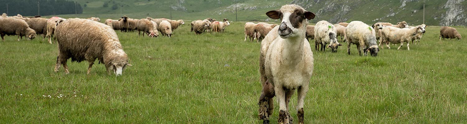
- Ovine and caprine anaplasmosis
- Vectors: Ticks
- Vectors: Tsetse flies
- Vectors: Muscidae
- Vectors: Tabanidae
- Vectors: Culicoides spp.
- Vectors: Mosquitoes
- Classification, epidemiology and control of arthropod-borne viruses
- Special factors affecting the control of livestock diseases in sub-Saharan Africa
- The control of infectious diseases of livestock: Making appropriate decisions in different epidemiological and socioeconomic conditions
- Infectious diseases of animals in sub-Saharan Africa: The wildlife⁄livestock interface
- Vaccination: An approach to the control of infectious diseases
- African animal trypanosomoses
- Dourine
- Trichomonosis
- Amoebic infections
- GENERAL INTRODUCTION: COCCIDIA
- Coccidiosis
- Cryptosporidiosis
- Toxoplasmosis
- Besnoitiosis
- Sarcocystosis
- Balantidiosis
- Leishmaniosis
- Neosporosis
- Equine protozoal myeloencephalitis
- GENERAL INTRODUCTION: BABESIOSES
- Bovine babesiosis
- Equine piroplasmosis
- Porcine babesiosis
- Ovine babesiosis
- GENERAL INTRODUCTION: THEILERIOSES OF CATTLE
- East Coast fever
- Corridor disease
- Zimbabwe theileriosis
- Turning sickness
- Theileria taurotragi infection
- Theileria mutans infection
- Theileria annulata theileriosis
- Theileriosis of sheep and goats
- Theileria buffeli⁄orientalis infection
- Non-pathogenic Theileria species in cattle
- GENERAL INTRODUCTION: RICKETTSIAL, CHLAMYDIAL AND HAEMOTROPIC MYCOPLASMAL DISEASES
- Heartwater
- Lesser known rickettsial infections in animals and humans
- Chlamydiosis
- Q fever
- Eperythrozoonosis
- Bovine Haemobartonellosis
- Potomac horse fever
- GENERAL INTRODUCTION: ANAPLASMOSES
- Bovine anaplasmosis
- Ovine and caprine anaplasmosis
Ovine and caprine anaplasmosis
This content is distributed under the following licence: Attribution-NonCommercial CC BY-NC  View Creative Commons Licence details here
Ovine and caprine anaplasmosis
W H STOLTSZ
Introduction
Anaplasmosis of sheep and goats is an arthropod-borne disease caused by the intra-erythrocytic rickettsial organism, Anaplasma ovis. It is usually a subclinical or mild condition, but moderate to severe clinical disease is generally characterized by fever and a variable degree of anaemia and icterus that may occasionally lead to death. Recovered animals remain carriers of the organism, resulting in premunity, which persists for life.
Anaplasmosis in sheep was first reported in 1912 in Zimbabwe. 4 Although a number of reports of Anaplasma infections in sheep and goats in Africa soon followed,51, 52, 62 the report by Di Domizio in 19198 is considered to be the first authentic description of anaplasmosis in non-bovine hosts. In 1924 Sergent et al.49 reported an Anaplasma infection in sheep that was not transmissible to cattle, and Lestoquard22 differentiated the causative organism from the Anaplasma spp. of cattle, describing and naming it A. ovis.
Ovine and caprine anaplasmosis occurs in many parts of Africa, North and South America, Asia (including the Middle and Far East), the southern and central parts of eastern and western Europe, and the former USSR.2, 13, 19, 28, 32, 35, 43, 53, 54, 56, 66, 69 However, the disease may be more widely distributed than present data indicate.
The condition in southern Africa is of relatively little economic importance. Because of its often insidious nature, the deleterious effects caused by A. ovis infection, such as weight loss and unthriftiness, are probably erroneously ascribed to other causes. Although mortality due to anaplasmosis in sheep and goats is seldom observed in southern Africa, it would appear that under certain circumstances, overt disease, abortion and mortality may occur.1, 3, 35
Aetiology and life cycle
Anaplasma ovis is the main cause of ovine and caprine anaplasmosis. It is more pathogenic for goats than for sheep56, 63, 69 and only very rarely is it infective to cattle.20, 48
Anaplasma mesaeterum, the name proposed for a hitherto unrecognized anaplasm of sheep and goats,63 is closely related to A. ovis and may also cause clinical disease in sheep and goats, but, unlike A. ovis, it appears to be less pathogenic for goats than for sheep and is not infective for cattle.63 Anaplasma marginale, a species pathogenic for cattle, may also, under certain circumstances, cause latent infection in sheep and goats.9, 16, 24, 29, 34, 36
In Giemsa-stained blood smears A. ovis is morphologically indistinguishable from A. marginale,56, 57 and appears as irregularly shaped, almost spherical, intra-erythrocytic granules staining a deep purple colour. The organisms (inclusion bodies) vary in diameter from approximately 0,4 to 0,8 μm,22, 35, 57 with an average of 0,5 μm.35 Within erythrocytes, 60 to 70 per cent of the inclusion bodies are located marginally and 30 to 40 per cent submarginally or centrally.22, 34, 56, 57 In comparison, up to 90 per cent of the inclusion bodies of A. marginale are situated marginally.34 Differences between A. ovis and A. marginale with regard to the intra-erythrocytic location of organisms have been found to be statistically significant.54
The ultrastructure of the marginal bodies of A. ovis in sheep erythrocytes is very similar to that of A. marginale in bovine erythrocytes.15, 30, 44 Development of A. ovis in sheep and goat erythrocytes, and in the tick vector, is presumably similar to the cycle of development proposed for A. marginale. 16, 17, 45, 57 The inclusion bodies of A. mesaeterum are morphologically indistinguishable by both light and electron microscopy from A. ovis, but a distinctive feature is that fewer than 30 per cent of the inclusion bodies are situated marginally in erythrocytes.
Apart from the morphological similarities, complete serological cross-reactivity has been demonstrated between A. ovis and A. marginale.57 They are nevertheless immunologically distinct as infection with one does not provide immunity against the other.56 Anaplasma ovis and A. mesaeterum, on the other hand, appear to be immunologically more closely related, but cross-immunity is nevertheless incomplete, a situation somewhat reminiscent of the relationship between A. centrale and A. marginale in cattle.63
Epidemiology
Anaplasmosis in sheep and goats has been reported from most tropical and subtropical parts of the world.2, 19, 28, 35, 43, 53, 54, 56, 66, 69 With the exception of Namibia, A. ovis has been reported from all the countries in southern Africa and is believed to be prevalent in most sheep- and goat-farming areas. In these areas, small numbers of parasites may frequently be detected during routine microscopic examination of blood smears from sheep and goats. Considering that microscopic examination of blood smears and serological techniques fail to distinguish between A. ovis and A. marginale,20, 56 the fact that sheep and goats exposed to A. marginale may yield serologically positive results without being immune to A. ovis may be of epidemiologic significance. Similarly, calves infected with A. ovis have been noted to react positively to A. marginale antigens in the complement fixation and rapid card agglutination tests, and yet were fully susceptible to A. marginale infection.20 Thus far A. mesaeterum has only been recorded on the Dutch island, Ameland, but circumstantial evidence seems to indicate a possible wider occurrence in northern and north-eastern Europe.63
The prevalence rates of A. ovis infection in sheep or goats vary considerably within endemic areas, and may be influenced by management factors such as the control measures employed and the movement of livestock from nonendemic to endemic areas.35 Although no extensive surveys have been conducted to determine the prevalence of A. ovis in South Africa, limited microscopic observations in sheep have revealed a prevalence ranging from 33 to 90 per cent in endemic areas.59, 64 Similarly, in Mozambique, its prevalence ranges from 0 to 85 per cent in sheep and from 0 to 90 per cent in goats.2 In India it has been shown that approximately 60 per cent of sheep and 70 per cent of goats are infected, 27 while in Kenya the prevalence of infection in goats from different localities ranges from 22 to 87 per cent.54 Although sheep and goats of all ages are susceptible to A. ovis infection,1, 54, 56, 60 older animals suffer from a greater reduction in haematocrit values.56, 69
Overt clinical disease and mortality are rare in nonsplenectomized animals,11, 35 but splenectomy drastically increases the susceptibility of animals and usually results in a recrudescence of parasitaemia (relapse) in carrier animals.6, 7, 19, 20, 28, 34, 47, 56 It may thus be assumed that any condition that results in suppression of immunity or decreased resistance to disease in animals, such as debilitating diseases, concurrent infections (particularly of other arthropod- borne rickettsial and protozoal infections such as Babesia spp., Theileria spp., Trypanosoma spp. and Eperythrozoon ovis,2, 5, 34, 57, 59, 60 heavy helminth or tick parasitism,31 malnutrition,13 and other stress factors (such as pregnancy), as well as variations in the pathogenicity of different parasite strains and individual susceptibility, could lead to a higher prevalence of clinical disease and mortality.
The tick vectors of A. ovis in southern Africa have not been determined. Based on the observations elsewhere that A. ovis and A. marginale may share common tick vectors,48, 58 one or more of the tick vectors of A. marginale or A. centrale reported from South Africa38, 39 may also prove to be vectors of A. ovis. However, during experimental studies on the tick transmission of Theileria separata,59 Rhipicephalus evertsi evertsi and Rhipicephalus evertsi mimeticus transmitted only T. separata from splenectomized sheep infected with both T. separata and A. ovis.
Experimental studies have indicated transstadial transmission of A. ovis by Ornithodoros lahorensis,41 Rhipicephalus bursa40 and Rhipicephalus turanicus.56 Transovarial transmission in Dermacentor silvarum has also been reported. 42 Adults of Dermacentor andersoni have been found to be naturally infected,48 and intrastadial transmission by adults of this tick species has been demonstrated.58 Other tick species that have been incriminated as vectors include Hyalomma plumbeum,37 Haemaphysalis otophila,37 Dermacentor albipictus, D. occidentalis and D. variabilis.58 Haemaphysalis punctata and Ixodes ricinus are suspected of being vectors of A. mesaeterum.63
Considering the ease with which A. ovis can be transmitted by the subinoculation of blood, transmission by iatrogenic means or mechanical transmission by biting flies would appear to be distinct possibilities. However, experimental attempts to transmit the parasite by means of bites of the sheep ked, Melophagus ovinus, a likely candidate as a mechanical vector of A. ovis in sheep, have failed, even when sheep with high parasitaemias were used as the source of the infection.68
Intra-uterine transmission has been reported in sheep10 and in goats.3 In sheep, transplacental infection has been found to occur during the second and third trimester of pregnancy, but no lesions have been observed in foetuses or neonatal lambs.67 In contrast, a high percentage of artificially infected female goats aborted or resorbed their foetuses following acute infection.3
Sheep and goats that have recovered from A. ovis infection remain carriers of the parasite, and in areas where both sheep and goats occur, sheep may be particularly important as reservoir hosts because of the lower pathogenicity of A. ovis in this host.55
White-tailed deer (Odocoileus virginianus), eland (Taurotragus oryx) and blesbok (Damaliscus dorcas phillipsi ) are susceptible to experimental infection with A. ovis,12, 18, 34, 36 and these hosts could act as potential reservoir hosts of infection for sheep and goats.12, 34 Although wild ruminants usually develop only mild or subclinical infection, severe disease may occur after splenectomy.18, 34
A variety of North American, Asian and African wild ruminant species has been found to be susceptible to A. marginale infection (see Bovine anaplasmosis). Naturally acquired Anaplasma infections isolated from blue wildebeest (Connochaetes taurinus), Coke’s hartebeest (Alcelaphus buselaphus cokii) and Thompson’s gazelle (Gazella thompsonii ) resulted in mild infections in calves and provided little or no immunity to subsequent A. marginale challenge.25
Positive serological reactions to A. marginale antigens have also been recorded in impala (Aepyceros melampus), waterbuck (Kobus ellipsiprymnus) Grant’s gazelle (Gazella grantii ) and eland,25, 26 but attempts to recover Anaplasma spp. from impala and Grant’s gazelle by inoculation of their blood into cattle have failed. These results seem to indicate the possible existence of hitherto unrecognized Anaplasma spp. in wild ruminants, some of which may be more closely related to A. ovis,21 as has been demonstrated in the case of a sable antelope (Hippotragus niger) in South Africa.61 Circumstantial evidence would similarly seem to indicate the susceptibility of wild ruminant hosts for A. mesaeterum.63
Naturally occurring infections with A. marginale in sheep and goats have not been reported although these species may become latently infected with A. marginale after experimental infection.30 Prolonged repeated passage by subinoculation of blood in sheep may lead to the gradual adaptation of A. marginale to this host, which results in a higher parasitaemia during infection and an attenuation of the organism for cattle.9, 14, 16, 20, 24, 36, 43, 46
Sheep and goats cannot be consistently infected experimentally with field isolates of A. marginale,7, 16, 29 indicating that there are possible strain differences of A. marginale with regard to its ability to infect these hosts. Furthermore, the carrier state of A. marginale in sheep and goats may be of short duration.11, 29 In splenectomized goats this transient carrier state has been found not to exceed 208 days.29
Anaplasma ovis has only rarely been shown to be infective for cattle. Numerous attempts to transmit it to non-splenectomized48 and splenectomized cattle have failed,7, 9, 30, 34, 47, 56 and this characteristic of A. ovis is often used to differentiate it from A. marginale. This difference would seem to indicate a greater host specificity in A. ovis than in A. marginale. However, short-term survival of A. ovis in splenectomized calves has been demonstrated,48 but confirmation of infection requires the subinoculation of blood into splenectomized sheep. In one study,20 A. ovis could not be recovered from a splenectomized calf at 17 days post-infection, but the animal was subsequently found to have retained the infection for up to 262 days. No loss of virulence of A. ovis for sheep has been observed following maintenance in splenectomized calves.20, 48
Pathogenesis
The pathogenesis of ovine and caprine anaplasmosis appears to be similar to that of A. marginale infection in cattle ( see Bovine anaplasmosis) and, as in bovine anaplasmosis, anaemia results from erythrophagocytosis. 37 Unlike the situation in bovine anaplasmosis, there appears to be no marked variation in age susceptibility to A. ovis infection in sheep and goats, and both young and adult animals usually develop only a mild form of the disease.35, 56
Various stress factors and splenectomy increase individual susceptibility and predispose to acute disease. 5, 13, 34, 48, 56
Splenectomy of chronically infected (carrier) animals is frequently followed by a severe recrudescence of parasitaemia and manifestation of clinical disease, indicating the importance of the spleen in controlling the infection and maintaining immunity.5–7, 19, 20, 47, 56, 59
In experimental transmission studies, in which A. ovis infected blood is used as the inoculum, the prepatent period may vary from 5 to 40 days, with an average of about two to three weeks.20, 22, 35, 56, 57 Generally, the duration of the prepatent period is inversely proportional to the number of parasites in the infective inoculum.23 Similarly, following transmission by a single pair of adult Dermacentor andersoni, a prepatent period of slightly more than six weeks has been recorded in a splenectomized sheep,48 whereas the prepatent periods are shorter when there are larger numbers of ticks.58
The parasitaemia gradually increases to a maximum level one or two weeks after the parasites have first become microscopically detectable.3, 19, 56, 57 In non-splenectomized animals, the maximum parasitaemia may range from approximately 0,1 to 4 per cent in sheep, and approximately 1 to 12 per cent in goats, but in splenectomized animals the maximum parasitaemia is more variable and may range from 2 to 90 per cent.19, 20, 28 The course of A. ovis infection in splenectomized sheep is identical to that of A. marginale in cattle, with the number of parasites doubling approximately every 24 hours.57
During the period of rising parasitaemia, parasites are removed from the circulation by phagocytosis of infected erythrocytes. This results in a gradual decline in the number of parasites and a concomitant development of a progressive anaemia. The haematocrit value gradually declines and reaches its lowest level a few days after the peak of parasitaemia, when parasite numbers are already reduced. 19, 69
The severity of the anaemia is usually greater than can be accounted for by simple removal of parasitized erythrocytes alone. Severe anaemia in the absence of a high parasitaemia31, 63 suggests an autoimmune response which results in the phagocytosis of infected as well as uninfected erythrocytes by the reticuloendothelial system.63 In nonsplenectomized sheep and goats, with average maximum parasitaemias of 0,8 and 2 per cent respectively, the corresponding average reductions in haematocrit values were approximately 40 and 50 per cent.56 However, in individual animals, the reduction in haematocrit values may be considerably greater. Similar reductions in haematocrit values were observed in A. mesaeterum infection, despite the presence of a low parasitaemia.63 In splenectomized animals, haematocrit values may be reduced to well below 10 per cent.30
The corresponding drop in the haemoglobin concentration, 3, 31, 48, 56, 69 if severe, will lead to hypoxic damage to various organs, which may ultimately result in the death of the animal.
Sheep and goats that recover from A. ovis infection remain carriers of the parasite.22, 34, 35
Clinical signs
The clinical or patent phase of the disease lasts approximately one to two weeks.19, 30 In animals that survive this phase, convalescence begins with increased erythropoiesis, which is characterized by reticulocytosis, anisocytosis, polychromatophilia, basophilic stippling of immature erythrocytes and the presence of Howell-Jolly bodies. Convalescence is usually slow and may last from several weeks to a few months.69 During this time, the haematological and clinical signs gradually return to normal.
Anaplasma ovis infection in sheep and goats usually results in subclinical or mild disease, but occasionally severe clinical disease and mortality may occur. When mortality does occur, its rate is seldom very high and may be due, in part, to concurrent infections with other haemoparasites.11 Because of the greater pathogenicity of A. ovis for goats, clinical signs of infection are more frequently observed in them than in sheep.3, 31, 56, 69
Fever is not always present and temperature reactions are quite variable. A slight rise in body temperature may occur at the onset of patent parasitaemia, followed by a return to normal for a few days before it again becomes elevated. 3 The maximum rise in body temperature usually coincides with the peak of parasitaemia,22, 56 and may range from 40 to 42 °C. In some animals the temperature may fluctuate during the clinical phase of the disease,22 while in others it may remain elevated for more than a week, before returning to normal.3, 35
The clinical signs are usually most pronounced at the height of anaemia, which occurs a few days after the parasitaemic peak and approximately one to two weeks after parasites first become microscopically detectable. The severity of clinical disease is to some extent dependent on the degree of the maximum parasitaemia,22 but severe anaemia may occur in the absence of a high parasitaemia.31, 56 However, even in animals with low haematocrit values, no obvious clinical signs of disease may be observed,1, 3, 56 but signs of rapid fatigue and severe respiratory distress, indicative of lung oedema, may become evident when they are herded or forcefully exercised.3, 56 Occasionally, animals may die acutely upon exertion.1
Generally, only slight listlessness and weakness,22, 56, 69 or a loss of condition5 are noticeable. The appetite usually remains unaffected, but reduced appetite or anorexia, rumen stasis and constipation may be observed in some severely affected animals.3, 56, 60, 69
On closer clinical examination of affected animals signs of anaemia, such as pale mucous membranes, thin watery blood, and increased heart and respiration rates are evident.5, 22, 69
Icterus is not consistently observed, even in cases which end fatally.35, 69 Submandibular oedema of the jowl and ventral part of the neck may be noticed.35 In milch goats, a marked drop in milk yield may occur, and this may persist for several weeks.35
The return of normal appetite and an improvement in the general condition of the animal may serve to indicate recovery.31 However, recovery from clinical disease is usually slow,67 and anaemia and emaciation may persist for several weeks when the nutrition of the animals is inadequate.35
Abortion and resorption of embryos have been reported in domestic goats infected experimentally with A. ovis.3 These phenomena occurred particularly in those animals which had developed a high fever and severe anaemia. Anaplasma ovis was transmitted transplacentally and frequently caused anaemia and death of the foetus in utero. In foetuses and live or stillborn kids, the A. ovis-parasitaemia ranged from 1 to 12 per cent.
Pathology
The severity of the lesions vary according to the duration and the severity of the disease.35 Numerous reports include descriptions of necropsy findings.1, 3, 22, 28, 35, 56, 60, 69
The gross lesions to a large extent resemble those found in cattle that die of anaplasmosis. The carcass generally appears pale because of the anaemia, and may be slightly or moderately icteric. The blood is thin and watery, and emaciation, serous atrophy of fatty tissues, and anasarca or dehydration may also be apparent. Increased amounts of straw-coloured fluid may be present in body cavities. The lungs are pale or congested, and oedematous. The liver is enlarged and yellowish-brown and the gall bladder may be enlarged and filled with viscous yellow-green bile. Nephrosis and enlargement of lymph nodes and adrenals may also be evident.
Microscopically in blood smears, evidence of a regenerative anaemia, characterized by the presence of reticulocytosis, anisocytosis, polychromasia, basophilic stippling and Howell-Jolly bodies, is found. In animals suffering from severe anaemia a monocytosis with erythrophagocytosis (as evidenced by the presence of parasitized erythrocytes, or their remnants, in macrophages) and eosinophilia (after a number of remissions of severe anaemia) may be observed.5
Changes in parenchymal organs characteristic of anaemic and hypoxic states may be seen in various organs (e.g. centrilobular degeneration and necrosis of varying intensity in the liver, and degeneration especially in cells of the convoluted tubules of the kidneys). The canaliculi in the liver may be distended as a result of the bile stasis. The red bone marrow is usually hyperplastic, but in chronic cases may show evidence of depletion. Haemosiderin accumulation in reticuloendothelial cells is usually most extensive in the spleen.
Diagnosis
The possibility of anaplasmosis should be considered in all cases of anaemia in sheep and goats in the tropics and subtropics.
During the acute stage of the disease, a diagnosis in the individual animal is usually made on the basis of clinical signs, the presence of the organism in blood smears and haematological evidence of infection. However, when clinical signs are most pronounced the degree of parasitaemia may have already declined considerably and confirmation of the diagnosis may be difficult.56 In addition, the presence of a regenerative anaemia during this stage may complicate the microscopic detection of parasites (Anaplasma inclusions at times being difficult to differentiate from basophilic stippling in reticulocytes and Howell-Jolly bodies or other stained particulate matter in erythrocytes). Microscopic detection of infection during the chronic or carrier stage is also often extremely difficult because of the exceedingly low parasitaemia. In doubtful clinical cases and during epidemiological surveys, serological tests, subinoculation of blood into susceptible animals, and DNA hybridization techniques may provide additional confirmation.
The subinoculation of blood from suspected carrier animals into susceptible splenectomized sheep or goats is an effective way of detecting A. ovis infection, but the technique is too expensive and time-consuming for routine application. 54 Various serological techniques including the complement fixation (CF) test,30, 56, 67 capillary tube agglutination (CT) test,27, 31 direct- and indirect fluorescent antibody (FA and IFA) tests18 and the rapid card agglutination (CA) test30, 67 have been used. Due to the high degree of serological cross-reactivity between A. ovis and A. marginale,30, 56 either of these may be used as antigen, but A. marginale antigen is most frequently employed because of its greater availability.
Sheep and goats, on average, develop positive reactions in the CF test from a few days before the onset of patent parasitaemia.48, 56 Antibody titres are highest during patent parasitaemia and lowest during the carrier phase but, generally, sheep and goats develop lower antibody titres than do cattle infected with A. marginale. The majority of sheep and goats remain serologically positive after one year, but some known carriers have been observed to revert to negativity against A. marginale and A. ovis antigen.48, 56 The CA test similarly seems to lack the sensitivity to detect all carrier infections in sheep and goats.30, 59
Uninfected lambs born of infected ewes may react positively in the CA test and the CF test for variable periods, but antibody levels diminish or disappear at the age of approximately three or four months.67
The CT test has been found to be a reliable technique for the detection of infection in goats.31
Similarly, a competitive inhibition enzyme-linked immunosorbent assay (CI-ELISA) based on a major surface protein 5 (MSP 5) B-cell epitope which is conserved among Anaplasma spp.65 was shown to reliably detect different isolates of A. ovis in both acute and chronically infected goats.33 Results suggested that the sensitivity and specificity of the test was high.
Serological techniques fail to distinguish between A. ovis and A. marginale and such differentiation may be of epidemiologic significance. DNA hybridization techniques, using either radio-labelled or biotin-labelled species-specific probes,54, 64 can differentiate between these species and appear to be more sensitive than the examination of blood smears by light microscopy for the detection of A. ovis infection in sheep and goats.
Differential diagnosis
The clinical signs of anaplasmosis in sheep and goats are non-specific and the disease, therefore, should be differentiated from other infectious and non-infectious causes of such signs as fever, anaemia, icterus, listlessness, inappetance, loss of condition, constipation, and submandibular oedema of the ventral region of the neck.
Fever, listlessness, inappetance, and loss of condition may be caused by a variety of infectious and non-infectious diseases including babesiosis, eperythrozoonosis, ehrlichiosis [Ehrlichia ovina and Ehrlichia (Cytoecetes) phagocytophila infections], theileriosis, trypanosomosis and severe internal or external parasitism. Of these, eperythrozoonosis and verminosis (e.g. haemonchosis) are probably the most likely to be confused with anaplasmosis in southern Africa,and either may occur concurrently in the same host. Eperythrozoon ovis frequently occurs as part of a mixed infection with A. ovis,12, 18, 34, 57 but the former is distinguishable by its generally smaller size and extracellular location.
Malnutrition, plant (e.g. Brassica spp.) or mineral (e.g. copper) intoxications, or deficiencies (e.g. cobalt and copper) may also cause anaemia. Icterus may to a greater or lesser extent also occur in certain plant and mineral (e.g. copper) intoxications. In addition to icterus, inappetance and weakness, chronic copper poisoning is also characterized by haemoglobinuria,47 which is not a sign associated with anaplasmosis.
It should also be borne in mind that the usually mild clinical signs of anaplasmosis may be exacerbated by concurrent infections or non-infectious conditions.
Control
Both specific and supportive therapy should be initiated as soon as possible, preferably before the onset of severe anaemia, in order to avoid mortality.31
Chlortetracycline and oxytetracycline are effective for the treatment of A. ovis infections.35 Anaplasmosis can be successfully treated by the intramuscular administration of oxytetracycline at a rate of 8 to 10 mg/kg body weight daily, administered in equally divided doses at 12-hour intervals for four to five days.31 The administration of divided doses at 12- hourly intervals is desirable in order to maintain higher blood levels throughout the period of treatment. However, two to three daily intravenous or intramuscular administrations of oxytetracycline at a rate of 10 mg/kg have also proved to be effective for the treatment of acute anaplasmosis in sheep.59 A single intramuscular administration of a long-acting formulation of oxytetracycline, at a rate of 20 mg/kg body weight, is also effective in preventing mortality in sheep.67
Symptomatic treatment and rest are beneficial.35 Supportive therapy may consist of the administration of haematinics, tonics and fluid.31 Gastrointestinal disturbances such as inappetence, constipation and rumenal stasis may complicate recovery and, where indicated, attempts should be made to correct such disorders by the administration of appetite stimulants, rumenotorics and mild laxatives. In valuable animals suffering from severe anaemia, blood transfusion may be indicated.
Adequate protection against the elements and easy access to palatable feed and water should be provided. In addition, anaemic animals should not be subjected to unnecessary exertion and rams should be rested before being used for breeding.35
Judicious control of external and internal parasites should be practised.35 Since intercurrent viral, bacterial, rickettsial and protozoal infections may precipitate disease,35, 56 these should also be controlled wherever possible.
Neither oxytetracycline (12 daily intravenous administrations at a rate of 10 mg/kg body weight) nor a combination of oxytetracycline (four intramuscular administrations of a long-acting formulation at a rate of 20 mg/kg body weight at seven-day intervals) and imidocarb (two intramuscular administrations at a rate of 5 mg/kg body weight at an interval of 14 days) eliminates A. ovis carrier infections in splenectomized sheep, although these treatment regimens do result in a transient suppression of parasitaemia to a level where parasites are no longer microscopically detectable in blood smears.59
In practice, sheep and goats are not usually vaccinated against anaplasmosis. Animals could be immunized by artificially infecting them with A. ovis-infected blood35 possibly followed by tetracycline treatment to control the infection. Care should be taken when selecting a suitable donor animal to avoid the accidental transmission of other blood-borne diseases. One or two administrations (at an interval of seven days) of a long-acting oxytetracycline formulation, administered during the prepatent period in experimentally induced A. ovis infection in sheep, has been shown to prolong the incubation period and to reduce the severity of the clinical reaction.50
References
- ANON., 1990. Anaplasma ovis. Monthly Report of the Regional Veterinary Laboratories for the month of January, Department of Veterinary Services, Republic of South Africa.
- ARNOLD, R.M. & TRAVASSOS SANTOS DIAS, J.A., 1983. Ticks and tickborne haemoparasites of sheep and goats. Seasonal distribution in Mozambique. World Animal Review, 45, 28–35.
- BARRY, D.M. & VAN NIEKERK, C. H., 1990. Anaplasmosis in improved Boer goats in South Africa artificially infected with Anaplasma ovis. Small Ruminant Research, 3, 191–197.
- BEVAN, L.E.W., 1912. Anaplasmosis of sheep. Veterinary Journal, 68, 400–401.
- DE KOCK, G., 1930. A short note on chronic anaplasmosis and gonderiosis in small ruminants after splenectomy. Sixteenth Report of the Director of Veterinary Services and Animal Industry, Union of South Africa, 3–10.
- DE KOCK, G. & QUINLAN, J., 1924. A short preliminary communication on anaplasmosis of sheep as observed in South Africa. Bulletin de la Société de Pathologie exotique, 17, 651–653.
- DE KOCK, G. & QUINLAN, J., 1926. Splenectomy in domesticated animals and its sequelae, with special reference to anaplasmosis in sheep. Eleventh and Twelfth Reports of the Director of Veterinary Education and Research, Union of South Africa, 369–480.
- DI DOMIZIO, G., 1919. Dell’ Anaplasma marginale. (Corpi di Jolly nel sangue anemico. Forme anaplasmatiche di piroplasmi). Clínica veterinaria e rassegna di polizia sanitaria di igiena, 42, 203–311.
- DONATIEN, A. & LESTOQUARD, F., 1930. Les anaplasmoses des ruminants. Revue Vétérinaire et Journal de Médecine vétérinaire et de zootechnie, 82, 125–139.
- DONATIEN, A., LESTOQUARD, F. & MILCHER-MANCOURT, A., 1934. Passage à travers le placenta de Babesiella ovis et de Anaplasma ovis. Algérie Médicale, 38, 320–321.
- DU TOIT, P.J., 1934. Anaplasmosis (abstract). The Veterinary Record, 14, 1266–1267.
- ENIGK, K., 1942. Die Empfänglichkeit der Elenantilope fu¨r Anaplasma ovis und Eperythrozoon ovis. Deutsche Tropenmedizinische Zeitschrift, 46, 48–52.
- GILBERT, S.J., 1926. A preliminary communication on the occurrence of anaplasmosis in sheep and goats in Palestine. Journal of Comparative Pathology and Therapeutics, 34, 246–247.
- HENNING, M.W., 1956. Animal Diseases in South Africa. 3rd edn. Pretoria: Central News Agency Limited.
- JATKAR, P.R., 1969. Electron microscopy study of Anaplasma ovis. American Journal of Veterinary Research, 30, 1891–1892.
- KESSLER, R.H. & RISTIC, M., 1979. In vitro cultivation of Anaplasma marginale: Invasion of and development in noninfected erythrocytes. American Journal of Veterinary Research, 40, 1774–1776.
- KOCAN, K.M., 1986. Development of Anaplasma marginale in ixodid ticks: Coordinated development of a rickettsial organism and its host tick. In: SAUER, J. & HAIR, J.A., (eds). Morphology, Physiology and Behavioural Ecology of Ticks. England: Ellis Horwood Ltd.
- KREIER, J.P. & RISTIC, M., 1963. Anaplasmosis. VII. Experimental Anaplasma ovis infection in a white-tailed deer (Dama virginiana). American Journal of Veterinary Research, 24, 567–572.
- KUIL, H. & FOLKERS, C., 1967. Studies on Anaplasma ovis infection. I. Course of spontaneous infections in splenectomised Nigerian sheep and goats. Bulletin of Epizootic Diseases of Africa, 16, 65–70.
- KUTTLER, K.L., 1981. Infection of splenectomized calves with Anaplasma ovis. American Journal of Veterinary Research, 42, 2094–2096.
- KUTTLER, K.L., 1984. Anaplasma infections in wild and domestic ruminants: A review. Journal of Wildlife Diseases, 20, 12–20.
- LESTOQUARD, F., 1924. Deuxième note sur les Piroplasmoses du mouton en Algérie. L’anaplasmose: Anaplasma ovis nov. sp. Bulletin de la Société de Pathologie exotique, 17, 784–787.
- LESTOQUARD, F., 1926. Les piroplasmoses du mouton et de la chèvre. Archives l’Institut Pasteur d’Algérie, 4, 222–317.
- LIGNIERES, J., 1919. La vaccination des bovides contre l’anaplasmose. L’Anaplasma inoculé au mouton et à la chèvre s’atténue dans l’organisme de ces espèces animales et leur sang est alors un excellent vaccin pour les bovides contre l’anaplasmose la plus grave. Bulletin de la Société de Pathologie exotique, 12, 765–774.
- LÖHR, K.F. & MEYER, H., 1973. Game anaplasmosis: The isolation of Anaplasma organisms from antelope. Zeitschrift fu¨r Tropenmedizin und Parasitologie, 24, 192–197.
- LÖHR, K.F., ROSS, J.P.J. & MEYER, H., 1974. Detection in game of fluorescent and agglutination antibodies in intraerythrocytic organisms. Zeitschrift fu¨r Tropenmedizin und Parasitologie, 25, 217–226.
- LOSOS, G.J., 1986. Anaplasmosis. Infectious Tropical Diseases of Domestic Animals. Harlow, UK: Longman Scientific and Technical.
- LU, W.S., LU ,W.X., ZHANG, Q.C., DOU, H.F., YU, F., YIN, H., JI, Z.D., HU, T.Y., SUN, B.G., HAN, J.S., GAO, D.F. & BAO, Y.J., 1987. Study on anaplasmosis in goats. Identification of the pathogen and observation of the clinical signs. Chinese Journal of Veterinary Science and Technology, 3, 5–8.
- MAAS, J. & BUENING, G.M., 1981. Characterization of Anaplasma marginale infection in splenectomized domestic goats. American Journal of Veterinary Research, 42, 142–145.
- MAGONIGLE, R.A., ECKBLAD, W.P., LINCOLN, S.D. & FRANK, F.W., 1981. Anaplasma ovis in Idaho sheep. American Journal of Veterinary Research, 42, 199–201.
- MALLICK, K.P., DWIVEDI, S.K. & MALHOTRA, M.N., 1979. Anaplasmosis in goats: Report on clinical cases. Indian Veterinary Journal, 56, 693–694.
- MOHAN, R.N. & PATHAK, R.C., 1966. Prevalence of anaplasmosis in ruminants in Northern India. Indian Veterinary Journal, 43, 685–689.
- NDUNG’U, L.W., AGUIRRE, C., RURANGIRWA, F.R., MCELWAIN, T.F., MCGUIRE, T.C., KNOWLES, D.P. & PALMER, G.H., 1995. Detection of Anaplasma ovis infection in goats by major surface protein 5 competitive inhibition enzyme-linked immunosorbent assay. Journal of Clinical Microbiology, 33, 675–679.
- NEITZ, W.O., 1939. Ovine anaplasmosis: The transmission of Anaplasma ovis and Eperythrozoon ovis to the blesbuck (Damaliscus albifrons). Onderstepoort Journal of Veterinary Science and Animal Industry, 13, 9–16.
- NEITZ, W.O., 1968. Anaplasma ovis infection. Bulletin de L’Office International des Epizooties, 70, 359–365.
- NEITZ, W.O. & DU TOIT, P.J., 1932. Bovine anaplasmosis: A method of obtaining pure strains of Anaplasma marginale and Anaplasma centrale by transmission through antelopes. Eighteenth Report of the Director of Veterinary Services and Animal Industry, Union of South Africa, 3–20.
- PIPANO, E., 1966. Piroplasmosis—A review. Refuah Veterinarith, 23, 52–47.
- POTGIETER, F.T., 1979. Epizootiology and control of anaplasmosis in South Africa. Journal of the South African Veterinary Association, 50, 367–372.
- POTGIETER, F.T., 1981. Tick transmission of anaplasmosis in South Africa. Proceedings of the International Conference on Tick Biology and Control, Grahamstown, Republic of South Africa, 27–29 January 1981.
- RASTEGAIEFF, E.F., 1933. Zur Frage der Übertrager der Schafpiroplasmosen in Azerbaidschan (Transkaukasien). Archiv fu¨r wissenschaftliche und praktische Tierheilkunde, 67, 176–186.
- RASTEGAIEFF, E.F., 1935. Un nouveau vecteur dans la transmission des hémoparasites des animaux domestiques: Ornithodorus lahorensis, Neumann 1908. Annales de l’Institut Pasteur, 54, 250–258.
- RASTEGAIEFF, E.F., 1937. Dermacentor silvarum, vecteur des hemoparasites du mouton: Anaplasma ovis et Theileria recondita. Bulletin de la Société de Pathologie exotique, 30, 479–480.
- RISTIC, M., 1960. Anaplasmosis. Advances in Veterinary Science, 6, 112–179.
- RISTIC, M., 1968. Anaplasmosis. In: WEINMAN, D. & RISTIC, M., (eds). Infectious Blood Diseases of Man and Animals, Vol. II. New York, London: Academic Press.
- RISTIC, M., 1981. Anaplasmosis. In: RISTIC, M. & MCINTYRE, I., (eds). Diseases of Cattle in the Tropics. The Hague, Boston, London: Martinus Nijhoff Publishers.
- RISTIC, M., SIBINOVIC, S. & WELTER, C.J., 1969. An attenuated Anaplasma marginale vaccine. Proceedings of the United States Livestock Sanitary Association, 72, 56–69.
- RYFF, J.F., GILBERT, C.S., WEIBEL, J.L. & BREEN, H., 1958. Anaplasmosis and concurrent copper intoxication in sheep. Journal of the American Veterinary Medical Association, 133, 312–315.
- RYFF, J.F., WEIBEL, J.L. & THOMAS, G.M., 1964. Relationship of ovine to bovine anaplasmosis. Cornell Veterinarian, 54, 407–414.
- SERGENT, E., DONATIEN, A., PARROT, L., LESTOQUARD, F., PLANTUREUX, E. & ROUGEBIEF, H., 1924. Inoculation au mouton de l’anaplasme du boeuf. Bulletin de la Société de Pathologie exotique, 17, 295–298.
- SHARMA, S.P. & BANSAL, G.C., 1985. Chemoprophylaxis of Anaplasma ovis infection in sheep with a long-acting oxytetracycline. Veterinary Parasitology, 17, 255–258.
- SCHELLHASE, W., 1912. Eine Beobachtung u¨ber das Vorkommen von Marginal-points (Anaplasma marginale) im Blut von Schafen in Deutch-Ostafrika. Berliner und Mu¨nchener tierärtzliche Wochenschrift, 28, 511–512.
- SCHELLHASE, W., 1913. Beobachtungen u¨ber die Anaplasmosis und Piroplasmosis der Schafe und Ziegen in Deutch-Ostafrika. Zeitschrift fu¨r Infektionskrankheiten, parasitäre Krankheiten und Hygiene der Haustiere, 13, 349–352.
- SHIMSHONI, A., 1967. Anaplasmosis in East-Fresian lambs. Refuah Veterinarith, 24, 213–218.
- SHOMPOLE, S., WHAGELA, S.D., RURANGIRWA, F.R. & MCGUIRE, T.C., 1989. Cloned DNA probes identify Anaplasma ovis in goats and reveal a high prevalence of infection. Journal of Clinical Microbiology, 27, 2730–2735.
- SINHA, G.K. & PATHAK, R.C., 1966. Anaplasmosis in goats and sheep. Indian Veterinary Journal, 43, 490–493.
- SPLITTER, E.J., ANTHONY, H.D. & TWIEHAUS, M.J., 1956. Anaplasma ovis in the United States. Experimental studies with sheep and goats. American Journal of Veterinary Research, 17, 487–491.
- SPLITTER, E.J., TWIEHAUS, M.J. & CASTRO, E.R., 1955. Anaplasmosis in sheep in the United States. Journal of the American Veterinary Medical Association, 127, 244–245.
- STILLER, D., GOFF, W.L., SHOMPOLE, S.P., JOHNSON, L.W., GORHAM, J.R. & MCGUIRE, T.C., 1989. Dermacentor andersoni Stiles: A natural vector of Anaplasma ovis Lestoquard on sheep in Idaho. Proceedings of the Eighth National Hemoparasite Disease Conference, St. Louis, Missouri, 10–12 April 1989.
- STOLTSZ, W.H., 1988–1990. Veterinary Research Institute, Onderstepoort. Unpublished data.
- SURENDRAN, N.S. & BHASKARA RAO, 1989. Anaplasmosis in sheep in Andhra Pradesh. Indian Veterinary Journal, 66, 672.
- THOMAS, S.E., WILSON, D.E. & MASON, T.E., 1982. Babesia, Theileria and Anaplasma spp. infecting sable antelope, Hippotragus niger (Harris, 1838) in southern Africa. Onderstepoort Journal of Veterinary Research, 49, 163–166.
- TRAUTMANN, O., 1913. Anaplasmosis der Schafe in Deutsch-Ost-Afrika. Berlin Tierärtzliche Wochenschrift, 29, 593–594.
- UILENBERG, G., VAN VORSTENBOSCH, C.J.A.H.V. & PERIÉ, N.M., 1979. Blood parasites of sheep in the Netherlands I: Anaplasma mesaeterum sp.n. (Rickettsiales, Anaplasmataceae). Veterinary Quarterly, 1, 14–22.
- VISSER, E.S., AMBROSIO, R.E. & DE WAAL, D.T., 1991. An Anaplasma centrale DNA probe that differentiates between Anaplasma ovis and Anaplasma marginale DNA. Veterinary Microbiology, 28, 313–325.
- VISSER, E.S., MCGUIRE, T.C., PALMER, G.H., DAVIS, W.C., SHKAP, V., PIPANO, E. & KNOWLES, D.P. JR., 1992. The Anaplasma marginale msp5 gene encodes a 19-kilodalton protein conserved in all recognized Anaplasma species. Infection and Immunity, 60, 5139–5144.
- YOUSIF, Y.A., DIMITRI, R.A., DWIVEDI, S.K., AHMED, N.J., 1983. Anemia due to anaplasmosis in Iraqi goats. I. Clinical and haematological features under field conditions. Indian Veterinary Journal, 60, 576–578.
- ZAUG, J.L., 1987. Ovine anaplasmosis: In utero transmission as it relates to stages of gestation. American Journal of Veterinary Research, 48, 100– 103.
- ZAUG, J.L. & COAN, M.E., 1986. Test of the sheep ked Melophagus ovinus (L) as a vector of Anaplasma ovis Lestoquard. American Journal of Veterinary Research, 47, 1060–1062.
- ZWART, D. & BUYS, J., 1968. Anaplasma ovis infection. II. Pathogenicity of a Nigerian goat strain for Dutch sheep and goats. Bulletin of Epizootic Diseases of Africa, 16, 73–80.


