- Infectious Diseases of Livestock
- Part 3
- Actinobacillus equuli infections
- GENERAL INTRODUCTION: SPIROCHAETES
- Swine dysentery
- Borrelia theileri infection
- Borrelia suilla infection
- Lyme disease in livestock
- Leptospirosis
- GENERAL INTRODUCTION: AEROBIC ⁄ MICRO-AEROPHILIC, MOTILE, HELICAL ⁄ VIBROID GRAM-NEGATIVE BACTERIA
- Genital campylobacteriosis in cattle
- Proliferative enteropathies of pigs
- Campylobacter jejuni infection
- GENERAL INTRODUCTION: GRAM-NEGATIVE AEROBIC OR CAPNOPHILIC RODS AND COCCI
- Moraxella spp. infections
- Bordetella bronchiseptica infections
- Pseudomonas spp. infections
- Glanders
- Melioidosis
- Brucella spp. infections
- Bovine brucellosis
- Brucella ovis infection
- Brucella melitensis infection
- Brucella suis infection
- Brucella infections in terrestrial wildlife
- GENERAL INTRODUCTION: FACULTATIVELY ANAEROBIC GRAM NEGATIVE RODS
- Klebsiella spp. infections
- Escherichia coli infections
- Salmonella spp. infections
- Bovine salmonellosis
- Ovine and caprine salmonellosis
- Porcine salmonellosis
- Equine salmonellosis
- Yersinia spp. infections
- Haemophilus and Histophilus spp. infections
- Haemophilus parasuis infection
- Histophilus somni disease complex in cattle
- Actinobacillus spp. infections
- Actinobacillus equuli infections
- Gram-negative pleomorphic infections: Actinobacillus seminis, Histophilus ovis and Histophilus somni
- Porcine pleuropneumonia
- Actinobacillus suis infections
- Pasteurella and Mannheimia spp. infections
- Pneumonic mannheimiosis and pasteurellosis of cattle
- Haemorrhagic septicaemia
- Pasteurellosis in sheep and goats
- Porcine pasteurellosis
- Progressive atrophic rhinitis
- GENERAL INTRODUCTION: ANAEROBIC GRAM-NEGATIVE, IRREGULAR RODS
- Fusobacterium necrophorum, Dichelobacter (Bacteroides) nodosus and Bacteroides spp. infections
- GENERAL INTRODUCTION: GRAM-POSITIVE COCCI
- Staphylococcus spp. infections
- Staphylococcus aureus infections
- Exudative epidermitis
- Other Staphylococcus spp. infections
- Streptococcus spp. infections
- Strangles
- Streptococcus suis infections
- Streptococcus porcinus infections
- Other Streptococcus spp. infections
- GENERAL INTRODUCTION: ENDOSPORE-FORMING GRAM-POSITIVE RODS AND COCCI
- Anthrax
- Clostridium perfringens group infections
- Clostridium perfringens type A infections
- Clostridium perfringens type B infections
- Clostridium perfringens type C infections
- Clostridium perfringens type D infections
- Malignant oedema⁄gas gangrene group of Clostridium spp.
- Clostridium chauvoei infections
- Clostridium novyi infections
- Clostridium septicum infections
- Other clostridial infections
- Tetanus
- Botulism
- GENERAL INTRODUCTION: REGULAR, NON-SPORING, GRAM-POSITIVE RODS
- Listeriosis
- Erysipelothrix rhusiopathiae infections
- GENERAL INTRODUCTION: IRREGULAR, NON-SPORING, GRAM-POSITIVE RODS
- Corynebacterium pseudotuberculosis infections
- Corynebacterium renale group infections
- Bolo disease
- Actinomyces bovis infections
- Trueperella pyogenes infections
- Actinobaculum suis infections
- Actinomyces hyovaginalis infections
- GENERAL INTRODUCTION: MYCOBACTERIA
- Tuberculosis
- Paratuberculosis
- GENERAL INTRODUCTION: ACTINOMYCETES
- Nocardiosis
- Rhodococcus equi infections
- Dermatophilosis
- GENERAL INTRODUCTION: MOLLICUTES
- Contagious bovine pleuropneumonia
- Contagious caprine pleuropneumonia
- Mycoplasmal pneumonia of pigs
- Mycoplasmal polyserositis and arthritis of pigs
- Mycoplasmal arthritis of pigs
- Bovine genital mycoplasmosis
- Neurotoxin-producing group of Clostridium spp.
- Contagious equine metritis
- Tyzzer's disease
- MYCOTIC AND ALGAL DISEASES: Mycoses
- MYCOTIC AND ALGAL DISEASES: Pneumocystosis
- MYCOTIC AND ALGAL DISEASES: Protothecosis and other algal diseases
- DISEASE COMPLEXES / UNKNOWN AETIOLOGY: Epivag
- DISEASE COMPLEXES / UNKNOWN AETIOLOGY: Ulcerative balanoposthitis and vulvovaginitis of sheep
- DISEASE COMPLEXES / UNKNOWN AETIOLOGY: Ill thrift
- Eperythrozoonosis
- Bovine haemobartonellosis
Actinobacillus equuli infections
This content is distributed under the following licence: Attribution-NonCommercial CC BY-NC  View Creative Commons Licence details here
Actinobacillus equuli infections
M M HENTON
Introduction and aetiology
Actinobacillus equuli is the cause of a peracute to chronic septicaemic condition of new-born foals, referred to as sleepy foal disease. Localization of the bacteria may occur in many tissues or organ systems, including the joints, lungs, heart and kidneys. This results in lesions such as purulent polyarthritis, bronchopneumonia, valvular endocarditis, myocarditis and embolic suppurative nephritis. It may also occasionally be the cause of abortions in mares and septicaemia and arthritis in new-born piglets.
Diseases caused by A. equuli probably occur in most countries, but in South Africa they are sporadic. Only 39 cases of disease caused by A. equuli in horses and one case in pigs were diagnosed bacteriologically at the Onderstepoort Veterinary Institute between 1987 and 1998.5
Actinobacillus equuli was first described by Meyer in 1910 in South Africa, who isolated the bacterium from kidney abscesses in foals and named it Shigella nephritidis equi.4 In the older literature a variety of other names were used for the organism, such as Bacterium viscosum equi and Shigella equirulis.1
The cultural, morphological and other characteristics of A. equuli are outlined in the introduction to Actinobacillus spp. infections. A number of closely related strains such as the apparently non-pathogenic Taxon 11 should be distinguished from A. equuli.18, 1, 2
Epidemiology
Actinobacillus equuli infection and resultant disease occur particularly in horses and occasionally in pigs. Healthy mares and sows may be carriers of the bacterium in their genital tracts (cervix and vagina) and less often in their upper respiratory tracts, particularly the tonsils,11, 20 but the most common site in healthy horses is the oral cavity.19 Horses may be carriers of different strains of A. equuli; it is not known whether all the strains are pathogenic to foals.3, 19
Foals most commonly become infected during or soon after parturition, by direct contact with the saliva19 or infected genital tracts of carrier mares. Transplacental infections and infection via the umbilicus may also occur. Foals less than two weeks old are usually affected, in particular those with a deficient colostral immunity.17
Although piglets are rarely affected by A. equuli, septicaemia complicated by arthritis and meningoecephalitis occurs most commonly in them during the first week of life and immediately after weaning.12 Actinobacillus equuli may also rarely cause metritis and abortion in sows.9, 10
Clinical signs
Signs of septicaemia are usually evident in foals suffering from peracute or acute sleepy foal disease, while those that are affected by the subacute disease generally reveal suppurative lesions in various tissues.
Foals afflicted by peracute disease are born weak, refuse to suck, or do so sluggishly, and are feverish, dull, listless and lethargic. They stand with heads hanging and ears drooping before they become recumbent and die. They seldom live for more than 24 hours.16
The clinical signs of the acute disease in foals are characterized by listlessness, lethargy, inappetence and fever one to three days after birth. Affected foals either abstain from suckling or may suck for a short while before apparently ‘falling asleep’ with the teat in their mouth (from whence the name ‘sleepy foal disease’). Some foals may show swelling and inflammation of the umbilicus, evidence of abdominal pain, shallow and laboured respiration, diarrhoea and frequent micturition before death supervenes usually within a week.1, 7, 11
Foals affected with the subacute disease reveal signs of purulent arthritis and/or bronchopneumonia, which usually develop two or three weeks after birth, but these signs may appear sooner or be delayed for several weeks. The first sign of illness is usually lameness, associated with a variable amount of swelling of one or more joints. The lameness soon increases in severity. Foals often lie down, rise with difficulty, and progressively become weak and emaciated.

In some cases a suppurative omphalophlebitis is present. Many affected foals also manifest signs of purulent bronchopneumonia, such as an increased respiratory rate, painful cough and purulent nasal discharge. The organisms are frequently localized in other organs or tissues, particularly the kidneys and myocardium, and evidence of this may be manifested clinically.1, 7, 11 The course of the subacute disease is protracted and the mortality rate usually not as high as in the peracute and acute disease. The older the foal when clinical signs appear, the greater its chances of recovery. In foals that survive, convalescence is slow and the animals may be emaciated for a protracted period before improvement becomes apparent.4
Actinobacillus equuli is a rare cause of abortion in mares at any stage of gestation. The organism reaches the genital tract either haematogenously or, more frequently, as an ascending infection. Other manifestations of infection are purulent rhinitis, pneumonia and polyarthritis, and tendovagitis in older foals. Wound infections, abscessations and peritonitis may occur in adult horses.3, 7, 8
Clinical signs of the septicaemic disease in piglets are similar to those in foals;12 the bacterium may localize in the joints, brain and kidneys. Some piglets may only show clinical signs of nervous involvement.
Pathology
Foals that die of peracute or acute disease show lesions of septicaemia, including petechiae in serosal surfaces and mucous membranes, generalized congestion, splenomegaly, and oedema and congestion of lymph nodes. There may be a slight increase in the volume of serous fluids in the pericardial and pleural cavities and bronchopneumonia may be evident in some of the foals that die of the acute disease.
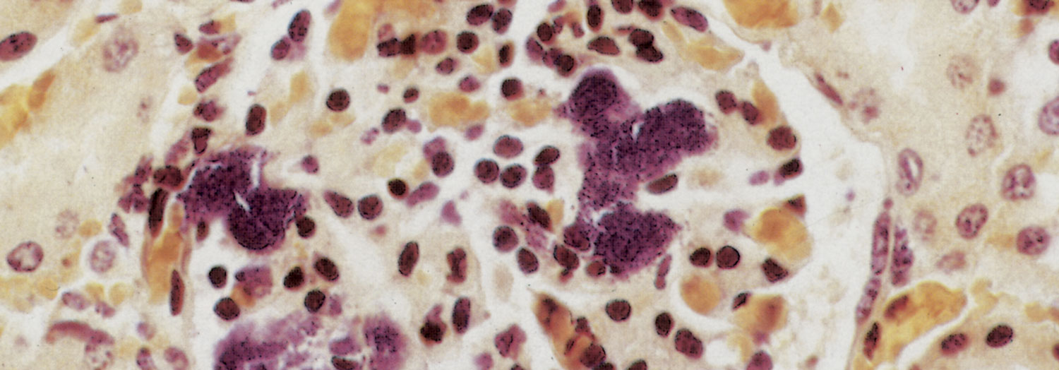
In the subacute disease, following a bacteraemic phase,A. equuli localizes in many organs and tissues, the outstanding lesions being purulent polyarthritis (particularly of the hock and knee joints), tendovaginitis and bronchopneumonia. In some animals miliary abscessation is present in the cortex of the kidneys and in the myocardium, the abscesses varying in size from microscopic to less than 3mm in diameter. Suppurative omphalophlebitis may be present. The microscopic lesions in the kidneys are those of a purulent embolic glomerulonephritis. Colonies of bacteria are usually evident in glomerular and other cortical capillaries (Figure 159.1).
In piglets infected with A. equuli lesions similar to those in foals may be evident.
Diagnosis and differential diagnosis
Because the clinical signs of sleepy foal disease may be confused with those of other infections, the diagnosis should be confirmed by the isolation of A. equuli from the blood and kidneys of septicaemic foals or from other affected tissues (pneumonic lesions, kidney abscesses) or exudates from foals in which the course of the disease is more prolonged. There is usually no difficulty in culturing A. equuli on blood agar medium and growth is characteristically profuse from septicaemic cases, but sparse from chronic cases.5 In newborn foals that die of the disease, microscopic lesions compatible with purulent embolic glomerular and interstitial nephritis are highly suggestive of sleepy foal disease.
Neonatal septicaemia in foals caused by A. equuli may be confused with septicaemia caused by Escherichia coli, Streptococcus spp. and Klebsiella spp.
Control
Apart from maintaining a high level of general hygiene and disinfecting the umbilicus at birth, very little can be done to prevent infection of new-born foals. Actinobacillus equuli is sensitive to streptomycin, tetracycline, chloramphenicol, ampicillin and trimethoprim-sulphonamide combinations. Affected animals should be treated parenterally at the recommended dosages for these antibiotics for three to five days.3
References
- BISGAARD, M., PIECHULLA, K., YING, Y-T., FREDERIKSEN, W. & MANNHEIM, W., 1984. Prevalence of organisms described as Actinobacillus suis or haemolytic Actinobacillus equuli in the oral cavity of horses. Comparative investigations of strains obtained and porcine strains of A. suis sensu stricto. Acta Pathologica et Microbiologica et Immunologica Scandinavica Section B, 92, 291–298.
- BLACKALL, P.J., BISGAARD, M. & MCKENZIE, R.A., 1997. Characterization of Australian isolates of Actinobacillus capsulatus, Actinobacillus equuli, Pasteurella caballi and Bisgaard Taxa 9 and 11. Australian Veterinary Journal, 75, 52–55.
- BLACKALL, P.J., CHRISTENSEN, J.P. & BISGAARD, M., 1998. Diversity among isolates of Actinobacillus equuli and related organisms as revealed by ribotyping. Australian Veterinary Journal, 76, 423–425.
- COTTEW, G.S. & RYLEY, J.W., 1952. Shigella equirulis infection in a foal. Australian Veterinary Journal, 28, 302–306.
- DIMOCK, W.W., EDWARDS, P.R. & BRUNER, D.W., 1947. Infections of fetuses and foal. Kentucky Agricultural Experimental Station Bulletin, 509.
- GAY, C.C. & LORDING, P.M., 1980. Peritonitis in horses associated with Actinobacillus equuli. Australian Veterinary Journal, 56, 296–300.
- GOLLAND, L.C., HODGSON, D.R., HODGSON, J.L., BROWNLOW, M.A., HUTCHINS, D.R., RAWLINSON, R.J., COLLINS, M.B., MCCLINTOCK, S.A. & RAISIS, A.L., 1994. Peritonitis associated with Actinobacillus equuli in horses: 15 cases (1982–1992). Journal of the American Veterinary Medical Association, 205, 340–343.
- HENNING, M.W., 1956. Animal Diseases in South Africa. 3rd edn. South Africa: Central News Agency Ltd.
- HENTON, M.M., 1993. Veterinary Research Institute, Onderstepoort. Unpublished observations.
- KAMADA, M., KUMANOMIDO, T., KANEMARU, T., YOSHIHARA, T., TOMIOKA, Y., KANEKO, M., SENBA, H. & OHISHI, H., 1985. Isolation of Actinobacillus equuli from neonatal foals with death in colostrum-deficiency or failure of maternal immunity transfer. Bulletin of Equine Research Institute, 22, 38–42.
- LITTLEJOHN, A., 1959. Sleepy foal disease in Natal. Journal of the South African Veterinary Medical Association, 30, 143–147.
- MAGUIRE, L.C., 1958. The role of Bacterium viscosum equi in the causation of equine disease. The Veterinary Record, 70, 989–991.
- PEDERSEN, K.B., 1977. Actinobacillus infections in swine. Nordisk Veterinaer Medizin, 29, 137–140.
- PHILLIPS, J.E., 1984. Actinobacillus. In: krieg, n.r. & holt, j.g., (eds). Bergey’s Manual for Systematic Bacteriology, Vol. I. 570–575.
- PLATT, H., 1973. Septicaemia in the foal: A review of 61 cases. British Veterinary Journal, 129, 221–229.
- RAISIS, A.L., HODGSON, J.L. & HODGSON, D.R., 1996. Equine neonatal septicaemia: 24 cases. Australian Veterinary Journal, 73, 137–140.
- ROBINSON, J.A., ALLEN, G.K., GREEN, E.M., FALES, W.H., LOCH, W.E. & WILKERSON, C.G., 1993. A prospective study of septicaemia in colostrum-deprived foals. Equine Veterinary Journal, 25, 214–219.
- SNEATH, P.H.A. & STEVENS, M., 1985. A numerical taxonomic study of Actinobacillus, Pasteurella and Yersinia. Journal of General Microbiology, 131, 2711–2738.
- STERNBERG, S., 1998. Isolation of Actinobacillus equuli from the oral cavity of healthy horses and comparison of isolates by restriction enzyme digestion and Pulsed-Field Gel Electrophoresis. Veterinary Microbiology, 59, 147–156.
- WARD, C.L., WOOD, J.L., HOUGHTON, S.B., MUMFORD, J.A. & CHANTER, N., 1998. Actinobacillus and Pasteurella sp. Isolated from horses with lower airway disease. The Veterinary Record, 143, 277–279.
- WINDSOR, R.S., 1973. Actinobacillus equuli infection in a litter of pigs and a review of previous reports on similar infections. The Veterinary Record, 92, 178–180.


