- Infectious Diseases of Livestock
- Part 3
- Corynebacterium pseudotuberculosis infections
- GENERAL INTRODUCTION: SPIROCHAETES
- Swine dysentery
- Borrelia theileri infection
- Borrelia suilla infection
- Lyme disease in livestock
- Leptospirosis
- GENERAL INTRODUCTION: AEROBIC ⁄ MICRO-AEROPHILIC, MOTILE, HELICAL ⁄ VIBROID GRAM-NEGATIVE BACTERIA
- Genital campylobacteriosis in cattle
- Proliferative enteropathies of pigs
- Campylobacter jejuni infection
- GENERAL INTRODUCTION: GRAM-NEGATIVE AEROBIC OR CAPNOPHILIC RODS AND COCCI
- Moraxella spp. infections
- Bordetella bronchiseptica infections
- Pseudomonas spp. infections
- Glanders
- Melioidosis
- Brucella spp. infections
- Bovine brucellosis
- Brucella ovis infection
- Brucella melitensis infection
- Brucella suis infection
- Brucella infections in terrestrial wildlife
- GENERAL INTRODUCTION: FACULTATIVELY ANAEROBIC GRAM NEGATIVE RODS
- Klebsiella spp. infections
- Escherichia coli infections
- Salmonella spp. infections
- Bovine salmonellosis
- Ovine and caprine salmonellosis
- Porcine salmonellosis
- Equine salmonellosis
- Yersinia spp. infections
- Haemophilus and Histophilus spp. infections
- Haemophilus parasuis infection
- Histophilus somni disease complex in cattle
- Actinobacillus spp. infections
- Actinobacillus equuli infections
- Gram-negative pleomorphic infections: Actinobacillus seminis, Histophilus ovis and Histophilus somni
- Porcine pleuropneumonia
- Actinobacillus suis infections
- Pasteurella and Mannheimia spp. infections
- Pneumonic mannheimiosis and pasteurellosis of cattle
- Haemorrhagic septicaemia
- Pasteurellosis in sheep and goats
- Porcine pasteurellosis
- Progressive atrophic rhinitis
- GENERAL INTRODUCTION: ANAEROBIC GRAM-NEGATIVE, IRREGULAR RODS
- Fusobacterium necrophorum, Dichelobacter (Bacteroides) nodosus and Bacteroides spp. infections
- GENERAL INTRODUCTION: GRAM-POSITIVE COCCI
- Staphylococcus spp. infections
- Staphylococcus aureus infections
- Exudative epidermitis
- Other Staphylococcus spp. infections
- Streptococcus spp. infections
- Strangles
- Streptococcus suis infections
- Streptococcus porcinus infections
- Other Streptococcus spp. infections
- GENERAL INTRODUCTION: ENDOSPORE-FORMING GRAM-POSITIVE RODS AND COCCI
- Anthrax
- Clostridium perfringens group infections
- Clostridium perfringens type A infections
- Clostridium perfringens type B infections
- Clostridium perfringens type C infections
- Clostridium perfringens type D infections
- Malignant oedema⁄gas gangrene group of Clostridium spp.
- Clostridium chauvoei infections
- Clostridium novyi infections
- Clostridium septicum infections
- Other clostridial infections
- Tetanus
- Botulism
- GENERAL INTRODUCTION: REGULAR, NON-SPORING, GRAM-POSITIVE RODS
- Listeriosis
- Erysipelothrix rhusiopathiae infections
- GENERAL INTRODUCTION: IRREGULAR, NON-SPORING, GRAM-POSITIVE RODS
- Corynebacterium pseudotuberculosis infections
- Corynebacterium renale group infections
- Bolo disease
- Actinomyces bovis infections
- Trueperella pyogenes infections
- Actinobaculum suis infections
- Actinomyces hyovaginalis infections
- GENERAL INTRODUCTION: MYCOBACTERIA
- Tuberculosis
- Paratuberculosis
- GENERAL INTRODUCTION: ACTINOMYCETES
- Nocardiosis
- Rhodococcus equi infections
- Dermatophilosis
- GENERAL INTRODUCTION: MOLLICUTES
- Contagious bovine pleuropneumonia
- Contagious caprine pleuropneumonia
- Mycoplasmal pneumonia of pigs
- Mycoplasmal polyserositis and arthritis of pigs
- Mycoplasmal arthritis of pigs
- Bovine genital mycoplasmosis
- Neurotoxin-producing group of Clostridium spp.
- Contagious equine metritis
- Tyzzer's disease
- MYCOTIC AND ALGAL DISEASES: Mycoses
- MYCOTIC AND ALGAL DISEASES: Pneumocystosis
- MYCOTIC AND ALGAL DISEASES: Protothecosis and other algal diseases
- DISEASE COMPLEXES / UNKNOWN AETIOLOGY: Epivag
- DISEASE COMPLEXES / UNKNOWN AETIOLOGY: Ulcerative balanoposthitis and vulvovaginitis of sheep
- DISEASE COMPLEXES / UNKNOWN AETIOLOGY: Ill thrift
- Eperythrozoonosis
- Bovine haemobartonellosis
Corynebacterium pseudotuberculosis infections
This content is distributed under the following licence: Attribution-NonCommercial CC BY-NC  View Creative Commons Licence details here
NJ Maclachlan and M-L Penrith (Editors). MW Paton, MG Collett and M Pepin, Corynebacterium pseudotuberculosis infections, 2019.
Corynebacterium pseudotuberculosis infections
Previous authors: M W PATON, M G COLLETT, M PÉPIN AND G F BATH
Current authors:
M W PATON - Retired Senior Veterinary Officer, BVSc, MANZCVS, Grad Cert Animal Welfare, PhD, 31 Clipson Mundaring, Western Australia, 6073, Australia
M G COLLETT - Senior Lecturer, BVSc, MMedVet (Path), Med (CAI), School of Veterinary Science, Massey University, Private Bag 112222, Palmerston North, Manawatu, 4442, New Zealand
T M ELLIS - Retired Specialist Veterinary Consultant in Microbiology and Pathobiology, BVSc, MSc, 30 St Leonards Street, Mosman Park, Western Australia, 6012, Australia
M PÉPIN - Professor of Microbiology / Immunology and Infectious Diseases, DVM, PhD, VETAGRO SUP / Campus Vétérinaire de Lyon, Marcy-L’Etoile, F-69280, France
Introduction
Corynebacterium pseudotuberculosis was first described in 1888 by the French veterinarian Edward Nocard, who isolated it from a case of bovine farcy (ulcerative lymphangitis). Three years later the same organism was isolated from a renal abscess in a sheep by the Bulgarian bacteriologist Hugo Von Preïsz,14, 36, 51 hence the original designation ”Preïsz-Nocard” bacillus. Subsequently, it was called Bacillus pseudotuberculosis. The organism was renamed C. ovis in 1923, and in 1948 the name was changed to C. pseudotuberculosis.14, 36
Table 1 Summary of natural infections caused by C. pseudotuberculosis
| Species | Specific lesions and diseases | Other |
| Ovine and caprine | Caseous lymphadenitis (CLA), bacterial icterus | Lymph node and non-lymph node abscesses in the lungs, liver, subcutaneous tissues, kidneys, mammary gland, testis, joints, plus abortion, stillbirth, perinatal death |
| Equine | Ulcerative lymphangitis, contagious acne | Folliculitis, furunculosis, pectoral and ventral abdominal abscesses, otitis media-interna, meningitis, abortion, mastitis |
| Bovine | Ulcerative lymphangitis | Deep subcutaneous abscesses, mastitis, heel dermatitis |
| Water buffalo | Oedematous skin disease | Lymph node abscesses, suppurative lymphangitis |
| Other Bovidae (mouflon and bighorn sheep, ibex, various antelope) |
| Subcutaneous and internal abscesses |
| Cervidae (deer, elk) |
| Subcutaneous and internal abscesses |
| Camelid (camel, alpaca) |
| Superficial and visceral lymphadenitis, mammary abscesses |
| Porcine |
| Purulent lymphadenitis |
| Erinaceidae (hedgehog) |
| Internal abscesses |
| Human |
| Lymphadenitis |
Corynebacterium pseudotuberculosis causes different conditions and lesions in animals and humans (see Table 1). Caseous lymphadenitis (CLA), caused by C. pseudotuberculosis, is a chronic disease of sheep and goats that occurs worldwide wherever small ruminants are farmed. Caseous lymphadenitis characteristically involves superficial or internal lymph nodes. Affected animals may also have non-lymph node abscesses in subcutaneous tissues and/or internal organs. Several other Bovidae species can acquire C. pseudotuberculosis infections, notably cattle (ulcerative lymphangitis) and water buffalo (“oedematous skin disease”), the latter being important in Egypt, the Middle East and South-East Asia. Certain wild Bovidae, Cervidae and even domestic pigs can be infected. Corynebacterium pseudotuberculosis is also the cause of ulcerative lymphangitis, pectoral abscesses, folliculitis and furunculosis (referred to as "contagious acne"), mastitis and abortion in horses. Pectoral abscesses and contagious acne have only ever been recorded in the Western USA and Canada. Caseous lymphadenitis can be zoonotic and affected people usually acquire infection following contact with sheep or goats.
The nature of CLA is of an insidious disease that is difficult to investigate and control. The research done on vaccine development and epidemiology in Australia has led to significant advances in the understanding and control of CLA. However, because of the non-clinical nature of CLA in most Australian sheep, the greatest challenge for the sheep industry is to convince farmers that they have a problem worth dealing with. The research done into CLA immunopathology in France has made major advances in the understanding of the behaviour of the sheep immune system in response to C. pseudotuberculosis. In the future, this may lead to improved tests and vaccines to fight this organism.
In Africa, UK, Netherlands, Canada, France, USA, South America, the Middle East and Scandinavia researchers and field veterinarians are dealing with outbreaks of CLA in sheep, goats, camelids and other species. In these flocks or herds, the production systems are vastly different from those in Australian sheep, so the epidemiology of CLA can be expected to have some differences. The challenges for those dealing with this disease are to learn from the methods or findings of the research done thus far and apply it to assist in solving the complex problems that C. pseudotuberculosis infection causes in their production systems.
Aetiology
Corynebacterium pseudotuberculosis is a short (0,5 to 0,6 x 1,0 to 3,0 micrometre), irregular, ovoid, non-sporulating, non-capsulated, Gram-positive rod almost resembling a coccus. It is facultatively intracellular and anaerobic. Although non-motile it has fimbriae.71 In smears made from lesions, the organisms show marked pleomorphism, with coccoid, bacillary and filamentous forms, invariably arranged in a palisade or “Chinese letter” pattern.14, 71 They are often clumped together and are more numerous in early lesions. The filamentous forms may exhibit a barred or beaded appearance when stained. Pleomorphism is not as marked in cultured organisms. After 48 hours' incubation at 37oC on blood tryptose agar, slow-growing, light cream to orange-coloured colonies are produced, surrounded by a narrow zone of β-haemolysis. The colonies are granular, opaque, and flat with a matt surface, do not attach to the medium and can be moved around on the surface of the agar.59, 71 All strains produce acid, but not gas, from a variety of sugars. The organism is phospholipase D (PLD) and catalase positive, but oxidase negative.71 It is closely related to C. ulcerans, which also produces PLD, and some strains of both organisms can produce diphtheria toxin.71 The Analytical Profile Index (API) Coryne system (bioMérieux, Inc., France) is a reliable and well-established biochemical test for coryneform bacteria identification.71
Although only a single species is recognized, isolates are classified into two genetically different biovars (biotypes) based on their ability to reduce nitrate:17, 27, 71, 237, 247 a nitrate reduction negative biovar ovis (biotype 1) infects sheep and goats and a nitrate reduction positive biovar equi (biotype 2) infects horses and cattle.64, 71, 155, 192, 246 Both biovars have been isolated from cattle17, 154, 233, 279 and camels252, 253 There are no biochemical or antigenic differences between ovine and caprine isolates and the genome is highly conserved on a multinational scale.20, 61 This categorisation based on nitrate reduction may be unsatisfactory since equine isolates from the UK were nitrate negative but differed from ovine and caprine isolates by producing alpha glucosidase and differed from each other in their production of alkaline phosphatase.62 Corynebacterium pseudotuberculosis biovar ovis infects various antelope in South Africa.97, 159 Isolates from a pig and a hedgehog have been characterized as biovar ovis.145, 168 In contrast, biovar equi is responsible for infecting water buffalo in the Middle East50, 154 and elk in Utah, USA.124 Biovar equi has apparently never been isolated in Australia and equine disease has not been reported there.22 Isolates from sheep and goats have a higher minimum inhibitory concentration for amikacin than isolates from horses and cattle.64 Under natural conditions, no cross species infection occurs with the respective biovars, although human infection with the nitrate-negative biotype has occurred following contact with infected sheep.107
More recently, considerable research has been conducted into molecular characterization of C. pseudotuberculosis, particularly in identifying different genotypes from sheep and goats and characterizing the extracellular proteins released or present during the infectious process.70 The enterobacterial repetitive intergenic consensus-PCR (ERIC-PCR) technique for random amplified polymorphic DNA (RAPD) analysis was applied to 127 C. pseudotuberculosis isolates from sheep lesions from 24 flocks from 13 municipalities in Brazil.91 Using different primer sets, 17 or 21 different genotypes were detected. Most flocks had 1-3 different genotypes but a few had between 4 and 9 genotypes present.91 The excretory proteins (exoproteome) and secretory proteins (secretome) of C. pseudotuberculosis from a goat strain from Brazil and a sheep strain from Australia were analysed by two dimensional gel electrophoresis (2-DE), then MALDI-TOF/TOF mass spectrometry of separated proteins, and then compared.236 In this study, 45 different extracellular proteins were identified, with 13 unique to the goat strain, 3 unique to the sheep strain and 29 common to both. In another study,172 93 different extracellular proteins were identified using three phase partitioning, liquid phase chromatography and mass spectrometry (TPP/LC-MS). Despite lower numbers of proteins detected, there were 11 unique proteins identified by 2-DE and MALDI-TOF/TOF and overall 104 extracellular proteins were detected from C. pseudotuberculosis cultures. A proteomics study of the horse biotype (biovar equi) has revealed excretory proteins that are key elements in the virulence, adaptation, adherence, intracellular growth and immune system evasion of C. pseudotuberculosis.235 Transcriptional profiles of biovar ovis200 and biovar equi201 have revealed candidates for resilience, virulence and pathogenicity. Work in Mexico has revealed two gene clusters capable of differentiating between biovars ovis and equi, namely Restriction Modification system and CRISPR-Cas.174 The elucidation of exactly which gene products of C. pseudotuberculosis are directly involved in organism survival and adaptation has yet to come.70
The complete genome sequences of some strains of C. pseudotuberculosis have been published.50, 97, 139, 174
Epidemiology
Caseous lymphadenitis is a highly prevalent and important disease of sheep and goats and, in some countries, is responsible for considerable financial losses as a result of the condemnation and downgrading of infected carcasses and skins at abattoirs (where the prevalence becomes most obvious) as well as reduced wool production.11, 20, 21, 23, 74, 89, 212, 222, 242 Sheep affected by CLA produce approximately four to seven per cent less wool in the year of initial infection compared to unaffected sheep.184, 186 The total annual (1987/88) economic loss as a result of CLA in Australia was estimated at A$25 million because of reduced wool production, condemnations and trimmings of carcasses, and the cost of meat inspection.177 In New Zealand CLA is a significant disease in sheep, particularly in the fine-woolled Merinos and Corriedales in the South Island. Because the New Zealand sheep meat industry relies largely on slaughtering and processing prime lamb carcasses for export, a very large proportion of the total sheep population is slaughtered each year.165 Because of this, the national prevalence of CLA is probably not as high compared to, for example, Australia.165 The cost of CLA in New Zealand was estimated at NZ$1 million in 1981.165
Caseous lymphadenitis is the most common infection in the so-called "thin-ewe syndrome" in the USA.88, 209 In a study in the western USA (1984), the average prevalence in adult ewes was 42,4 per cent.242 In Quebec, Canada, the prevalence of clinical CLA in culled adult sheep ranged from 21 to 36 per cent.9
In Australia, the overall prevalence in a study from Tasmania was 26 per cent,150 while in Western Australia, the average for 412 Western Australia sheep flocks was 45 per cent,178(Paton, 1997) with the highest (1986) being 61 per cent.20 The year 2002, over 50 per cent of carcass condemnations in Australia were due to CLA.269 Since then, and following the introduction of vaccination programmes in 1983 whereby the CLA antigen was included in clostridial vaccines, the prevalence had dropped by 2002 to 20, 23 and 29 per cent in Western Australia, Victoria and New South Wales, respectively.187 By 2009, because of the success of vaccination, the prevalence was estimated to be only 5,2 per cent.273
In South Africa, CLA was first reported by Jowett in 1909.121 It appears to be more common in the drier areas of South Africa, such as the Karoo, Eastern Cape and Free State Provinces; a prevalence of 2,4 per cent of CLA in Merino sheep less than one year old and 7,4 per cent in older sheep from the Karoo has been reported.160 During the period 1986 to 1988, 18,7 to 20,3 per cent of condemned carcasses at Cato Ridge Abattoir in the KwaZulu-Natal Province contained lesions of CLA.265 These condemnations represented 0,24 to 0,3 per cent of all the sheep and goats slaughtered at that abattoir during the period, and amounted to an annual loss of more than ZAR400 000 for carcass condemnations alone, to which should be added unmeasured but substantial trimmings from carcasses.265 Caseous lymphadenitis was the most important reason for carcass condemnation in sheep at South African abattoirs in 1990 and 1991.8
Research in North Africa and the Middle East has reported prevalences ranging from 24 per cent in Morocco,127 to 23 and 11 per cent in sheep and goats, respectively, in Egypt,4 47 per cent in older sheep in Iran,283 and eight per cent in camels in Jordan.98 In other countries, such as Brazil, the Patagonia region of Argentina and Chile, Venezuela, Malaysia, India and Spain, the disease is becoming more important in small stock and in ibex.56, 60, 84, 92, 93, 129, 130, 229
The disease occurs in all continents although it was absent from Britain until the first report in goats in 1990.90, 138, 146 The probable source of infection was Boer goats imported from the then West Germany.14, 62 Since then the disease has spread to sheep in the UK, where certain breeds may have an increased susceptibility due to the expression of different major histocompatibility ovine leukocyte antigens.225 The steady and seemingly relentless spread of infection in sheep and goats in the UK is of great concern to farmers and veterinarians.14 Cases of CLA in Northern Ireland142 and the Republic of Ireland166 have followed the importation of sheep from Scotland and England, respectively. Contagious lymphadenitis is a notifiable disease in both Northern Ireland and the Republic of Ireland, but restrictions have been removed in Great Britain since 1991.14, 142 In Ireland, a concurrent outbreak of tuberculosis caused by Mycobacterium bovis and caseous lymphadenitis in milking goats has been reported.231
Although CLA had not been reported in the UK before 1990, the disease became a political issue in the 1920s when mutton carcasses, imported from a number of countries (Argentina in particular), were found to have abscesses at import meat inspection, or even when on the dinner table.14 It is possible that the original dissemination of C. pseudotuberculosis was associated with the dual-purpose Merino breed, originally from Spain. Several countries, notably South Africa, Australia, and the Americas, imported Merinos during the 19th-century. Following concerns in Britain, these mutton-exporting countries had to take heed, institute abattoir surveillance, and conduct research into the nature of C. pseudotuberculosis.14 Notably, the Merino was not a breed farmed commercially in Britain at the time.14
Corynebacterium pseudotuberculosis has been isolated from subcutaneous and internal abscesses in an emaciated Rocky Mountain bighorn sheep.125 Routine meat inspection of carcasses of 33 per cent of 139 antelope (black and blue wildebeest, blesbok, red hartebeest and springbok) harvested on a commercial game reserve in South Africa revealed tuberculosis-like encapsulated necrogranulomas in lungs, liver and lymph nodes that yielded C. pseudotuberculosis and various non-tuberculous mycobacteria on culture.159 Subcutaneous abscesses in the Arabian oryx have also yielded C. pseudotuberculosis.250 Molecular methods have demonstrated co-infection with C. pseudotuberculosis, Mycobacterium bovis and M. avium ssp. paratuberculosis in red deer with granulomatous mesenteric lymphadenitis.143 In Pakistan, CLA has been diagnosed in chinkara deer (Indian gazelle), spotted deer, and mouflon sheep.114 Corynebacterium pseudotuberculosis can also infect other cervids, such as elk.124 Superficial lymphadenitis due to C. pseudotuberculosis has been described in camels from all parts of the world where they are found.252 The infection may affect more than 10 per cent of camels in a herd.3 In alpacas in Peru, C. pseudotuberculosis causes abscesses in superficial lymph nodes, renal lymph nodes, mammary gland, liver and lungs.30 The organism has been isolated from internal abscesses in a zoo hedgehog in the USA.145
Human lymphadenitis due to C. pseudotuberculosis was first described in 1966. Since then, a number of sporadic cases, mostly from Australia, involving superficial lymph nodes (especially axillary or inguinal), and with occupational (e.g. shearers, shepherds, abattoir workers, butchers) exposure to sheep a common feature, have been reported.14, 152, 189 A most unusual case involving infection of an ocular implant with this organism has also been described.137
Caseous lymphadenitis can be reproduced experimentally in sheep and goats by the administration of C. pseudotuberculosis by a number of different routes.1, 11, 32, 33, 37, 41, 49, 76, 86, 113, 161, 162, 193, 248 The typical incubation period from inoculation to abscessation is two to six months.270 Guinea pigs and mice are also susceptible and, provided small inocula are used, mice will develop a disease that is very similar to CLA.19, 47, 48, 156, 282
Several modes of transmission of C. pseudotuberculosis have been suggested as being important in the spread of CLA. Studies in Australian and Japanese sheep flocks181, 228 have indicated that most spread of CLA occurs shortly after shearing since the organism can enter its host through cuts in the skin.39, 226 Contamination of the environment, including shearing sheds and holding pens, by C. pseudotuberculosis in faeces, has been suggested as being important.226 This has led to attention being paid to shearing shed hygiene to prevent CLA spread, particularly in New Zealand. Alternatively, physical transfer of CLA pus from abscesses discharging at shearing by shearing combs, other fomites, in dips or by direct contact with other sheep is also a potentially important mode of infection.141, 160 Another study showed that sheep with lung infections might be a source of infection.83 These various infection routes would require different management changes to minimise CLA spread at shearing.
To investigate the main route of CLA spread, detailed observations were made at shearing to monitor the CLA incidence in individual sheep over a number of shearings.176 In this study, the CLA incidence was low for the first few shearings, then increased rapidly, similar to that observed in another study of natural infection in commercial sheep flocks.181 Contagious lymphadenitis spreads naturally from artificially infected to susceptible sheep run together. This resulted in a 76 per cent prevalence of sheep with CLA lesions in 85 surviving unprotected sheep. This spread occurred without any superficial CLA abscesses discharging at any of five shearings over two years.176, 180 The absence of discharging CLA abscesses during this period in the face of a high transmission rate of C. pseudotuberculosis clearly indicated that sources of the bacteria other than external discharging abscesses caused this high rate of spread. It was concluded that environmental contamination or direct spread from lung lesions were the two most likely explanations for the spread of this disease.176, 180
If CLA spreads by C. pseudotuberculosis contamination of fomites or dust in the shearing shed environment then there should be an equal opportunity for the disease to spread through cuts at all shearings. However, about 80 per cent of the spread of CLA in Australian commercial sheep flocks occurred at the second and third shearings.181 In this study, 77 per cent of the sheep that seroconverted, did so at the fourth and fifth shearings.176 In addition to this evidence, dusty conditions in yards after shearing were not associated with high CLA incidence in a risk factor study of CLA incidence.185 It is therefore unlikely that environmental contamination with C. pseudotuberculosis or pus from discharging superficial CLA abscesses are major sources of CLA spread in Australian sheep flocks.
This evidence clearly pointed to a major role for lung abscesses in the spread of CLA in commercial sheep flocks. By culturing C. pseudotuberculosis from the tracheas of sheep with lung abscesses, it has been observed that many lung abscesses in sheep discharge into airways.215 If a small number of sheep become infected by various routes before their second shearing, it would take some time for any lung lesions to develop sufficiently in these sheep to discharge into the airways. When this occurs these sheep may be capable of spreading CLA to a large number of sheep at one shearing by aerosol contamination of skin cuts on uninfected sheep.185
Treating sheep for lice by showering them in an insecticide solution in an enclosed recycling shower apparatus (shower dipping) increased the risk of having a high CLA incidence fivefold. Keeping sheep under cover for one hour or more after shearing increased the risk of high CLA incidence threefold.185 The influence of shower dipping on CLA incidence may be explained by the ability of the CLA organism to spread in dip wash. This could happen when bacteria from discharging lung abscesses are coughed up by infected sheep into the dipping fluid, which is then recirculated and sprayed over sheep with skin cuts. Further evidence suggests that sheep without skin cuts are also infected in these dips.178, 180
It has also been found that for every 10 per cent increase in the seroprevalence of CLA in an age group before shearing the risk of the CLA incidence in that age group is 2.6 times higher. For every ten per cent increase in the prevalence of CLA in cull ewes in a flock, the risk that one- or two-year-old sheep will have a high CLA incidence becomes only 1,4 times higher.185 Variations in CLA incidence were better explained by a model where the CLA incidence was divided into three groups. Those groups with a zero incidence were a group on their own in this model. This model better explained the data than a model where the CLA incidence was divided into two groups around its median of 5,5 per cent. This meant that age groups with a zero incidence were much more likely to have had a zero seroprevalence before shearing. This is further evidence that most CLA spread in Australian sheep occurs within groups of sheep shorn together, rather than from the environment or from sheep outside of the group.185
Pus from abscesses ruptured or accidentally “lanced” during shearing can be a source of cross-infection between animals, since the number of viable organisms varies between 1 x 10⁶ and 5 x 10⁷ cfu/g.36, 269 Merino sheep that have large neck wrinkles are particularly prone to iatrogenic shearing injuries.269 Abscess rupture, therefore, releases massive quantities of bacteria onto the skin and fleece, with contamination of the environment, neighbouring animals and fomites.86 The organism can survive on moist debris on pen floors at room temperature for at least ten days116 and for several months, or even more than a year, on fomites (e.g. hay or bedding), particularly if environmental temperatures are low.12 In the Netherlands, bales of hay purchased from another farm where CLA was highly prevalent in goats and where the goats were housed in the hay shed, resulted in an outbreak of CLA in a closed and accredited disease-free goat herd.69 It can also survive in organically rich soil for long periods, but it is not known whether it multiplies in soil.270 Despite this, dusty conditions are not believed to increase the prevalence of CLA.185 Corynebacterium pseudotuberculosis can be isolated from215 and can survive in commercial sheep-dipping fluids for at least 24 hours.162 It may be isolated from the faeces of some sheep.100 The primary habitat of C. pseudotuberculosis is probably infected animals.206
The epidemiology of CLA seems to vary among different production systems and falls into two broad forms. In one form, the disease is prevalent in adults but much less so in lambs (for example in Australia, South Africa, New Zealand and some USA wool producing flocks). This form appears to occur more frequently in extensive production systems. The other form is characterized by moderate prevalences in lambs and adults with some adults showing a chronic wasting disease (for example in Saudi Arabia, Canada, France, UK and more intensive production systems in the USA). This form seems more common where sheep are raised intensively or where some housing is involved. Infrastructure that can cut the skin has also been noted as a risk factor in these production systems and isolating animals being treated appears to be protective.92, 127, 178 Where rams are closely associated, fighting can lead to skin trauma, infection and facial cellulitis.225
Caseous lymphadenitis occurs more commonly in Merino sheep in South Africa, Australia and New Zealand than in other breeds.53, 160 In France, however, some dairy breeds have a higher prevalence.194
In goats, abrasions on the head caused by head-butting, ear-biting and browsing may predispose to infection.23, 204, 255 Ingestion of infective material has been reported to be a cause of abscess development in the mandibular lymph nodes of goats.11
Nodular worm (Oesophagostomum spp.) larvae burrowing into the wall of the intestines create a portal of entry for bacteria (commonly Trueperella (Arcanobacterium, Actinomyces) pyogenes or C. pseudotuberculosis) that cause miliary abscesses. Miliary subcutaneous abscessation may also be caused by C. pseudotuberculosis following skin damage by ticks, other parasites or plant material such as thorns or awns.
In horses in the USA and Canada, the presence of abscesses has been correlated with seasonal increases of biting insects in California and Texas.5, 111, 151, 268 Insects including houseflies have been evaluated as possible vectors.241 In Israel, infections in dairy cattle showed marked seasonality and houseflies possibly played a role in the spread of the infection.276 Corynebacterium pseudotuberculosis, transmitted by the blood-sucking fly, Hippobosca equina, causes oedematous skin disease in water buffalo in Egypt and the Middle East.227
Pathogenesis
Corynebacterium pseudotuberculosis is a facultative intracellular parasite; it has the capacity to replicate within and ultimately escape from the macrophage phagolysosomes.96, 120, 251, 261 Because of this, it is able to resist immune clearance by a naïve host and spread beyond the primary locus of infection.86No avirulent strains of this pathogen have been described to date.14
At least two virulence factors, a toxic cell-wall lipid and a protoplasmic exotoxin, which is a sphingomyelinase-specific phospholipase D (PLD),40, 42, 52, 73, 238 play essential roles in the development of CLA. The toxic cell-wall lipid, which comprises mycolic acids that are similar to the mycolic wax of Mycobacterium tuberculosis, is associated with the virulence of the organism and probably protects it mechanically and/or biochemically from the enzymes of lysosomes, enabling it to survive inside phagolysosomes and thus ensuring the chronicity of the infection.54, 95, 120, 204, 251, 270 The lipid is also pyogenic,282 but not immunogenic.54 Strains of C. pseudotuberculosis selected for their greater lipid content produce larger numbers of abscesses in mice.156
The PLD exotoxin is also an important virulence factor because a strain with a deletion in the PLD gene is unable to produce a persistent infection or CLA in sheep.101, 102 It acts on the sphingomyelin of erythrocyte and endothelial cell membranes, causing haemolysis, increased vascular permeability and enhanced bacterial invasion.25, 34, 35, 53, 71, 108, 119, 239, 249, 282The sphingomyelinase catalyses the dissociation of sphingomyelin into ceramide phosphate and choline.26, 239 It is presumably responsible for the development of "bacterial icterus"; intravenous or subcutaneous inoculation of culture material, exotoxin, or fresh, incompletely toxoided vaccine may cause dyspnoea, anaemia, icterus and haemoglobinuria in sheep and goat kids.48, 49, 51, 108, 204, 216, 217 In addition, the exotoxin can cause dermonecrosis,52, 157, 240 lethality,34, 52, 157 synergistic lysis26 and inhibition of staphylococcal lysin induced lysis of erythrocytes.240, 282 It activates complement and limits bacterial opsonisation.280 The PLD is approximately 31 kDa in size,135 and has been purified,73 and the C. pseudotuberculosis pld gene has been cloned and sequenced.101, 238
Another possible determinant of virulence is a putative C. pseudotuberculosis iron uptake gene cluster.28
Following their entry through the skin or a mucous membrane, C. pseudotuberculosis organisms are transported within macrophages via afferent lymphatics to the regional lymph nodes, in which lesions may then develop.77, 161 Early bacterial dissemination leads to colonisation of other lymph nodes and lungs by the haematogenous route.196 In some animals infection may spread via lymphogenous and haematogenous routes to the lungs and other parts of the body in the absence of involvement of lymph nodes regional to the portal of entry.162 Lesions at both primary and secondary sites may undergo resolution during the early stages of their development.22, 261The size and severity of lesions depend upon the initial number of organisms entering the tissues of the host, the rate of multiplication of the organisms, and the accessibility of the host defense mechanisms to the bacteria;22 they appear to have little relationship to the duration of the infection.162 Chronic, and frequently lifelong, disease is the rule rather than the exception, since viable bacteria can be recovered from abscesses several years after initial infection.14
A more recent sheep study72 examined acute phase protein (APP, comprising haptoglobin [Hp], serum amyloid A [SAA], and α1 acid glycoprotein [AGP]) responses to C. pseudotuberculosis experimental challenge, and found that serum concentration to all three APP were raised during acute infection and an extended AGP response occurred in sheep transitioning from acute to the chronic phase of infection. In another C. pseudotuberculosis field observational study, sheep in an infected flock that became seropositive but that did not develop abscesses had significantly higher Hp concentration and lower monocyte counts during the acute phase of the disease than those that subsequently developed abscesses.18
Sequence studies of a C. pseudotuberculosis isolate from a human case identified a distinct gene set promoting its survival under unfavourable environmental conditions encountered in the mammalian host.257 From this study and others63, 221, 243 targeted several genes thought likely to be associated with virulence of C. pseudotuberculosis infection. Using quantitative real-time PCR with reverse transcription (qRT-PCR), they compared in vivo and in vitro expression of the genes phospholipase D (pld), CP40 (cpp), neuraminidase H (nanH), and superoxide dismutase C (sodC) and spaC gene (C. pseudotuberculosis adherent pili protein) and found that gene expression of all was 6-8 times higher in abscesses than in vitro cultures suggesting links to virulence.63
Humoral and cell-mediated immunity to C. pseudotuberculosis play a role in protection against the development of lesions.46, 47, 49, 78, 113 The humoral response is primarily antitoxic, limiting the progression of the disease,73, 117, 163, 245 whereas cell-mediated immunity restricts bacterial proliferation.36, 87, 102, 117, 118 Humoral immunity may be ineffective in halting the progression of CLA, however, as the organisms are intracellular.80, 81 Selection of certain mutant strains of C. pseudotuberculosis, such as one that is iron-acquisition-deficient, hold promise for better vaccines that elicit potent humoral and cellular responses.211
ELISAs have been developed to study the humoral response to the immunodominant PLD in experimentally infected sheep.196-198 Antibodies develop from the fifth day following inoculation and reach a plateau around the third week after which the titre slowly decreases.196, 198
Some variability in sensitivity of ELISA tests for measurement of immune response to C. pseudotuberculosis infection has been observed specifically regarding variation in performance for sheep and goat sera. Recent studies103 examined ATCC reference strains of C. pseudotuberculosis biovar ovis, biovar equi, caprine field isolates and ovine field isolates by SDS-PAGE as well as by immunoblotting of the sheep and goat field isolates with sheep and goat sera from C. pseudotuberculosis infected and free flocks. They observed major differences in SDS-PAGE profiles between sheep and goat field isolates and ATCC reference strains, with broader and more intense antigenic profiles detected in sheep than in goat strains. There were immunodominant antigens recognized equally by sheep and goats, and other immunodominant antigens recognized specifically by sheep or by goats. However, goat sera showed higher sensitivity than sheep sera for these immunodominant antigens, particularly for the PLD antigen. They concluded that serological tests for diagnosis of C. pseudotuberculosis infections should use a combination of immunodominant antigens in addition to PLD and also that the antigens are appropriate for sheep and goat sera.103
The development of pyogranulomas probably plays an important role in restricting bacterial dissemination to the local lymph nodes.196 The role of neutrophils appears to be to reduce the number of viable bacteria following inoculation, while macrophages are the main effector cells.193 Macrophages play a pivotal role in the formation of the focal granulomas, which have a central necrotic core surrounded by a layer mainly composed of activated macrophages, then a layer mainly comprising lymphocytes which is surrounded by a fibrous capsule.191 Lymphocyte subpopulations, especially CD4+ and CD8+ T cells, are distributed between the macrophage zone and the fibrous capsule and their proportions vary according to the age of the lesion.78, 199, 264 T-lymphocyte- and macrophage-derived inflammatory and fibrogenetic cytokines contribute to the maintenance of central necrosis and the stimulation of peripheral scarring.82, 190 The presence of pulmonary intravascular macrophages in sheep may explain the relative tropism of C. pseudotuberculosis for the lungs because of their role in the clearance of neutrophils containing bacteria.57, 266, 267
Co-infection studies with C. pseudotuberculosis and the Haemonchus contortus parasite in sheep were conducted to examine whether negative effects of the Th2 immune response to the parasite would affect the Th1 immune response cells that are important for the control of C. pseudotuberculosis infection.219 There was a trend for reduced ɣ- interferon (ɣ-IFN) in the dual infection compared with just C. pseudotuberculosis alone but no differences in class-specific antibodies.219
Clinical signs and pathology
The incubation period for the development of C. pseudotuberculosis abscesses ranges from 25 to 140 days.7, 11 The clinical signs manifested by affected animals depend on the site and extent of the lesions.
Generally, CLA in sheep and goats does not cause obvious clinical signs unless the lesions are progressive, very large or numerous, or are located where they can be clinically detected or where their presence compromises the normal function of an organ or tissue. Animals that are clinically normal may have large (up to 150 mm or more in diameter) internal abscesses that are only detected at necropsy. Because of this, CLA is often a “hidden disease” where the seriousness only becomes apparent at slaughter or necropsy examination.165 Sheep or goats suffering from large or numerous active lesions may show progressive weight loss, weakness, and terminal collapse, these signs being accompanied by dyspnoea and coughing if there is lung involvement.
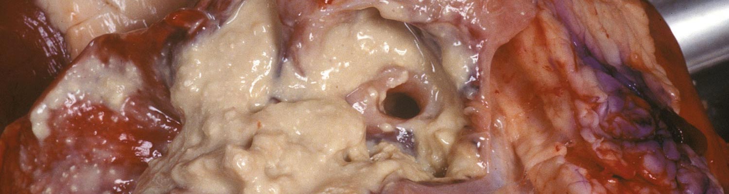
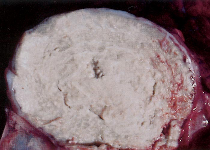
In sheep, the dorsal superficial cervical (prescapular) and subiliac (prefemoral) are the most commonly affected superficial lymph nodes,13, 121, 160 although those of the head and neck and those draining the buccal cavity are also often involved.147, 214, 225 Initially, affected lymph nodes are enlarged (40 to 50 mm in diameter), soft or doughy on palpation and, if superficially located, the skin that covers them may be thin and hairless. A tenacious, thick, semifluid to inspissated, yellowish-white to greenish-white pus (Figure 1) may be discharged should the abscess rupture which, in some, will result in the formation of persistently draining fistulae. The content of more long-standing abscesses is usually severely inspissated and will have lost the greenish colour.13, 165, 209, 261 Most abscesses are enveloped by well-developed connective tissue capsules and, although not evident in all cases, successive necrosis and redevelopment of the capsule impart a concentrically lamellated ("onion ring") pattern to the inspissated content – a characteristic of the lesion that becomes particularly prominent when the layers become partially mineralized (Figure 2).86 There is generally no evidence of infection at the site of entry of C. pseudotuberculosis.
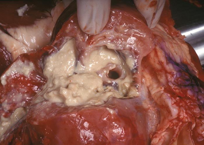
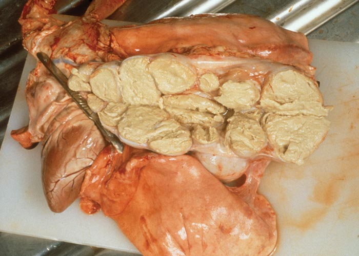
Of the internal organs and other tissues, the bronchial and mediastinal lymph nodes and lungs are most frequently involved. Single or multiple abscesses of varying size, but which may become quite large, or caseopurulent bronchopneumonia, which in some animals can be extensive, may be found in the lungs, frequently in the absence of lesions elsewhere.121, 209, 242 If an abscess or bronchopneumonic lesion abuts on the pleura, localized fibrinous or fibrous pleural adhesions generally develop.165, 209, 261 Lung abscesses may rupture into airways.242 The pulmonary lesions are often accompanied by lesions in the bronchial or mediastinal lymph nodes. (Figures 3 and 4) Abscesses in the mediastinal lymph nodes, however, often occur in the absence of lung abscesses. Large abscesses in these lymph nodes may cause pressure on, and partial occlusion of, the oesophagus, resulting in interference with deglutition and eructation, loss of weight and chronic ill-thrift.261
Corynebacterium pseudotuberculosis infections are occasionally the cause of abscesses in the liver, kidneys, spleen, brain, spinal cord, vertebral bodies, eyes, diaphragm, muscles, heart, tongue, uterus, mammary gland, testes, bones and joints.65, 85, 112, 126, 140, 144, 173, 206, 209, 261 A variety of other conditions can occur in sheep, including reproductive disorders (such as abortion, stillbirth, and neonatal infection) in ewes,1, 7, 68 suppurative orchitis in rams,134, 271 suppurative mastitis in ewes, arthritis and bursitis,206 and rapid death due to toxaemia was reported in experimentally inoculated neonatal lambs and kids.108 Corynebacterium pseudotuberculosis has been isolated from the preputial cavity and from the accessory sex organs and epididymis of clinically normal rams.116
In a 1953 survey in sheep slaughtered in the USA, the frequency of CLA lesions was as follows: bronchial lymph nodes (89 per cent), mediastinal lymph nodes (81 per cent), lungs (76 per cent), prescapular lymph nodes (24 per cent), prefemoral lymph nodes (15 per cent), hepatic lymph nodes (14 per cent), liver (12 per cent), superficial inguinal or supramammary lymph nodes (nine per cent), castrated scrotum (eight per cent), mammary gland (six per cent), kidney (five per cent), spleen and muscle (each four per cent) and popliteal lymph nodes (two per cent).140
Prior to the development of improved vaccines, several outbreaks of haemolytic anaemia and icterus occurred after sheep had been vaccinated against C. pseudotuberculosis. These outbreaks of "bacterial icterus" were associated with high mortality, and a high proportion of icteric carcasses was found in those sheep that were sent to slaughter.48, 49
In goats, lung lesions are uncommon. Suppurative lesions or abscesses of the subcutaneous tissues and lymph nodes of the head are more common, and often more severe than in sheep. This is probably because of the combative behaviour of goats and because of injuries by ticks or thorns to the facial skin or buccal mucosa.10, 13, 23, 89, 138, 147, 165, 261 In South Africa, however, CLA and lung abscesses are quite common in Angora goats.262 Abscesses caused by C. pseudotuberculosis in goats usually develop slowly, are encapsulated, and contain odourless, yellowish-white or greenish-white pus that is pasty, dry and inspissated or semifluid and viscous.11, 23, 44, 89, 136, 138, 222 Inspissation, calcification and the "onion-ring" appearance, so typical of CLA in sheep, are rarely encountered in goats.23, 165 Rupture of superficial lymph node abscesses frequently results in fistulae to the exterior.261
Histologically, the earliest inflammatory changes in the lymph nodes of sheep and goats comprise multiple micro-abscesses and a massive infiltration of neutrophils and, to some extent, eosinophils. The latter impart a greenish tinge to the pus.261( Centrally there is liquefactive necrosis and this is surrounded by a zone of coagulative (caseous) necrosis with multiple foci of loosely arranged concentric mineralization.14, 86 As the abscesses enlarge they may coalesce, the pus being encompassed by an inflammatory reaction consisting of neutrophils, macrophages, epithelioid cells and lymphocytes.14, 193 They soon become encapsulated by immature connective tissue but they continue to enlarge due to necrosis of the peripheral tissues and reformation of the capsule.14 The successive layers of necrotic tissue that develop as the lesion expands undergo dystrophic mineralization, which is responsible for the macroscopic lamellated and pale cream-white appearance of some lesions.14 Eventually an entire lymph node may be greatly enlarged (to several times its normal dimensions) and consist only of necrotic debris surrounded by a mature fibrous connective tissue capsule infiltrated by epithelioid cells, macrophages, lymphocytes, and occasional plasma cells and neutrophils.14, 32, 78, 105, 106 Gram-positive bacteria are frequently discernible at the periphery of the necrotic tissue.14, 37, 77, 121 In the lungs, abscesses compress the adjacent parenchyma, which is infiltrated by inflammatory cells and shows a varying degree of interstitial fibrosis.78 Changes in and around the bronchial tree include mild to moderate peribronchiolar lymphoid cuffing and fibrosis, and hyperplasia of bronchiolar epithelial cells.78 Lesions may be accompanied by neutrophilia.
Infection of horses with C. pseudotuberculosis is rare but conditions such as ulcerative lymphangitis,24, 175 folliculitis and furunculosis ("contagious acne", Canadian horsepox, equine contagious pustular dermatitis),14, 99, 175 acute or chronic abscesses of the pectoral ("pigeon fever", Wyoming strangles, false strangles) and ventral abdominal regions5, 14, 110, 255, 274 and, more rarely, mastitis,2 abortion,202 and a generalized form with visceral abscessation5, 111 are associated with this organism. Rare cases of otitis media-interna and meningitis have also been reported.207, 256
Ulcerative lymphangitis in horses, and more rarely in mules and donkeys, is a chronic contagious disease characterized by inflammation of subcutaneous lymphatic vessels, resulting in abscess formation and ulceration along their course. It may occur in outbreak form and spread rapidly, particularly where animals are congregated in large numbers under unhygienic conditions. Transmission from animal to animal by contaminated grooming utensils or other means may also occur. Corynebacterium pseudotuberculosis gains entrance to the tissues through wounds and abrasions of the skin, particularly of the extremities. The lesions occur most frequently in the skin of a hind limb between the fetlock and hock joints.51, 206, 218, 255 Clinically, the disease has a sudden onset and is characterized initially by pain, oedema and increase in temperature of the affected part. Later, abscesses form along the course of lymphatic vessels, and may rupture externally to discharge a thick, greenish-yellow, blood-tinged pus, before developing into granulating ulcers. These may heal in one to two weeks, leaving areas of depilated, depigmented skin, after which new abscesses and ulcers form. There is cord-like thickening of associated lymphatics.206, 218, 255 Before bursting, the abscesses may be up to about 30 mm in diameter. The regional lymph nodes are usually not affected. Lesions in untreated cases may persist for many months and may eventually progress to a stage where euthanasia of the animal has to be considered. Foals may be severely affected and die.51, 175, 218
"Contagious acne" is spread when contaminated blankets, grooming equipment or saddles and harnesses are used.206 Non-pruritic papules and pustules, which may be painful on palpation, are usually found in areas where the skin comes into contact with a saddle or harness. The pustules contain greenish pus.206
Abscesses due to C. pseudotuberculosis infections of the pectoral and ventral abdominal regions in horses result from contamination of superficial skin wounds. No sex, age or breed predilections have been found.94 These abscesses eventually rupture to liberate viscid, odourless and cream to light tan-coloured pus.65, 110, 111, 274 Other clinical signs include fever, ventral oedema, pain on palpation and lameness,151 and, when the viscera are involved, anorexia, lethargy, obtundation, respiratory signs, abdominal pain and emaciation.111, 203
In cattle, ulcerative lymphangitis occurs rarely but there have been reports of outbreaks in Kenya,123, 164, 205 Denmark,213 southern California65 and in Israel.233, 279 The cutaneous disease resembles that in horses, but initial lesions tend to develop on the neck or trunk rather than on the legs, and regional lymph nodes become progressively suppurative. Subcutaneous abscesses, which may be deep, eventually rupture leaving large (up to 200 mm in diameter) granulating ulcers that heal with or without treatment.65, 233, 279 The pus in these abscesses is greyish-yellow, semi-liquid and lardaceous.123, 164, 205, 218 In Israel, sporadic or even epidemic outbreaks have affected dairy cattle only, with subclinical to severe clinical mastitis occurring, as well as abscessation of the upper and lower respiratory tract and/or mediastinal lymph nodes.233, 277 Another syndrome involving C. pseudotuberculosis infection in Israel is characterized by necrotic and ulcerative dermatitis of the heel in heifers with a morbidity of 91 per cent in one outbreak.278
In water buffalo, affected animals develop oedematous swellings of the limbs, the ventral abdomen and dewlap, and large abscesses in lymph nodes.6, 227
Diagnosis
A presumptive diagnosis of CLA can be made on the history, clinical examination of superficially located abscesses, and the characteristic greenish-yellow, inspissated exudate they contain, which may have a lamellated appearance on cut surface, especially in sheep.162 Radiography and/or ultrasonographic imaging can be useful in identifying internal organ and tissue involvement.203, 270
Bacterial culture and identification
Confirmation of C. pseudotuberculosis infection requires bacterial culture and identification. Specimens of the content of abscesses for bacteriological examination should preferably be collected aseptically from the edge of the abscesses and can be obtained in an exposed lesion by light curettage of the capsular surface. Some specimens may yield no growth, especially when specimens of the exudate have been collected from the centre of an abscess, or from inspissated pus. It is usually possible to isolate the organism from lesions no matter how old they are.14, 86
Specific identification of C. pseudotuberculosis is necessary in diagnostic laboratories that support CLA control programmes. New rapid identification techniques are increasingly used including biochemical methods like the API Coryne Strip (bioMérieux), which was compared with the standard biochemical pattern of Cowan and Steel (BPCS) and using 16S rRNA sequencing as reference and showed the commercial test strip was equivalent to BPCS.109 PCR methods have also been developed and evaluated for rapid detection and confirmation of C. pseudotuberculosis from sheep and goats.171 A multiplex PCR assay that targets three C. pseudotuberculosis genes (16S rRNA, RNA polymerase β subunit [rpoB] and phospholipase D [pld]) was evaluated on 40 isolates that had been confirmed by biochemical testing and on taxonomically related species. The assay was sensitive, specific and rapid and its detection limit was 103 cfu of C. pseudotuberculosis in pus samples from infected sheep and goats.171 An alternative PCR-RFLP procedure to identify and confirm C. pseudotuberculosis isolates involved amplification of a 446-bp fragment of the rpoB gene from a crude colony lysate followed by restriction enzyme cleavage of the PCR product by Mse1 enzyme (98 + 348 bp products) or Stu1 (191 + 255 bp products) and detection by agarose gel electrophoresis.188 This technique is relatively simple to perform and can rapidly identify and confirm sheep and goat isolates, and differentiate C. pseudotuberculosis from 62 other Corynebacterium spp., including the closely related C. ulcerans, as well as from Trueperella pyogenes, an ovine pathogen with similar clinical characteristics.188
Histopathological examination of biopsy specimens can be a useful tool in the confirmation of the diagnosis of ulcerative lymphangitis in horses or cattle.
Serological tests
Although isolation and identification of C. pseudotuberculosis remains the gold standard in diagnosis, sometimes this may not be advantageous or possible. Because of this, there has been a lot of research into serological testing as an alternative to bacteriology.14Most serological diagnostic tests for CLA are based on the detection of a humoral response to the PLD exotoxin, and the ELISA has shown particular promise.14Numerous serological tests, including ELISA, have been developed for the detection of antibodies against C. pseudotuberculosis exotoxin and cell-wall antigens.37, 38, 43, 49, 55, 79, 83, 128, 131, 132, 141, 147, 149, 184, 197, 244, 248, 254, 281 Commercial ELISA kits for detection of antibody to C. pseudotuberculosis in sheep and goats are now available [CLA IgG ELISA kit (USA) www.creative-diagnostics.com; ELITEST CLA (France) www.hyohen-biomed.com]. Results of serological tests are often difficult to interpret, especially for carrier adult animals or young animals where infection with C. pseudotuberculosis is not always associated with a significant antibody response. In horses, for example, animals that have had large abscesses lanced frequently have low or no titres.65 Another difficulty arises from the persistence of antibodies in animals that have been in contact with C. pseudotuberculosis and have eliminated it, thereby producing false positives. In addition, the sensitivity and specificity of most of these tests are relatively low150 and there is no significant relationship between the extent of development of the lesions and antibody titre.79 Also, cross-reactions with Mycobacterium avium ssp. paratuberculosis, the cause of Johne’s disease, may occur.147, 195
Despite this, some authors have used serological tests when implementing measures to prevent the spread of CLA in their country.254 The use of the ELISA as a tool in the eradication of CLA in goat herds in the Netherlands has been successful.69 The use of this test in sheep is less successful, but it is useful as a flock test or research tool where the sheep's ELISA test history is known.176 Moreover, a more recent double antibody sandwich ELISA conducted on four Suffolk sheep flocks with clinical cases of CLA in Ireland had a sensitivity of 88 per cent for detecting C. pseudotuberculosis culture positive sheep and detected 87.5 per cent that had CLA lesions restricted to internal organs.142
As an additional tool to detect or diagnose sheep and goats infected with C. pseudotuberculosis, measurement of cell- mediated immunity, specifically assays for ɣ-IFN, has been used on blood and milk specimens from sheep and goats.169, 170, 208 Comparing ɣ-IFN levels produced by peripheral blood leukocytes from sheep and goats from CLA free areas, CLA seropositives without clinical disease and seropositives with clinical CLA after exposure to C. pseudotuberculosis culture extracts (secretory/excretory antigens) revealed that the ɣ-IFN assay had a relatively low sensitivity of 55.8 and 56 per cent for goats and sheep but a high specificity of 100 and 93 per cent, respectively.208 Recognizing that no diagnostic test could identify all cases or stages of the disease,169 proposed the development of a diagnostic plan incorporating indirect ELISA and ɣ-IFN assays based on more than one immunodominant antigen and the best antigen combination for sheep and goat testing.169The indirect ELISA based on a combination of immunodominant antigens has better sensitivity but the ɣ-IFN assays have better specificity and are unaffected by vaccination with PLD-based vaccines. Another useful tool for flock based testing is the ɣ-IFN assay in milk from lactating sheep that has performed with similar sensitivity and specificity to ɣ-IFN assays on blood samples.170
In horses, the synergistic haemolysis inhibition test proved useful for diagnosing internal abscesses203 due to C. pseudotuberculosis but was unreliable in diagnosing external ones.5
Differential diagnosis
Caseous lymphadenitis in sheep and goats should be differentiated from pyo- or necrogranulomatous lesions found in diseases such as actinobacillosis, tuberculosis and melioidosis (caused by Burkholderia pseudomallei),77, 121, 261 and from abscessation caused by Trueperella pyogenes89 or Pasteurella multocida.206 Injection site reactions, sebaceous gland cysts65 and epidermal inclusion cysts in sheep and goats58 can sometimes be confused with lesions of CLA.
In parts of Europe, CLA and another similar condition, Morel's disease, are often grouped together and called "abscess disease".212 Morel's disease is caused by Staphylococcus aureus subsp. anaerobius and is characterized by abscesses in superficial lymph nodes, subcutaneous tissue and sometimes between muscles. It differs from CLA in that it affects young animals and disappears from the adult flock.16, 29, 66
Wasting or ill-thrift due to visceral CLA may clinically resemble chronic parasitism,77 emaciation due to abnormal dental wear, alveolar periodontitis, malnutrition, and chronic diseases such as jaagsiekte,258 maedi232 or Johne’s disease.65 In New Zealand, other important causes of ill-thrift in older sheep include chronic sporidesmin-induced hepatotoxicity (facial eczema) and small intestinal carcinoma. Secondary pneumonic lesions due to C. pseudotuberculosis (as well as T. pyogenes or Pasteurella spp.) may obscure those of jaagsiekte.121, 258 In rams, orchitis or epididymitis caused by C. pseudotuberculosis should be differentiated from similar lesions caused by Brucella ovis, Gram-negative pleomorphic organisms (such as Actinobacillus seminis, Histophilus ovis, and Pasteurella spp.), spermatic granulomas and other conditions.
The haemolytic and icteric syndromes in sheep may be confused with other conditions associated with haemolysis and/or icterus. Examples of these are many and varied, but they include eperythrozoonosis (Mycoplasma ovis infection), leptospirosis, chronic copper poisoning ("enzootic icterus"), facial eczema, and poisonings by a range of hepatotoxic plants (e.g. Tribulus terrestris [tribulosis, "geeldikkop"], certain Asteraceae, and others). Outbreaks of a form of bacterial icterus in sheep caused by a Gram-negative bacterium which has never been positively identified were encountered in the early 1960s in South Africa.259 A disease with icterus was experimentally produced in some of the sheep inoculated with the organism.
Ulcerative lymphangitis caused by C. pseudotuberculosis in horses should be differentiated from similar lesions caused by Histoplasma farciminosum (the cause of "epizootic lymphangitis"), Sporothrix schenkii, Rhodococcus equi, and Burkholderia (Pseudomonas) mallei (the skin form of glanders, also known as "farcy"). Ulcerative lymphangitis in cattle can also be caused by Rhodococcus equi164 and Nocardia farcinica.275
The crusting, purulent or alopecic skin lesions of "contagious acne" in horses should be differentiated from lesions associated with dermatophytoses, dermatophilosis, onchocerciasis, bacterial pyoderma, and lesions caused by insect bites (e.g. Culicoides spp.).99 Streptococcus equi is the most common cause of abscesses in the muscles of horses, and should not be confused with those caused by C. pseudotuberculosis112 and staphylococci ("breast boil"). Other causes of internal abdominal abscesses in horses are S. equi and S. zooepidemicus.220 Abscesses in the pectoral region should be distinguished from seromas and haematomas due to trauma.65
Control
Since affected and even apparently recovered animals serve as reservoirs of infection, the most practical method of control of CLA in sheep or goat flocks, apart from the use of an effective vaccine, is to cull all animals that have palpable lesions.121, 209, 270 In woolled sheep these are best detected at shearing. If no attempts at controlling the infection are made, the disease may eventually infect the majority of animals in a flock or herd.14
In general, attention should be paid to hygiene, specifically the prevention of unnecessary wounds, prevention of wound infection, and limiting the source of wound contamination.269 Management practices that aim at the reduction of organism transmission include isolating young recently shorn sheep from older animals, shearing young sheep first, and reducing the time during which sheep are held together under cover.272 Shorn sheep should be moved to clean paddocks as soon as possible after shearing. Two-tier holding pens where sheep in the lower tier become fouled by those above should be avoided. Shearing equipment and the shearing floor should be cleaned and disinfected regularly. Overcrowding should be avoided where possible, and sheep should not be dipped for two weeks after shearing to allow for healing of any wounds that may have arisen.269
Environmental hazards such as protruding nails or wire, and contaminated water or feed troughs or yards should be removed, repaired or cleaned. Care should be taken to avoid iatrogenic spread (e.g. needles). Pour-on preparations rather than plunge dips should be used for the control of biting and sucking ectoparasites (especially lice and ticks).270
Sheep with abscesses should be isolated and disposed of. New additions to a herd or flock should be carefully inspected for swollen lymph nodes or scars suggestive of CLA. Serology may also be used pre-purchase to lessen the likelihood of subclinical animals being introduced.15, 270
Most common disinfectants, including calcium hypochlorite, formalin and cresol appear effective at killing the organism. A longer exposure time is required when organic material is present.115
Treatment of sheep and goats suffering from CLA of the peripheral lymph nodes is usually neither economically justifiable nor prudent.77 Superficially located abscesses in valuable breeding stock, however, can be lanced or preferably removed surgically, provided strict aseptic techniques are employed, infective material is destroyed, and contaminated equipment and the surroundings suitably disinfected.65 Abscesses frequently recur, especially in sheep and goats, and the infection repeats itself throughout the life of the animal.65 If the abscess or infection cannot be precisely located or is in a position that is unfavourable for surgery, a prolonged course of antimicrobial treatment using lipophilic drugs, such as one of the macrolides, at high dosage rates may be effective. Despite the in vitro sensitivity of C. pseudotuberculosis to penicillin, tetracyclines and cephalosporins,122, 284 the use of these drugs is generally ineffective because their ability to diffuse through the capsule of abscesses is poor and because the bacteria are located intracellularly.10, 89
Despite this evidence, a study has demonstrated a reduced prevalence after control measures using penicillin at day zero of shearing in addition to disinfection of shearing instruments and wounds.4 However, only two flocks were studied and no other risk factors were examined. In another report, treatment with kanamycin had an apparent effect on CLA lesion scores in goats in a herd where infection had been present for 3 years, indicating that the drug could aid clinical recovery.260 However, no further monitoring of goats experiencing this recovery was mentioned.
In horses, applications of hot-packs, surgical lancing, and flushing with antiseptics may be attempted for the treatment of abscesses.65, 110, 151 Equine isolates of C. pseudotuberculosis are sensitive to penicillin G, ampicillin, chloramphenicol, oxytetracyclines and trimethoprim-sulphadoxine.2, 268 In valuable horses with deep-seated or internal abscesses, the following treatments, which may have to be continued for a minimum of six weeks, can be attempted: intramuscular procaine penicillin G (20 000 to 50 000 units/kg body weight/day, divided and administered twice daily); intravenous trimethoprim (40 mg/mλ) and sulphadoxine (200 mg/mλ) combination at 1 mλ/15 kg daily; intravenous erythromycin (4 to 15 mg/kg twice daily); or oral rifampin (2,5 to 5 mg/kg twice daily) (since resistance to the latter drug develops rapidly, penicillin, erythromycin or sulphatrimethoprim should be given concurrently).65, 175, 203 Horses with external abscesses respond better to conventional treatment than those with internal abscesses.5The mean healing time of abscesses is 77 days.94 A colt with C. pseudotuberculosis otitis media-interna responded well to cefataxime and chloramphenicol and anti-inflammatory treatment with flunixin meglumine.207 Autogenous bacterins have been used with some success but injection site granulomas and associated pain are disadvantages.65 Ulcerative lymphangitis and "contagious acne" can be treated with the same drugs. Ulcers, however, should also be dressed daily with iodophors.99, 175 Grooming equipment, blankets and saddles or harnesses should be disinfected frequently.206
Commercial vaccines
Although several proprietary vaccines have been developed to protect sheep and goats against CLA, none of them offer complete protection to immunized animals.133 A specific diagnosis is essential prior to vaccination. Most vaccines are based on the inactivated PLD exotoxin and are called toxoid vaccines.71 It has been demonstrated that anti-exotoxin immunity can prevent CLA.75, 183 Well-planned vaccination programmes with a toxoid vaccine effectively decrease CLA prevalence in flocks to a very low level.178
Multicomponent vaccines to protect against CLA as well as two (Glanvac 3) or five clostridial diseases (Glanvac 6)272 are marketed worldwide by Zoetis. In the USA, Case-Bac (C. pseudotuberculosis toxoid only) and Caseous D-T (C. pseudotuberculosis bacterin and toxoid as well as Cl. perfringens type D and Cl. tetani toxoids) are available from Colorado Serum.270 A formalinised, aluminium hydroxide gel adsorbed suspension of C. pseudotuberculosis (bacterin) is available from Onderstepoort Biological Products in South Africa. Although the production of autogenous vaccines against CLA is permitted in the UK, proprietary vaccines that are used elsewhere in the world are not licensed for use without authorisation by the UK Veterinary Medicines Directorate.15 In Brazil, a live attenuated vaccine using C. pseudotuberculosis strain 1002 is licensed and provides good protection in goats.71 Not all vaccines licensed for use in sheep can be used to vaccinate goats.71
To ensure adequate protection, it is recommended that lambs be vaccinated twice before their first shearing.269 They should receive the first at about 12 weeks of age (or at docking/marking), at the stage when maternally derived (colostral) immunity has waned.7, 182 A second should be administered four to six weeks later (or at weaning) followed by annual boosters. For optimal results, boosters should be given two weeks before shearing. If the CLA prevalence in lambs is very low then vaccination at six and 12 weeks will provide a good foundation for boosting immunity in adults.178, 272 Vaccination causes a transient local swelling. In goats, revaccination is recommended every six months.71 These commercial vaccines mainly activate the humoral immune responses and thus can limit the dissemination of an existing infection rather than eliminate it completely.270 In naïve animals, vaccines can prevent the establishment of infection.270
Persistent vaccination for CLA improves the health and welfare of sheep.273 Vaccination programmes have been evaluated in a controlled pen trial, in sheep flocks run on research establishments, and in commercial flocks. In controlled trials, the incidence of CLA infection decreased from 95 per cent in controls to five per cent in vaccinates. In five research station flocks the prevalence of CLA decreased from 44 per cent to one per cent in ten years, a decrease of 98 per cent. In commercial flocks, the prevalence of CLA in flocks using complete vaccination programmes was three per cent compared to 26 per cent in flocks not using CLA vaccine, a decrease of 88 per cent.178
Experimental vaccines
There are five main types of vaccine used in CLA control studies, namely bacterin, toxoid, combined bacterin and toxoid, attenuated live, and DNA vaccines.14, 133, 272 Toxoid vaccines produced in Australia74-76, 184 afford good protection against C. pseudotuberculosis infections while inactivated cell-wall or whole-cell (bacterin) vaccines in the USA afford variable protection32, 148 and reduce the number of abscesses in vaccinated sheep.199 In South Africa, inactivated whole-cell vaccines, with an aluminium hydroxide adjuvant containing saponin and devoid of exotoxin, have provided better protection in sheep than early toxoid vaccines.46 While the inclusion of levamisole in C. pseudotuberculosis vaccines appears to have no potentiating effect on immunity,45, 104 the use of a mycobacterial component (muramyl dipeptide) as adjuvant has proved beneficial.31 Live vaccines are no better than inactivated vaccines and the local reaction they cause at the injection site is unacceptable.46, 148The antigenic purity of many of the early toxoid vaccines has been questioned, as numerous other soluble antigens are present in vaccines made from C. pseudotuberculosis culture filtrates.80, 81, 158
Some vaccination studies have made the mistake of using serology (regardless of the type of test) to measure protection provided by a vaccine. The complexity of the immune response to C. pseudotuberculosis makes it difficult to draw conclusions about the level of protection from one type of immunological response.179 Studies in Brazil used an attenuated vaccine strain of C. pseudotuberculosis (T1), and compared it to vaccines from different antigen extracts mixed with various adjuvants, in goats. After challenge with 2 x 10⁵ cfu of a virulent C. pseudotuberculosis strain, the attenuated vaccine gave 33 per cent protection despite weak humoral responses, whereas the adjuvanted antigen extract vaccines gave strong humoral responses but did not stop spread.153
In recent years, studies have been undertaken to identify more optimal immunodominant antigen targets of C. pseudotuberculosis for enhanced diagnostics and potentially for improved vaccine development.210, 230, 234 Proteome analysis of exoproteins from cultures of C. pseudotuberculosis strains from a goat (1002) and a sheep (C231) were analysed using 2D electrophoresis with Western blotting and serum pools from CLA positive and negative sheep and goats. Immunodominant target proteins were then characterized by liquid chromatography, then mass spectroscopy, (Nano LC-ESI-MS/MS) and 16 immune-reactive proteins (11 common to both, 3 sheep strain specific and 2 goat strain specific) were identified from goat and sheep strains that may include promising targets for vaccines.230 Another technique to identify potential diagnostic and vaccine targets used a computational target method on DNA sequences to identify C. pseudotuberculosis exoproteins with a high density of MHC 1 epitopes (mature epitope density [MED] analysis).210 This was followed by cloning these proteins into E. coli to express target proteins for evaluation by immunoblotting and indirect ELISA to enable diagnostic testing using sheep sera of known positive and negative status for CLA.210 Two proteins, CP0369 and CP1957, each showed 92.5 per cent specificity and 90 or 85 per cent sensitivity, respectively, in the ELISA.210 In mice, protection studies using these target recombinant proteins rCP01850 (= CP0369) or rCP09720 (= CP1957), with recombinant PLD as a vaccine, the rCP01850 + rPLD vaccine > rCP09720 + rPLD vaccine > rPLD vaccine gave greater protection against challenge (50, 40 and 30 per cent, respectively) and the rCP01850 + rPLD vaccine gave a greatly enhanced Th1 immune response (higher total IgG, IgG1, IgG2a and ɣ-IFN) than the other vaccines.234
The efficacy of experimental DNA vaccines has been less successful than conventional toxoid vaccines, and is strongly influenced by the route of administration.67, 272
The use of serological tests in control and eradication
Despite the limitations of most of the serological tests, they are of value in countries - such as those in north-west Europe - where CLA in sheep and goats is rare or absent and where relaxation of border controls (as a result of the establishment of the European Community) may lead to less stringent control of animal movement.146, 223, 254 For example, the disease appears to be more common in goats in some parts of Europe but is not present in others,104 and the 1987 importation of infected Boer goats into Great Britain14, 62 emphasizes the importance of pre-importation serological screening.90 A test and slaughter programme using a double-antibody sandwich ELISA has proved successful in the eradication of CLA in goat herds and sheep flocks in the Netherlands.69, 224 Because CLA vaccines containing PLD antigen produce seroconversion detectable by ELISA tests, the use of vaccination in a test-and-cull eradication programme is not recommended.15 Accurate serodiagnosis, combined with segregation and culling of infected animals, is believed to be the best approach for the complete eradication of CLA in the UK.14
A modelling study concluded that serological testing could lead to the elimination of infection after five tests, but was highly dependent upon the diagnostic test sensitivity and specificity and management options used.167 A test sensitivity and specificity of >0.90 is highly successful in the elimination of CLA.142 A commercially available ELISA test kit (ELITEST CLA, Hyphen, France) was used in an attempt to eradicate CLA from a commercial hill sheep flock.263 The test-and-cull protocol resulted in the seroprevalence being reduced to 0.4 per cent.263 Using a regimen of regular clinical examinations, as well as an antibody ELISA and western blot assays to detect antibodies to the PLD exotoxin to identify subclinically infected sheep was successful in the complete eradication of infection for two flocks in the UK.15
References
- ADDO, P. B., 1979. Pathology and bacteriology of abortion in sheep experimentally infected with Corynebacterium pseudotuberculosis. Bulletin of Animal Health and Production in Africa, 27, 257-262.
- ADDO, P. B., WILCOX, G. E. & TAUSSIG, R., 1974. Mastitis in a mare caused by Corynebacterium ovis. Veterinary Record, 95(9), 193.
- AFZAL, M., SAKIR, M. & HUSSAIN, M. M., 1996. Corynebacterium pseudotuberculosis infection and lymphadenitis (taloa or mala) in the camel. Tropical Animal Health and Production, 28(2), 158-162. doi: 10.1007/bf02299568.
- AL-GAABARY, M. H., OSMAN, S. A. & OREIBY, A. F., 2009. Caseous lymphadenitis in sheep and goats: Clinical, epidemiological and preventive studies. Small Ruminant Research, 87(1-3), 116-121. doi: 10.1016/j.smallrumres.2009.10.008.
- ALEMAN, M., SPIER, S. J., WILSON, W. D. & DOHERR, M., 1996. Corynebacterium pseudotuberculosis infection in horses: 538 cases (1982-1993). Journal of the American Veterinary Medical Association, 209(4), 804-809.
- ALI, H. S. & ZAITOUN, A. M., 1999. Studies on cutaneous suppurative lymphangitis in buffaloes at Assiut Governorate-Egypt. Assiut Veterinary Medical Journal, 41(81), 208-222.
- ALONSO, J. L., SIMON, M. C., GIRONES, O., MUZQUIZ, J. L., ORTEGA, C. & GARCIA, J., 1992. The effect of experimental infection with Corynebacterium pseudotuberculosis on reproduction in adult ewes. Research in Veterinary Science, 52, 267-272.
- ANONYMOUS., 1991. Livestock and Meat Statistics. PO Box 40051, Arcadia 0007, South Africa.
- ARSENAULT, J., GIRARD, C., DUBREUIL, P., DAIGNAULT, D., GALARNEAU, J. R., BOISCLAIR, J., SIMARD, C. & BELANGER, D., 2003. Prevalence of and carcass condemnation from maedi-visna, paratuberculosis and caseous lymphadenitis in culled sheep from Quebec, Canada. Preventive Veterinary Medicine, 59(1-2), 67-81. doi: 10.1016/s0167-5877(03)00060-6.
- ASHFAQ, M. K. & CAMPBELL, S. G., 1979. A survey of caseous lymphadenitis and its aetiology in goats in the United States. Veterinary Medicine Small Animal Clinician, 74, 1161-1165.
- ASHFAQ, M. K. & CAMPBELL, S. G., 1980. Experimentally induced caseous lymphadenitis in goats. American Journal of Veterinary Research, 41, 1789-1792.
- AUGUSTINE, J. L. & RENSHAW, H. W., 1986. Survival of Corynebacterium pseudotuberculosis in axenic purulent exudate on common barnyard fomites. American Journal of Veterinary Research, 47(4), 713-715.
- AYERS, J. L., 1977. Caseous lymphadenitis in goats and sheep: a review of diagnosis, pathogenesis, and immunity. Journal of the American Veterinary Medical Association, 171(12), 1251-1254.
- BAIRD, G. J. & FONTAINE, M. C., 2007. Corynebacterium pseudotuberculosis and its role in ovine caseous lymphadenitis. Journal of Comparative Pathology, 137(4), 179-210. doi: 10.1016/j.jcpa.2007.07.002.
- BAIRD, G. J. & MALONE, F. E., 2010. Control of caseous lymphadenitis in six sheep flocks using clinical examination and regular ELISA testing. Veterinary Record, 166(12), 358-362. doi: 10.1136/vr.b4806.
- BAJMOCY, E., FAZEKAS, B. & TANYI, J., 1984. An outbreak of Morel's disease (a contagious sheep disease accompanied by abscess formation) in Hungary. Acta Veterinaria Hungaricae, 32, 9-13.
- BARAKAT, A. A., SELIM, S. A., ATEF, A., SABER, M. S., NAFIE, E. K. & EL-EBEEDY, A. A., 1984. Two serotypes of Corynebacterium pseudotuberculosis isolated from different animal species. Revue Scientifique et Technique Office International des Épizooties, 3, 151-163.
- BASTOS, B. L., MEYER, R., GUIMARÃES, J. E., AYRES, M. C., GUEDES, M. T., MOURA-COSTA, L. F., DE BURGHGRAVE, U. S., SENA, L., AZEVEDO, V. & PORTELA, R. W., 2011. Haptoglobin and fibrinogen concentrations and leukocyte counts in the clinical investigation of caseous lymphadenitis in sheep. Veterinary Clinical Pathology, 40(4), 496-503. doi: 10.1111/j.1939-165X.2011.00355.x.
- BATEY, R. G., 1986a. Aspects of pathogenesis in a mouse model of infection by Corynebacterium pseudotuberculosis. Australian Journal of Experimental Biology and Medical Science, 64, 237-249.
- BATEY, R. G., 1986b. The effect of caseous lymphadenitis on body condition and weight of merino mutton carcases. Australian Veterinary Journal, 63, 268.
- BATEY, R. G., 1986c. Frequency and consequence of caseous lymphadenitis in sheep and lambs slaughtered at a Western Australian abattoir. American Journal of Veterinary Research, 47, 482-485.
- BATEY, R. G., 1986d. Pathogenesis of caseous lymphadenitis in sheep and goats. Australian Veterinary Journal, 63, 269-272.
- BATEY, R. G., SPEED, C. M. & KOBES, C. J., 1986. Prevalence and distribution of caseous lymphadenitis in feral goats. Australian Veterinary Journal, 63, 33-36.
- BENHAM, C. L., SEAMAN, A. & WOODBINE, M., 1962. Corynebacterium pseudotuberculosis and its role in diseases of animals. Veterinary Bulletin, 32, 645-657.
- BERNHEIMER, A. W., CAMPBELL, B. J. & FORRESTER, L. J., 1985. Comparative toxinology of Loxosceles reclusa and Corynebacterium pseudotuberculosis. Science, 228, 590-591.
- BERNHEIMER, A. W., LINDER, R. & AVIGAD, L. S., 1980. Stepwise degradation of membrane sphingomyelin by corynebacterial phospholipases. Infection and Immunity, 29, 123-131.
- BIBERSTEIN, E. L., KNIGHT, H. D. & JANG, S., 1971. Two biotypes of Corynebacterium pseudotuberculosis. Veterinary Record, 89(26), 691-692.
- BILLINGTON, S. J., ESMAY, P. A., SONGER, J. G. & JOST, B. H., 2002. Identification and role in virulence of putative iron acquisition genes from Corynebacterium pseudotuberculosis. FEMS Microbiology Letters, 208(1), 41-45. doi: 10.1111/j.1574-6968.2002.tb11058.x.
- BOUKERROU, A., GANIERE, J.-P., EL SOLH, N., ANDRÉ, G. & GARROS, D., 1985. Analyse bactériologique d'un coque gram positif (microcoque de Morel) isolé chez des ovins atteints de la maladie des abcès. Revue Médecine Vétérinaire, 136, 391-397.
- BRAGA, W. U., CHAVERA, A. & GONZALEZ, A., 2006. Corynebacterium pseudotuberculosis infection in highland alpacas (Lama pacos) in Peru. Veterinary Record, 159(1), 23-24. doi: 10.1136/vr.159.1.23.
- BROGDEN, K. A., CHEDID, L., CUTLIP, R. C., LEHMKUHL, H. D. & SACKS, J., 1990a. Effect of muramyl dipeptide on immunogenicity of Corynebacterium pseudotuberculosis whole-cell vaccines in mice and lambs. American Journal of Veterinary Research, 51, 200-202.
- BROGDEN, K. A., CUTLIP, R. C. & LEHMKUHL, H. D., 1984a. Comparison of protection induced in lambs by Corynebacterium pseudotuberculosis whole cell and cell wall vaccines. American Journal of Veterinary Research, 45(11), 2393-2395.
- BROGDEN, K. A., CUTLIP, R. C. & LEHMKUHL, H. D., 1984b. Experimental Corynebacterium pseudotuberculosis infection in lambs. American Journal of Veterinary Research, 45(11), 1532-1534.
- BROGDEN, K. A. & ENGEN, R. L., 1990. Alterations in the phospholipid composition and morphology of ovine erythrocytes after intravenous inoculation of Corynebacterium pseudotuberculosis. American Journal of Veterinary Research, 51(6), 874-877.
- BROGDEN, K. A., ENGEN, R. L., SONGER, J. G. & GALLAGHER, J., 1990b. Changes in ovine erythrocyte morphology due to sphingomyelin degradation by Corynebacterium pseudotuberculosis phospholipase D. Microbial Pathogenesis, 8(2), 157-162.
- BROWN, C. C. & OLANDER, H. J., 1987. Caseous lymphadenitis of goats and sheep: a review. Veterinary Bulletin, 57, 1-12.
- BROWN, C. C., OLANDER, H. J., BIBERSTEIN, E. L. & MORENO, D., 1985. Serologic response and lesions in goats experimentally infected with Corynebacterium pseudotuberculosis of caprine and equine origins. American Journal of Veterinary Research, 46(11), 2322-2326.
- BROWN, C. C., OLANDER, H. J., ZOMETA, C. & ALVES, S. F., 1986. Serodiagnosis of inapparent caseous lymphadenitis in goats and sheep, using the synergistic hemolysis-inhibition test. American Journal of Veterinary Research, 47(11), 1461-1463.
- BULL, L. B. & DICKINSON, C. G., 1934. The influence of skin wounds made during shearing on the incidence of caseous lymphadenitis in sheep. Journal of the Council for Scientific and Industrial Research, 7, 201-202.
- BULL, L. B. & DICKINSON, C. G., 1935. Studies on infection by and resistance to the Preisz-Nocard bacillus. IV. Notes on the toxin, the pyogenic action, and the lipoid content of the bacillus. Australian Veterinary Journal, 11, 126-138.
- BURRELL, D. H., 1978. Experimental induction of caseous lymphadenitis in sheep by intralymphatic inoculation of Corynebacterium ovis. Research in Veterinary Science, 24, 269-276.
- BURRELL, D. H., 1979. Conditions for in vitro haemolytic activity by Corynebacterium ovis exotoxin Research in Veterinary Science, 26, 333-338.
- BURRELL, D. H., 1980. A haemolysis inhibition test for detection of antibody to Corynebacterium ovis exotoxin. Research in Veterinary Science, 28, 190-194.
- BURRELL, D. H., 1981. Caseous lymphadenitis in goats. Australian Veterinary Journal, 57(3), 105-110.
- CAMERON, C. M., 1977. Effect of levamisole on immunity to Corynebacterium pseudotuberculosis in mice and sheep. Onderstepoort Journal of Veterinary Research, 44, 47-48.
- CAMERON, C. M. & BESTER, F. J., 1984. An improved Corynebacterium pseudotuberculosis vaccine for sheep. Onderstepoort Journal of Veterinary Research, 51(4), 263-267.
- CAMERON, C. M. & ENGELBRECHT, M. M., 1971. Mechanism of immunity to Corynebacterium pseudotuberculosis (Buchanan, 1911) in mice using inactivated vaccine. Onderstepoort Journal of Veterinary Research, 38, 73-82.
- CAMERON, C. M. & MINNAAR, J. L., 1969. Immunization of mice against Corynebacterium pseudotuberculosis infection. Onderstepoort Journal of Veterinary Research, 36, 207-210.
- CAMERON, C. M., MINNAAR, J. L., ENGELBRECHT, M. M. & PURDOM, M. R., 1972. Immune response of Merino sheep to inactivated Corynebacterium pseudotuberculosis vaccine. Onderstepoort Journal of Veterinary Research, 39(1), 11-24.
- CANARIO VIANA, M. V., FIGUEIREDO, H., RAMOS, R., GUIMARAES, L. C., PEREIRA, F. L., DORELLA, F. A., SELIM, S. A. K., SALAHELDEAN, M., SILVA, A., WATTAM, A. R. & AZEVEDO, V., 2017. Comparative genomic analysis between Corynebacterium pseudotuberculosis strains isolated from buffalo. PLoS One, 12(4). doi: 10.1371/journal.pone.0176347.
- CARNE, H. R., 1939. A bacteriological study of 134 strains of Corynebacterium ovis. Journal of Pathology and Bacteriology, 49, 313-328.
- CARNE, H. R., 1940. The toxin of Corynebacterium ovis. Journal of Pathology and Bacteriology, 51, 199-212.
- CARNE, H. R. & ONON, E. O., 1978. Action of Corynebacterium ovis exotoxin on endothelial cells of blood vessels. Nature, 271, 246-248.
- CARNE, H. R., WICKHAM, N. & KATER, J. C., 1956. A toxic lipid from the surface of Corynebacterium ovis. Nature, 178, 701-702.
- CHIKAMATSU, S., ZHAO, H.-K., KIKUCHI, N. & HIRAMUNE, T., 1989. Seroepidemiological survey of Corynebacterium pseudotuberculosis infection in sheep in Japan using enzyme-linked immunosorbent assay and immunodiffusion. Japanese Journal of Veterinary Science, 51, 887-890.
- CHIRINO-ZÁRRAGA, C., REY-VALEIRÓN, C., SCARAMELLI, A. & CARRERO, L., 2009. Diagnosis of caseous lymphadenitis by ELISA in naturally infected goats from Venezuela. Small Ruminant Research, 87(1-3), 92-95. doi: 10.1016/j.smallrumres.2009.09.031.
- CHITKO-MCKOWN, C. G. & BLECHA, F., 1992. Pulmonary intravascular macrophages: a review of immune properties and functions. Annales de Recherches Veterinaires, 23, 201-214.
- COLLETT, M. G., 1999. Institute of Veterinary, Animal and Biomedical Sciences, Massey University, Palmerston North, New Zealand.
- COLLINS, M. D. & CUMMINS, C. S., 1986. Genus Corynebacterium. In: SNEATH, P. H. A., MAIR, N. S., SHARPE, M. E. & HOLT, J. G., (eds.). Bergey's Manual of Systematic Bacteriology. Baltimore, London: Williams & Wilkins.
- COLOM-CADENA, A., VELARDE, R., SALINAS, J., BORGE, C., GARCA-BOCANEGRA, I., SERRANO, E., GASS, D., BACH, E., CASAS-DAZ, E., LPEZ-OLVERA, J. R., LAVN, S., LEN-VIZCANO, L. & MENTABERRE, G., 2014. Management of a caseous lymphadenitis outbreak in a new Iberian ibex (Capra pyrenaica) stock reservoir. Acta Veterinaria Scandinavica, 56, 83. doi: 10.1186/s13028-014-0083-x.
- CONNOR, K. M., FONTAINE, M. C., RUDGE, K., BAIRD, G. J. & DONACHIE, W., 2007. Molecular genotyping of multinational ovine and caprine Corynebacterium pseudotuberculosis isolates using pulsed-field gel electrophoresis. Veterinary Research, 38(4), 613-623. doi: 10.1051/vetres:2007013.
- CONNOR, K. M., QUIRIE, M. M., BAIRD, G. & DONACHIE, W., 2000. Characterization of United Kingdom isolates of Corynebacterium pseudotuberculosis using pulsed-field gel electrophoresis. Journal of Clinical Microbiology, 38(7), 2633-2637.
- CORRÊA, J. I., STOCKER, A., TRINDADE, S. C., VALE, V., BRITO, T., BASTOS, B., RAYNAL, J. T., DE MIRANDA, P. M., DE ALCANTARA, A. C., FREIRE, S. M., COSTA, L. M. & MEYER, R., 2018. In vivo and in vitro expression of five genes involved in Corynebacterium pseudotuberculosis virulence. AMB Express, 8. doi: 10.1186/s13568-018-0598-z.
- COSTA, L. R., SPIER, S. J. & HIRSH, D. C., 1998. Comparative molecular characterization of Corynebacterium pseudotuberculosis of different origin. Veterinary Microbiology, 62(2), 135-143.
- DAVIS, E. W., 1990. Corynebacterium pseudotuberculosis infections in animals. In: SMITH, B. P., (ed.). Large Animal Internal Medicine. St Louis, Baltimore, Philadelphia, Toronto: The C.V. Mosby Company.
- DE LA FUENTE, R., SUAREZ, G. & SCHLEIFER, K. H., 1985. Staphylococcus aureus subsp. anaerobius subsp. nov., the causal agent of abscess disease of sheep. International Journal of Systemic Bacteriology, 35, 99-102.
- DE ROSE, R., TENNENT, J., MCWATERS, P., CHAPLIN, P. J., WOOD, P. R., KIMPTON, W., CAHILL, R. & SCHEERLINCK, J. P. Y., 2002. Efficacy of DNA vaccination by different routes of immunisation in sheep. Veterinary Immunology and Immunopathology, 90(1-2), 55-63. doi: 10.1016/s0165-2427(02)00221-0.
- DENNIS, S. M. & BAMFORD, V. W., 1966. The role of corynebacteria in perinatal lamb mortality. Veterinary Record, 79(4), 105-108.
- DERCKSEN, D. P., TER LAAK, E. A. & SCHREUDER, B. E. C., 1996. Eradication programme for caseous lymphadenitis in goats in the Netherlands. Veterinary Record, 138(10), 237.
- DORELLA, F. A., GALA-GARCIA, A., PINTO, A. C., SARROUH, B., ANTUNES, C. A., RIBEIRO, D., ABURJAILE, F. F., FIAUX, K. K., GUIMARÃES, L. C., SEYFFERT, N., EL-AOUAR, R. A., SILVA, R., HASSAN, S. S., CASTRO, T. L. P., MARQUES, W. S., RAMOS, R., CARNEIRO, A., DE SÁ, P., MIYOSHI, A., AZEVEDO, V. & SILVA, A., 2013. Progression of 'omics' methodologies for understanding the pathogenicity of Corynebacterium pseudotuberculosis: The Brazilian experience. Computational and Structural Biotechnology Journal, 6, e201303013-e201303013. doi: 10.5936/csbj.201303013.
- DORELLA, F. A., PACHECO, L. G. C., OLIVEIRA, S. C., MIYOSHI, A. & AZEVEDO, V., 2006. Corynebacterium pseudotuberculosis: microbiology, biochemical properties, pathogenesis and molecular studies of virulence. Veterinary Research, 37(2), 201-218. doi: 10.1051/vetres:2005056.
- ECKERSALL, P. D., LAWSON, F. P., BENCE, L., WATERSTON, M. M., LANG, T. L., DONACHIE, W. & FONTAINE, M. C., 2007. Acute phase protein response in an experimental model of ovine caseous lymphadenitis. BMC Veterinary Research, 19, 3-35. doi: 10.1186/1746-6148-3-35.
- EGEN, N. B., CUEVAS, W. A., MCNAMARA, P. J., SAMMONS, D. W., HUMPHREYS, R. & SONGER, J. G., 1989. Purification of the phospholipase D of Corynebacterium pseudotuberculosis by recycling isoelectric focusing. American Journal of Veterinary Research, 50(8), 1319-1322.
- EGGLETON, D. G., DOIDGE, C. V., MIDDLETON, H. D. & MINTY, D. W., 1991a. Immunisation against ovine caseous lymphadenitis: Efficacy of monocomponent Corynebacterium pseudotuberculosis toxoid vaccine and combined clostridial-corynebacterial vaccines. Australian Veterinary Journal, 68, 320-321.
- EGGLETON, D. G., HAYNES, J. A., MIDDLETON, H. D. & COX, J. C., 1991b. Immunisation against ovine caseous lymphadenitis: Correlation between Corynebacterium pseudotuberculosis toxoid content and protective efficacy in combined clostridial-corynebacterial vaccines. Australian Veterinary Journal, 68, 322-325.
- EGGLETON, D. G., MIDDLETON, H. D., DOIDGE, C. V. & MINTY, D. W., 1991c. Immunisation against ovine caseous lymphadenitis: Comparison of Corynebacterium pseudotuberculosis vaccines with and without bacterial cells. Australian Veterinary Journal, 68(10), 317-319.
- ELLIS, J. A., 1983. Ovine caseous lymphadenitis. Compendium on Continuing Education for the Practicing Veterinarian, 5(1), S504-S509.
- ELLIS, J. A., 1988. Immunophenotype of pulmonary infiltrates in sheep with caseous lymphadenitis. Veterinary Pathology, 25, 362-368.
- ELLIS, J. A., HAWK, D. A., HOLLER, L. D., MILLS, K. W. & PRATT, D. L., 1990. Differential antibody responses to Corynebacterium pseudotuberculosis in sheep with naturally acquired caseous lymphadenitis. Journal of the American Veterinary Medical Association, 196, 1609-1613.
- ELLIS, J. A., HAWK, D. A., MILLS, K. W. & PRATT, D. L., 1991a. Antigen specificity and activity of ovine antibodies induced by immunization with Corynebacterium pseudotuberculosis culture filtrate. Veterinary Immunology and Immunopathology, 28, 303-316.
- ELLIS, J. A., HAWK, D. A., MILLS, K. W. & PRATT, D. L., 1991b. Antigen specificity of antibody responses to Corynebacterium pseudotuberculosis in naturally infected sheep with caseous lymphadenitis. Veterinary Immunology and Immunopathology, 28, 289-301.
- ELLIS, J. A., LAIRMORE, M. D., O'TOOLE, D. T. & CAMPOS, M., 1991c. Differential induction of tumor necrosis factor alpha in ovine pulmonary alveolar macrophages following infection with Corynebacterium pseudotuberculosis, Pasteurella haemolytica, or lentiviruses. Infection and Immunity, 59(5), 3254-3260.
- ELLIS, T. M., SUTHERLAND, S. S., WILKINSON, F. C., MERCY, A. R. & PATON, M. W., 1987. The role of Corynebacterium pseudotuberculosis lung lesions in the transmission of this bacterium to other sheep. Australian Veterinary Journal, 64(10), 261-263.
- ESTEVAO BELCHIOR, S., GALLARDO, A., ABALOS, A., DÍAZ, Y., ALVAREZ, L., CALLEJO, R., PRIETO, M., JODOR, N. & JENSEN, O., 2007. Diagnosis of caseous lymphadenitis in sheep from Patagonia. Revista Argentina de Microbiologia, 39(1), 44-46.
- FERRER, L. M., LACASTA, D., CHACÓN, G., RAMOS, J. J., VILLA, A., GÓMEZ, P. & LATRE, M. V., 2009. Clinical diagnosis of visceral caseous lymphadenitis in a Salz ewe. Small Ruminant Research, 87(1-3), 126-127. doi: 10.1016/j.smallrumres.2009.10.011.
- FONTAINE, M. C. & BAIRD, G. J., 2008. Caseous lymphadenitis. Small Ruminant Research, 76(1-2), 42-48. doi: 10.1016/j.smallrumres.2007.12.025.
- GARG, D. N. & CHANDIRAMANI, N. K., 1985. Cellular and humoral immune responses in sheep experimentally injected with killed and live Corynebacterium pseudotuberculosis. Zentralblatt für Bakteriologie, Mikrobiologie und Hygiene, Series A, 260, 117-125.
- GATES, N. L., EVERSON, D. O. & HULET, C. V., 1977. Effects of thin ewe syndrome on reproductive efficiency. Journal of the American Veterinary Medical Association, 171(12), 1266-1267.
- GEZON, H. M., BITHER, H. D., HANSON, L. A. & THOMPSON, J. K., 1991. Epizootic of external and internal abscesses in a large goat herd over a 16-year period. Journal of the American Veterinary Medical Association, 198, 257–263.
- GILMOUR, N. J. L., 1990. Caseous lymphadenitis: a cause for concern. Veterinary Record, 126(23), 566.
- GUIMARÃES, A. D. S., SELES DORNELES, E. M., ANDRADE, G. I., LAGE, A. P., MIYOSHI, A., AZEVEDO, V., GUIMARAES GOUVEIA, A. M. & HEINEMANN, M. B., 2011a. Molecular characterization of Corynebacterium pseudotuberculosis isolates using ERIC-PCR. Veterinary Microbiology, 153(3-4), 299-306. doi: 10.1016/j.vetmic.2011.06.002.
- GUIMARÃES, A. S., CARMO, F. B., HEINEMANN, M. B., PORTELA, R. W. D., MEYER, R., LAGE, A. P., SEYFFERT, N., MIYOSHI, A., AZEVEDO, V. & GOUVEIA, A. M. G., 2011b. High sero-prevalence of caseous lymphadenitis identified in slaughterhouse samples as a consequence of deficiencies in sheep farm management in the state of Minas Gerais, Brazil. BMC Veterinary Research, 7, 68. doi: 10.1186/1746-6148-7-68.
- GUIMARÃES, A. S., SEYFFERT, N., BASTOS, B. L., PORTELA, R. W. D., MEYER, R., CARMO, F. B., CRUZ, J. C. M., MCCULLOCH, J. A., LAGE, A. P., HEINEMANN, M. B., MIYOSHI, A., AZEVEDO, V. & GOUVEIA, A. M. G., 2009. Caseous lymphadenitis in sheep flocks of the state of Minas Gerais, Brazil: Prevalence and management surveys. Small Ruminant Research, 87(1-3), 86-91. doi: 10.1016/j.smallrumres.2009.09.027.
- HALL, K., MCCLUSKEY, B. J. & CUNNINGHAM, W., 2001. Corynebacterium pseudotuberculosis infections (pigeon fever) in horses in western Colorado: An epidemiological investigation. Journal of Equine Veterinary Science, 21(6), 284-286. doi: 10.1016/s0737-0806(01)70058-5.
- HARD, G. C., 1969. Electron microscopic examination of Corynebacterium ovis. Journal of Bacteriology, 97(3), 1480-1485.
- HARD, G. C., 1972. Examination by electron microscopy of the interaction between peritoneal phagocytes and Corynebacterium ovis. Journal of Medical Microbiology, 5(4), 483-491.
- HASSAN, S. S., et al., 2012. Complete genome sequence of Corynebacterium pseudotuberculosis biovar ovis strain P54B96 isolated from antelope in South Africa obtained by rapid next generation sequencing technology. Standards in Genomic Sciences, 7(2), 189-199. doi:10.4056/sigs.3066455.
- HAWARI, A. D., 2008. Corynebacterium pseudotuberculosis infection (caseous lymphadenitis) in camels (Camelus dromedarius) in Jordan. American Journal of Animal and Veterinary Sciences, 3(3), 68-72.
- HEFFNER, K. A., WHITE, S. D., FREVERT, C. W. & JAKOWSKI, R., 1988. Corynebacterium folliculitis in a horse. Journal of the American Veterinary Medical Association, 193(1), 89-90.
- HENTON, M. M., 1989. Onderstepoort Veterinary Institute, Onderstepoort, South Africa.
- HODGSON, A. L. M., BIRD, P. & NISBET, I. T., 1990. Cloning, nucleotide sequence, and expression in Escherichia coli of the phospholipase D gene from Corynebacterium pseudotuberculosis. Journal of Bacteriology, 172(3), 1256-1261.
- HODGSON, A. L. M., KRYWULT, J., CORNER, L. A., ROTHEL, J. S. & RADFORD, A. J., 1992. Rational attenuation of Corynebacterium pseudotuberculosis: potential cheesy gland vaccine and live delivery vehicle. Infection and Immunity, 60(7), 2900-2905.
- HOELZLE, L. E., SCHERRER, T., MUNTWYLER, J., WITTENBRINK, M. M., PHILIPP, W. & HOELZLE, K., 2013. Differences in the antigen structures of Corynebacterium pseudotuberculosis and the induced humoral immune response in sheep and goats. Veterinary Microbiology, 164(3-4), 359-365. doi: 10.1016/j.vetmic.2013.02.031.
- HOLSTAD, G., 1989. Corynebacterium pseudotuberculosis infection in goats. IX. The effect of vaccination against natural infection. Acta Veterinaria Scandinavica, 30, 285-293.
- HOLSTAD, G. & TEIGE, J., 1988. Corynebacterium pseudotuberculosis infection in goats. VII. Clinical, pathological, serological and hematological changes after subcutaneous inoculation of the organism. Acta Veterinaria Scandinavica, 29, 287-294.
- HOLSTAD, G., TEIGE, J. & LARSEN, H. J., 1989. Corynebacterium pseudotuberculosis infection in goats. VIII. The effect of vaccination against experimental infection. Acta Veterinaria Scandinavica, 30(3), 275-283.
- HOUSE, R. W., SCHOUSBOE, M., ALLEN, J. P. & GRANT, C. C., 1986. Corynebacterium ovis (pseudotuberculosis) lymphadenitis in a sheep farmer: a new occupational disease in New Zealand. New Zealand Medical Journal, 99(809), 659-662.
- HSU, T.-Y., RENSHAW, H. W., LIVINGSTON, C. W., AUGUSTINE, J. L., ZINK, D. L. & GAUER, B. B., 1985. Corynebacterium pseudotuberculosis exotoxin: Fatal hemolytic anemia induced in gnotobiotic neonatal small ruminants by parenteral administration of preparations containing exotoxin. American Journal of Veterinary Research, 46(5), 1206-1211.
- HUERTA, B., GÓMEZ-GASCÓN, L., VELA, A. I., FERNÁNDEZ-GARAYZÁBAL, J. F., CASAMAYOR, A., TARRADAS, C. & MALDONADO, A., 2013. Comparison of two biochemical methods for identifying Corynebacterium pseudotuberculosis isolated from sheep and goats. The Veterinary Journal, 196(3), 552-554. doi: 10.1016/j.tvjl.2012.12.008.
- HUGHES, J. P. & BIBERSTEIN, E. L., 1959. Chronic equine absesses associated with Corynebacterium pseudotuberculosis. Journal of the American Veterinary Medical Association, 135, 559-562.
- HUGHES, J. P., BIBERSTEIN, E. L. & RICHARDS, W. P. C., 1962. Two cases of generalized Corynebacterium pseudotuberculosis infection in mares. Cornell Veterinarian, 52, 51-62.
- HULLAND, T. J., 1993. Muscle and Tendon. In: JUBB, K. V. F., KENNEDY, P. C. & PALMER, N. (eds.) Pathology of Domestic Animals. 4th ed. New York, London: Academic Press.
- HUSBAND, A. J. & WATSON, D. L., 1977. Immunological events in the popliteal lymph node of sheep following injection of live or killed Corynebacterium ovis into an afferent popliteal lymphatic duct. Research in Veterinary Science, 22, 105-112.
- HUSSAIN, R., KHALIQ, S. A., SIDDIQUE, A. B., KHAN, I. A., HASSAN, M. F. & YOUNUS, M., 2017. Clinico-pathological and bacteriological studies on caseous lymphadenitis in small ruminants. Pakistan Journal of Agricultural Sciences, 54(2), 437-442. doi: 10.21162/pakjas/17.2622.
- ISMAIL, A. A. & HAMID, Y. M. A., 1972. Studies on the effect of some chemical disinfectants used in veterinary practice in Corynebacterium ovis. Journal of the Egyptian Veterinary Medical Association, 32(3/4), 195-202.
- JANSEN, B. C., 1983. The epidemiology of bacterial infection of the genitalia in rams. Onderstepoort Journal of Veterinary Research, 50, 275-282.
- JOLLY, R. D., 1965a. Experimental infection of convalescent mice with Corynebacterium ovis. New Zealand Veterinary Journal, 13(6), 148-153.
- JOLLY, R. D., 1965b. The pathogenesis of experimental Corynebacterium ovis infection in mice. New Zealand Veterinary Journal, 13, 141-147.
- JOLLY, R. D., 1965c. The pathogenic action of the exotoxin of Corynebacterium ovis. Journal of Comparative Pathology, 75(4), 417-431.
- JOLLY, R. D., 1966. Some observations on surface lipids of virulent and attenuated strains of Corynebacterium ovis. Journal of Applied Bacteriology, 29, 189-196.
- JOWETT, W., 1909. Abscesses in the lungs and lymphatic glands of sheep (caseous lymphadenitis). Agricultural Journal of the Cape of Good Hope, 35, 733-735.
- JUDSON, R. & SONGER, J. G., 1991. Corynebacterium pseudotuberculosis: in vitro susceptibility to 39 antimicrobial agents. Veterinary Microbiology, 27, 145-150.
- KARIUKA, D. P. & POULTON, J., 1982. Corynebacterial infection of cattle in Kenya. Tropical Animal Health and Production, 14, 33-36.
- KELLY, E. J., ROOD, K. A. & SKIRPSTUNAS, R., 2012. Abscesses in captive elk associated with Corynebacterium pseudotuberculosis, Utah, USA. Journal of Wildlife Diseases, 48(3), 803-805. doi: 10.7589/0090-3558-48.3.803.
- KELLY, E. J., ROUG, A., JONES, C., SEAMONS, J. & HALL, J. O., 2018. Corynebacterium pseudotuberculosis and copper deficiency in a male rocky mountain bighorn sheep (Ovis canadensis canadensis) in Utah, USA. Journal of Wildlife Diseases, 54(1), 193-195. doi: 10.7589/2017-05-099.
- KENNEDY, P. C. & MILLER, R. B., 1993. The Female Genital System. In: JUBB, K. V. F., KENNEDY, P. C. & PALMER, N., (eds.). Pathology of Domestic Animals. 4th ed. New York: Academic Press.
- KICHOU, F., MECHAAL, A., BOUSLIKHANE, M., KADIRI, A., ZRO, K. & BERRADA, J., 2016. Facteurs de risque et caractéristiques cliniques et lésionnelles de la lymphadénite caséeuse ou maladie des abcès chez les ovins au Maroc. Revue d'élevage et de Médecine Vétérinaire des pays Tropicaux, 69(2), 79-86.
- KNIGHT, H. D., 1978. A serologic method for the detection of Corynebacterium pseudotuberculosis infections in horses. Cornell Veterinarian, 68(2), 220-237.
- KOMALA, T. S., RAMLAN, M., YEOH, N. N., SURAYANI, A. R. & SHARIFAH, H. S. M., 2008. A survey of caseous lymphadenitis in small ruminant farms from two districts in Perak, Malaysia - Kinta and Hilir Perak. Tropical Biomedicine, 25(3), 196-201.
- KUMAR, J., SINGH, F., TRIPATHI, B. N., KUMAR, R., DIXIT, S. K. & SONAWANE, G. G., 2012. Epidemiological, bacteriological and molecular studies on caseous lymphadenitis in Sirohi goats of Rajasthan, India. Tropical Animal Health and Production, 44(7), 1319-1322. doi: 10.1007/s11250-012-0102-8.
- KURIA, J. K. N. & HOLSTAD, G., 1989a. A seroepidemiological investigation of Corynebacterium pseudotuberculosis infection in sheep flocks in southern Norway. Acta Veterinaria Scandinavica, 30, 107-108.
- KURIA, J. K. N. & HOLSTAD, G., 1989b. Serological investigation of Corynebacterium pseudotuberculosis infection in sheep: Correlation between the hemolysis inhibition test and the ELISA test. Acta Veterinaria Scandinavica, 30, 109-110.
- LACASTA, D., FERRER, L. M., RAMOS, J. J., GONZÁLEZ, J. M., ORTÍN, A. & FTHENAKIS, G. C., 2015. Vaccination schedules in small ruminant farms. Veterinary Microbiology, 181(1-2), 34-46. doi: 10.1016/j.vetmic.2015.07.018.
- LADDS, P. W., 1993. Male Genital System. In: JUBB, K. V. F., KENNEDY, P. C. & PALMER, N. (eds.) Pathology of Domestic Animals. 4th ed. New York, London: Academic Press.
- LINDER, R. & BERNHEIMER, A. W., 1978. Effect on sphingomyelin-containing liposomes of phospholipase D from Corynebacterium ovis and the cytolysin from Stoichactis helianthus. Biochimica et Biophysica Acta, 530(2), 236-246.
- LINDSAY, H. J. & LLOYD, S., 1991. Diagnosis of caseous lymphadenitis in goats. Veterinary Record, 128(4), 86.
- LIU, D. T. L., CHAN, W. M., FAN, D. S. P. & LAM, D. S. C., 2005. An infected hydrogel buckle with Corynebacterium pseudotuberculosis. British Journal of Ophthalmology, 89(2), 245-246. doi: 10.1136/bjo.2004.051698.
- LLOYD, S., LINDSAY, H. J., SLATER, J. D. & JACKSON, P. G. G., 1990. Caseous lymphadenitis in goats in England. Veterinary Record, 127(19), 478.
- LOUREIRO, D., PORTELA, R. W., SOUSA, T. J., ROCHA, F., PEREIRA, F. L., DORELLA, F. A., CARVALHO, A. E., MENEZES, N., MACEDO, E. S., MOURA-COSTA, L. F., MEYER, R., LEAL, C. A. G., FIGUEIREDO, H. C. & AZEVEDOB, V., 2016. Complete genome sequence of Corynebacterium pseudotuberculosis viscerotropic strain N1. Microbiology Resource Announcements, 4(1). doi: 10.1128/genomeA.01673-15.
- MADDY, K. T., 1953. Caseous lymphadenitis of sheep. Journal of the American Veterinary Medical Association, 122(913), 257-259.
- MAKI, L. R., SHEN, S.-H., BERGSTROM, R. C. & STETZENBACH, L. D., 1985. Diagnosis of Corynebacterium pseudotuberculosis infections in sheep, using an enzyme-linked immunosorbent assay. American Journal of Veterinary Research, 46, 212-214.
- MALONE, F. E., FEE, S. A., KAMP, E. M., KING, D. C., BAIRD, G. J., O'REILLY, K. M. & MURDOCK, F. E. A., 2006. A serological investigation of caseous lymphadenitis in four flocks of sheep. Irish Veterinary Journal, 59(1), 19-21. doi: 10.1186/2046-0481-59-1-19.
- MATOS, A. C., DIAS, A. P., MORAIS, M., FIGUEIRA, L., MARTINS, M. H., MATOS, M., PINTO, M. L. & COELHO, A. C., 2015. Granuloma coinfection with Mycobacterium bovis, Mycobacterium avium subsp paratuberculosis, and Corynebacterium pseudotuberculosis in five hunted red deer (Cervus elaphus) in Portugal. Journal of Wildlife Diseases, 51(3), 793-794. doi: 10.7589/2014-09-240.
- MAXIE, M. G., 1993. Urinary System. In: JUBB, K. V. F., KENNEDY, P. C. & PALMER, N., (eds.). Pathology of Domestic Animals. 4th ed. New York, London: Academic Press.
- MCALLISTER, H. A. & KEAHEY, K. K., 1971. Infection of a hedgehog (Erinaceus albiventris) by Corynebacterium pseudotuberculosis. Veterinary Record, 89(10), 280. doi: 10.1136/vr.89.10.280.
- MELDRUM, K. C., 1990. Caseous lymphadenitis outbreak. Veterinary Record, 126(15), 369.
- MENZIES, P. I. & MUCKLE, C. A., 1989. The use of a microagglutination assay for the detection of antibodies to Corynebacterium pseudotuberculosis in naturally infected sheep and goat flocks. Canadian Journal of Veterinary Research, 53, 313-318.
- MENZIES, P. I., MUCKLE, C. A., BROGDEN, K. A. & ROBINSON, L., 1991. A field trial to evaluate a whole cell vaccine for the prevention of caseous lymphadenitis in sheep and goat flocks. Canadian Journal of Veterinary Research, 55, 362-366.
- MENZIES, P. I., MUCKLE, C. A., HWANG, Y. T. & SONGER, J. G., 1994. Evaluation of an enzyme-linked immunosorbent assay using an Escherichia coli recombinant phospholipase D antigen for the diagnosis of Corynebacterium pseudotuberculosis infection. Small Ruminant Research, 13, 193-198.
- MIDDLETON, M. J., EPSTEIN, V. M. & GREGORY, G. G., 1991. Caseous lymphadenitis on Flinders Island: prevalence and management surveys. Australian Veterinary Journal, 68(9), 311-312.
- MIERS, K. C. & LEY, W. B., 1980. Corynebacterium pseudotuberculosis infection in the horse: Study of 117 clinical cases and consideration of etiopathogenesis. Journal of the American Veterinary Medical Association, 177(3), 250-253.
- MILLS, A. E., MITCHELL, R. D. & LIM, E. K., 1997. Corynebacterium pseudotuberculosis is a cause of human necrotising granulomatous lymphadenitis. Pathology, 29(2), 231-233. doi: 10.1080/00313029700169944.
- MOURA-COSTA, L. F., BAHIA, R. C., CARMINATI, R., VALE, V. L. C., PAULE, B. J. A., PORTELA, R. W., FREIRE, S. M., NASCIMENTO, I., SCHAER, R., BARRETO, L. M. S. & MEYER, R., 2008. Evaluation of the humoral and cellular immune response to different antigens of Corynebacterium pseudotuberculosis in Canindé goats and their potential protection against caseous lymphadenitis. Veterinary Immunology and Immunopathology, 126(1-2), 131-141. doi: 10.1016/j.vetimm.2008.06.013.
- MOUSSA, I. M., ALI, M. S., HESSAIN, A. M., KABLI, S. A., HEMEG, H. A. & SELIM, S. A., 2016. Vaccination against Corynebacterium pseudotuberculosis infections controlling caseous lymphadenitis (CLA) and oedematous skin disease. Saudi Journal of Biological Sciences, 23(6), 718-723. doi: 10.1016/j.sjbs.2016.06.005.
- MUCKLE, C. A. & GYLES, C. L., 1982. Characterization of strains of Corynebacterium pseudotuberculosis. Canadian Journal of Comparative Medicine : Revue Canadienne de Medecine Comparee, 46(2), 206-208.
- MUCKLE, C. A. & GYLES, C. L., 1983. Relation of lipid content and exotoxin production to virulence of Corynebacterium pseudotuberculosis in mice. American Journal of Veterinary Research, 44, 1149-1153.
- MUCKLE, C. A. & GYLES, C. L., 1986. Exotoxic activities of Corynebacterium pseudotuberculosis. Current Microbiology, 13(2), 57-60.
- MUCKLE, C. A., MENZIES, P. I., LI, Y., HWANG, T. & VAN WESENBEECK, M., 1992. Analysis of the immunodominant antigens of Corynebacterium pseudotuberculosis. Veterinary Microbiology, 30(1), 47-58.
- MÜLLER, B., DE KLERK-LORIST, L.-M., HENTON, M. M., LANE, E., PARSONS, S., GEY VAN PITTIUS, N. C., KOTZE, A., VAN HELDEN, P. D. & TANNER, M., 2011. Mixed infections of Corynebacterium pseudotuberculosis and non-tuberculous mycobacteria in South African antelopes presenting with tuberculosis-like lesions. Veterinary Microbiology, 147(3-4), 340-345. doi: 10.1016/j.vetmic.2010.07.017.
- NAGY, G., 1971. Ticks and caseous lymphadenitis in sheep: preliminary observations. Journal of the South African Veterinary Medical Association, 42, 227-232.
- NAGY, G., 1976. Caseous lymphadenitis in sheep - methods of infection. Journal of the South African Veterinary Association, 47, 197-199.
- NAIRN, M. E. & ROBERTSON, J. P., 1974. Corynebacterium pseudotuberculosis infection of sheep: Role of skin lesions and dipping fluids. Australian Veterinary Journal, 50, 537-542.
- NAIRN, M. E., ROBERTSON, J. P. & MCQUADE, N. C., 1977. The control of caseous lymphadenitis in sheep by vaccination. In: ALLAN, G. S. & DONALD, J. M., eds. Proceedings of the 54th Annual Conference of the Australian Veterinary Association, 159-161.
- NEAVE, R. M. S., 1951. An outbreak of ulcerative lymphangitis in young heifers in Kenya. Veterinary Record, 63(10), 185.
- NUTTALL, W., 1988. Caseous lymphadenitis in sheep and goats in New Zealand. Surveillance, 15(1), 10-12.
- O'DOHERTY, S., PRENDERGAST, M., SCANLON, M., SCHOFIELD, W. L., DOHERTY, M. L., FRAME, M. & BASSETT, H. F., 2000. Corynebacterium pseudotuberculosis infection in an imported ewe. Irish Veterinary Journal, 53(12), 631-634.
- O'REILLY, K. M., MEDLEY, G. F. & GREEN, L. E., 2010. The control of Corynebacterium pseudotuberculosis infection in sheep flocks: A mathematical model of the impact of vaccination, serological testing, clinical examination and lancing of abscesses. Preventive Veterinary Medicine, 95(1-2), 115-126. doi: 10.1016/j.prevetmed.2010.02.012.
- OLIVEIRA, M., BARROCO, C., MOTTOLA, C., SANTOS, R., LEMSADDEK, A., TAVARES, L. & SEMEDO-LEMSADDEK, T., 2014. First report of Corynebacterium pseudotuberculosis from caseous lymphadenitis lesions in Black Alentejano pig (Sus scrofa domesticus). BMC Veterinary Research, 10. doi: 10.1186/s12917-014-0218-3.
- OREIBY, A. F., 2015. Diagnosis of caseous lymphadenitis in sheep and goat. Small Ruminant Research, 123(1), 160-166. doi: 10.1016/j.smallrumres.2014.11.013.
- OREIBY, A. F. & HEGAZY, Y. M., 2016. Diagnosis of ovine caseous lymphadenitis by blood and milk gamma interferon assays. Small Ruminant Research, 144, 109-112. doi: doi.org/10.1016/j.smallrumres.2016.08.005.
- PACHECO, L. G. C., PENA, R. R., CASTRO, T. L. P., DORELLA, F. A., BAHIA, R. C., CARMINATI, R., FROTA, M. N. L., OLIVEIRA, S. C., MEYER, R., ALVES, F. S. F., MIYOSHI, A. & AZEVEDO, V., 2007. Multiplex PCR assay for identification of Corynebacterium pseudotuberculosis from pure cultures and for rapid detection of this pathogen in clinical samples. Journal of Medical Microbiology, 56(4), 480-486. doi: 10.1099/jmm.0.46997-0.
- PACHECO, L. G. C., SLADE, S. E., SEYFFERT, N., SANTOS, A. R., CASTRO, T. L. P., SILVA, W. M., SANTOS, A. V., SANTOS, S. G., FARIAS, L. M., CARVALHO, M. A. R., PIMENTA, A. M. C., MEYER, R., SILVA, A., SCRIVENS, J. H., OLIVEIRA, S. C., MIYOSHI, A., DOWSON, C. G. & AZEVEDO, V., 2011. A combined approach for comparative exoproteome analysis of Corynebacterium pseudotuberculosis. BMC Microbiology, 11. doi: 10.1186/1471-2180-11-12.
- PALMER, N., 1993. Bones and Joints. In: JUBB, K. V. F., KENNEDY, P. C. & PALMER, N., (eds.). Pathology of Domestic Animals. 4th ed. New York: Academic Press.
- PARISE, D., PARISE, M. T. D., VIANA, M. V. C., MUNOZ-BUCIO, A. V., CORTES-PEREZ, Y. A., ARELLANO-REYNOSO, B., DIAZ-APARICIO, E., DORELLA, F. A., PEREIRA, F. L., CARVALHO, A. F., FIGUEIREDO, H. C. P., GHOSH, P., BARH, D., GOMIDE, A. C. P. & AZEVEDO, V. A. C., 2018. First genome sequencing and comparative analyses of Corynebacterium pseudotuberculosis strains from Mexico. Standards in Genomic Sciences, 13. doi: 10.1186/s40793-018-0325-z.
- PASCOE, R. R., 1984. Infectious skin diseases of horses. Veterinary Clinics of North America - Large Animal Practice, 6(1), 27-46.
- PATON, M., SUTHERLAND, S. S., ROSE, A. L., HART, R. A., MERCY, A. R. & ELLIS, T. M., 1995a. The spread of Corynebacterium pseudotuberculosis infection to unvaccinated and vaccinated sheep. Australian Veterinary Journal, 72, 266-269.
- PATON, M. W., 1989. The impact of caseous lymphadenitis on the wool and sheep meat industries. In: OUTTERIDGE, P. M. & RICHARDS, R. B., (eds.). Australian Advances in Veterinary Science.
- PATON, M. W., 1997. Caseous lymphadenitis. Proceedings of the Fourth International Congress for Sheep Veterinarians, Armidale, New South Wales, Australia, 121.
- PATON, M. W., 1999. Avon Districts Agriculture Centre, Northam, Western Australia.
- PATON, M. W., BULLER, N. B., ROSE, I. R. & ELLIS, T. M., 1995b. CLA infection from dipping not affected by time after shearing. Proceedings of Australia Sheep Veterinary Society, Australian Veterinary Association Conference, Melbourne, 63.
- PATON, M. W., MERCY, A. R., SUTHERLAND, S. S. & ELLIS, T. M., 1988a. The influence of shearing and age on the incidence of caseous lymphadenitis in Australian sheep flocks. In: WILLEBERG, P., AGGER, J. F. & RIEMANN, H. P., (eds.). Proceedings of the 5th International Symposium on Veterinary Epidemiology and Economics, Copenhagen, Denmark, 101.
- PATON, M. W., MERCY, A. R., SUTHERLAND, S. S., ELLIS, T. M. & DUDA, S. R., 1991. The effect of antibody to caseous lymphadenitis in ewes on the efficacy of vaccination in lambs. Australian Veterinary Journal, 68, 143-146.
- PATON, M. W., MERCY, A. R., SUTHERLAND, S. S., ELLIS, T. M., ROSE, I. R. & HART, R. A., 1993. Vaccination and post-shearing management to control CLA. In: ROLLS, P., (ed.). Proceedings of Australian Sheep Veterinary Society, Australian Veterinary Association Conference, 101.
- PATON, M. W., MERCY, A. R., WILKINSON, F. C., GARDNER, J. J., SUTHERLAND, S. S. & ELLIS, T. M., 1988b. The effects of caseous lymphadenitis on wool production and bodyweight in young sheep. Australian Veterinary Journal, 65, 117-119.
- PATON, M. W., ROSE, I. R., HART, R. A., SUTHERLAND, S. S., MERCY, A. R. & ELLIS, T. M., 1996. Post-shearing management affects the seroincidence of Corynebacterium pseudotuberculosis infection in sheep flocks. Preventative Veterinary Medicine, 26, 275-284.
- PATON, M. W., ROSE, I. R., HART, R. A., SUTHERLAND, S. S., MERCY, A. R., ELLIS, T. M. & DHALIWAL, J. A., 1994. New infection with Corynebacterium pseudotuberculosis reduces wool production. Australian Veterinary Journal, 72, 47-49.
- PATON, M. W., WALKER, S. B., ROSE, I. R. & WATT, G. F., 2003. Prevalence of caseous lymphadenitis and usage of caseous lymphadenitis vaccines in sheep flocks. Australian Veterinary Journal, 81(1-2), 91-95. doi: 10.1111/j.1751-0813.2003.tb11443.x.
- PAVAN, M. E., ROBLES, C., CAIRÓ, F. M., MARCELLINO, R. & PETTINARI, M. J., 2012. Identification of Corynebacterium pseudotuberculosis from sheep by PCR-restriction analysis using the RNA polymerase β-subunit gene (rpoB). Research in Veterinary Science, 92, 202-206. doi:10.1016/j.rvsc.2011.02.007.
- PEEL, M. M., PALMER, G. G., STACPOOLE, A. M. & KERR, T. G., 1997. Human lymphadenitis due to Corynebacterium pseudotuberculosis: Report of ten cases from Australia and review. Clinical Infectious Diseases, 24(2), 185-191. doi: 10.1093/clinids/24.2.185.
- PÉPIN, M., 1993. L'inflammation: mécanismes et conséquences pathologiques (Inflammation: mechanisms and pathological consequences). In: BOULARD, C., PÉPIN, M. & PARODI, A. L., (eds.). Veterinary Research. Elsevier/INRA.
- PÉPIN, M., BERNARD, S., MENANTEAU, P., OLIVIER, M., BERTHON, P. & LANTIER, F., 1993a. Reactivity of workshop monoclonal antibodies with normal and pathological lymph nodes. Veterinary Immunology and Immunopathology, 39, 249-267.
- PÉPIN, M., BOISRAMÉ, A. & MARLY, J., 1989. Corynebacterium pseudotuberculosis: Biochemical properties, production of toxin and virulence of ovine and caprine strains. Annales de Recherches Veterinaires, 20, 111-115.
- PÉPIN, M., FONTAINE, J.-J., PARDON, P., MARLY, J. & PARODI, A. L., 1991a. Histopathology of the early phase during experimental Corynebacterium pseudotuberculosis infection in lambs. Veterinary Microbiology, 29, 123-134.
- PÉPIN, M., LANTIER, F., PARDON, P. & MARLY, J., 1988a. Breed susceptibility to experimental Corynebacterium pseudotuberculosis infection in sheep. In: ORTAVANT, R., (ed.). Third World Congress on Sheep and Beef Cattle Breeding, INRA Publications, 646-648.
- PÉPIN, M., MARLY, J. & PARDON, P., 1987. Corynebacterium pseudotuberculosis infection in sheep and the complement fixation test for paratuberculosis. Veterinary Record, 120, 236.
- PÉPIN, M., PARDON, P., LANTIER, F., MARLY, J., LEVIEUX, D. & LAMAND, M., 1991b. Experimental Corynebacterium pseudotuberculosis infection in lambs: Kinetics of bacterial dissemination and inflammation. Veterinary Microbiology, 26, 381-392.
- PÉPIN, M., PARDON, P., MARLY, J. & LANTIER, F., 1988b. Corynebacterium pseudotuberculosis infection in adult ewes by inoculation in the external ear. American Journal of Veterinary Research, 49, 459-463.
- PÉPIN, M., PARDON, P., MARLY, J., LANTIER, F. & ARRIGO, J. L., 1993b. Acquired immunity after primary caseous lymphadenitis in sheep. American Journal of Veterinary Research, 54, 873-877.
- PÉPIN, M., PITTET, J. C., OLIVIER, M. & GOHIN, I., 1994. Cellular composition of Corynebacterium pseudotuberculosis pyogranulomas in sheep. Journal of Leukocyte Biology, 56(5), 666-670.
- PINTO, A. C., CARACCIOLO GOMES DE SA, P. H., RAMOS, R. T. J., BARBOSA, S., MELO BARBOSA, H. P., RIBEIRO, A. C., SILVA, W. M., ROCHA, F. S., SANTANA, M. P., DE PAULA CASTRO, T. L., MIYOSHI, A., SCHNEIDER, M. P. C., SILVA, A. & AZEVEDO, V., 2014. Differential transcriptional profile of Corynebacterium pseudotuberculosis in response to abiotic stresses. BMC Genomics, 15. doi: 10.1186/1471-2164-15-14.
- PINTO GOMIDE, A. C., IBRAIM, I. C., ALVES, J. T. C., DE SA, P. G., DE OLIVEIRA SILVA, Y. R., SANTANA, M. P., MARQUES SILVA, W., FOLADOR, E. L., MARIANO, D. C. B., DE PAULA CASTRO, T. L., BARBOSA, S., DORELLA, F. A., CARVALHO, A. F., PEREIRA, F. L., LEAL, C. A. G., FIGUEIREDO, H. C. P., AZEVEDO, V., SILVA, A. & CARNEIRO FOLADOR, A. R., 2018. Transcriptome analysis of Corynebacterium pseudotuberculosis biovar Equi in two conditions of the environmental stress. Gene, 677, 349-360. doi: 10.1016/j.gene.2018.08.028.
- POONACHA, K. B. & DONAHUE, J. M., 1995. Abortion in a mare associated with Corynebacterium-pseudotuberculosis infection. Journal of Veterinary Diagnostic Investigation, 7(4), 563-564. doi: 10.1177/104063879500700428.
- PRATT, S. M., SPIER, S. J., CARROLL, S. P., VAUGHAN, B., WHITCOMB, M. B. & WILSON, W. D., 2005. Evaluation of clinical characteristics, diagnostic test results, and outcome in horses with internal infection caused by Corynebacterium pseudotuberculosis: 30 cases (1995-2003). Journal of the American Veterinary Medical Association, 227(3), 441-448. doi: 10.2460/javma.2005.227.441.
- PRESCOTT, J. F. & MUCKLE, C. A., 1986. Corynebacterium. In: GYLES, C. L. & THOEN, C. O., (eds.). Pathogenesis of bacterial infections in animals. Ames: Iowa State University Press.
- PURCHASE, H. S., 1944. An outbreak of ulcerative lymphangitis in cattle caused by Corynebacterium ovis. Journal of Comparative Pathology and Therapeutics, 54, 238-244.
- RADOSTITS, O. M., BLOOD, D. C. & GAY, C. C., 1994. Veterinary Medicine, London, Baillière Tindall.
- RAND, C. L., HALL, T. L., ALEMAN, M. & SPIER, S. J., 2012. Otitis media-interna and secondary meningitis associated with Corynebacterium pseudotuberculosis infection in a horse. Equine Veterinary Education, 24(6), 271-275. doi: 10.1111/j.2042-3292.2011.00273.x.
- REBOUCAS, M. F., PORTELA, R. W., LIMA, D. D., LOUREIRO, D., BASTOS, B. L., MOURA-COSTA, L. F., VALE, V. L., MIYOSHI, A., AZEVEDO, V. & MEYER, R., 2011. Corynebacterium pseudotuberculosis secreted antigen-induced specific gamma-interferon production by peripheral blood leukocytes: Potential diagnostic marker for caseous lymphadenitis in sheep and goats. Journal of Veterinary Diagnostic Investigation, 23(2), 213-220. doi: 10.1177/104063871102300204.
- RENSHAW, H. W., GRAFF, V. P. & GATES, N. L., 1979. Visceral caseous lymphadenitis in thin ewe syndrome: Isolation of Corynebacterium, Staphylococcus and Moraxella spp. from internal abscesses in emaciated ewes. American Journal of Veterinary Research, 40, 1110-1114.
- REZENDE, A. D. F. S., BRUM, A. A., REIS, C. G., ANGELO, H. R., LEAL, K. S., DE OLIVEIRA SILVA, M. T., SIMIONATTO, S., AZEVEDO, V., SANTOS, A., PORTELA, R. W., DELLAGOSTIN, O. & BORSUK, S., 2016. In silico identification of Corynebacterium pseudotuberculosis antigenic targets and application in immunodiagnosis. Journal of Medical Microbiology, 65, 521-529. doi: 10.1099/jmm.0.000263.
- RIBEIRO, D., ROCHA, F. D. S., COSTA LEITE, K. M., SOARES, S. D. C., SILVA, A., DIAS PORTELA, R. W., MEYER, R., MIYOSHI, A., OLIVEIRA, S. C., AZEVEDO, V. & DORELLA, F. A., 2014. An iron-acquisition-deficient mutant of Corynebacterium pseudotuberculosis efficiently protects mice against challenge. Veterinary Research, 45. doi: 10.1186/1297-9716-45-28.
- RICHARD, Y., FONTAINE, M., OUDAR, J. & FONTAINE, M. P., 1979. Contribution à l'étude de l'épidémiologie et de la pathogénie de la maladie des abcès du mouton. Comparative Immunology and Microbiology of Infectious Diseases, 2(1), 125-148.
- RIISING, H. J. & HESSELHOLT, M., 1973. Lymphadenitis in Danish cattle caused by a Corynebacterium. Nordisk Veterinaermedicin, 25, 131-138.
- RIZVI, S., GREEN, L. E. & GLOVER, M. J., 1997. Caseous lymphadenitis: An increasing cause for concern. Veterinary Record, 140(22), 586-587.
- ROBERTSON, J. P., 1980. Studies on Diagnosis, Epidemiology and Immunity of Corynebacterium pseudotuberculosis Infection in Sheep. Dissertation/Thesis, Murdoch University, Western Australia.
- ROBINSON, E. M., 1928. Preliminary investigations into an icterus of sheep caused by a bacterium (bacterial icterus). 13th and 14th Reports of the Director of Veterinary Education and Research, Part 2. Pretoria, South Africa.
- ROBINSON, E. M., 1929. A note on Corynebacterium ovis (bacillus of Preisz Nocard). Pretoria, South Africa.
- ROBINSON, W. F. & MAXIE, M. G., 1993. Cardiovascular System. In: JUBB, K. V. F., KENNEDY, P. C. & PALMER, N., (eds.). Pathology of Domestic Animals. 4th ed. New York, London: Academic Press.
- RODRIGUES, G., VALE, V. L. C., NASCIMENTO, A. B., NASCIMENTO, A. B., SILVA, M. D. C., RAYNAL, J. T., COSTA, L. F. D. M., TRINDADE, S. C. & MEYER, R., 2018. Aspects of the immune response in sheep experimentally co-infected with Corynebacterium pseudotuberculosis and Haemonchus contortus. Pubvet, 12(5), 1-11. doi: 10.22256/pubvet.v12n5a99.1-11.
- RUMBAUGH, G. E., SMITH, B. P. & CARLSON, G. P., 1978. Internal abdominal abscesses in the horse: a study of 25 cases. Journal of the American Veterinary Medical Association, 172(3), 304-309.
- SANJAY, M. K., SHRIDESHIKAN, S. M., USHA, M. S., PHILIPRAJ, A., GADDAD, S. M. & SHIVANNAVAR, C. T., 2010. Detection, amplification & sequence homology of sodC in clinical isolates of Salmonella sp. Indian Journal of Medical Research, 131, 565-570.
- SANTA ROSA, J., JOHNSON, E. H., ALVES, F. S. F. & SANTOS, L. F. L., 1989. A retrospective study of hepatic abscesses in goats: pathological and microbiological findings. British Veterinary Journal, 145(1), 73-76.
- SCHREUDER, B. E. C., TER LAAK, E. A. & BAKKER, J., 1989. 'Caseous lymphadenitis' bij heideschapen in Nederland. Tijdschrift voor Diergeneeskunde, 114(23), 1189-1192.
- SCHREUDER, B. E. C., TER LAAK, E. A. & DERCKSEN, D. P., 1994. Eradication of caseous lymphadenitis in sheep with the help of a newly developed ELISA technique. Veterinary Record, 135(8), 174-176.
- SCOTT, P. R., COLLIE, D. D. S. & HUME, L. H., 1997. Caseous lymphadenitis in a commercial ram stud in Scotland. Veterinary Record, 141(21), 548-549. doi: 10.1136/vr.141.21.548.
- SEDDON, H. R., BELSCHNER, H. G., ROSE, A. L. & BLUMER, C., 1929. Further observations on the method of infection in caseous lymphadenitis of sheep. Australian Veterinary Journal, 5, 139-148.
- SELIM, S. A., 2001. Oedematous skin disease of buffalo in Egypt. Journal of Veterinary Medicine Series B-Infectious Diseases and Veterinary Public Health, 48(4), 241-258. doi: 10.1046/j.1439-0450.2001.00451.x.
- SERIKAWA, S., ITO, S., HATTA, T., KUSAKARI, N., SENNA, K., SAWARA, S., HIRAMUNE, T., KIKUCHI, N. & YANAGAWA, R., 1993. Seroepidemiological evidence that shearing wounds are mainly responsible for Corynebacterium pseudotuberculosis infection in sheep. Journal of Veterinary Medical Science, 55, 691-692.
- SEYFFERT, N., GUIMARÃES, A. S., PACHECO, L. G. C., PORTELA, R. W., BASTOS, B. L., DORELLA, F. A., HEINEMANN, M. B., LAGE, A. P., GOUVEIA, A. M. G., MEYER, R., MIYOSHI, A. & AZEVEDO, V., 2010. High seroprevalence of caseous lymphadenitis in Brazilian goat herds revealed by Corynebacterium pseudotuberculosis secreted proteins-based ELISA. Research in Veterinary Science, 88(1), 50-55. doi: 10.1016/j.rvsc.2009.07.002.
- SEYFFERT, N., SILVA, R. F., JARDIN, J., SILVA, W. M., DE PAULA CASTRO, T. L., TARTAGLIA, N. R., DE OLIVEIRA SANTANA, K. T., PORTELA, R. W., SILVA, A., MIYOSHI, A., LE LOIR, Y. & AZEVEDO, V., 2014. Serological proteome analysis of Corynebacterium pseudotuberculosis isolated from different hosts reveals novel candidates for prophylactics to control caseous lymphadenitis. Veterinary Microbiology, 174(1-2), 255-260. doi: 10.1016/j.vetmic.2014.08.024.
- SHARPE, A. E., BRADY, C. P., JOHNSON, A., BYRNE, W., KENNY, K. & COSTELLO, E., 2010. Concurrent outbreak of tuberculosis and caseous lymphadenitis in a goat herd. Veterinary Record, 166(19), 591-594. doi: 10.1136/vr.b4825.
- SHERMAN, D. M., 1983. Unexplained weight loss in sheep and goats. Veterinary Clinics of North America - Large Animal Practice, 5(3), 571-590.
- SHPIGEL, N. Y., ELAD, D., YERUHAM, I., WINKLER, M. & SARAN, A., 1993. An outbreak of Corynebacterium pseudotuberculosis infection in an Israeli dairy herd. Veterinary Record, 133(4), 89-94.
- SILVA, M. T. D. O., BRILHANTE BEZERRA, F. S., DE PINHO, R. B., BEGNINI, K. R., SEIXAS, F. K., COLLARES, T., PORTELA, R. D., AZEVEDO, V., DELLAGOSTIN, O. & BORSUK, S., 2018. Association of Corynebacterium pseudotuberculosis recombinant proteins rCP09720 or rCP01850 with rPLD as immunogens in caseous lymphadenitis immunoprophylaxis. Vaccine, 36(1), 74-83. doi 10.1016/j.vaccine.2017.11.029.
- SILVA, W. M., DE OLIVEIRA CARVALHO, R. D., DORELLA, F. A., FOLADOR, E. L., SOUZA, G. H. M. F., PIMENTA, A. M. C., FIGUEIREDO, H. C. P., LE LOIR, Y., SILVA, A. & AZEVEDO, V., 2017. Quantitative proteomic analysis reveals changes in the benchmark Corynebacterium pseudotuberculosis biovar equi exoproteome after passage in a murine host. Frontiers in Cellular and Infection Microbiology, 7. doi: 10.3389/fcimb.2017.00325.
- SILVA, W. M., SEYFFERT, N., SANTOS, A. V., CASTRO, T. L. P., PACHECO, L. G. C., SANTOS, A. R., CIPRANDI, A., DORELLA, F. A., ANDRADE, H. M., BARH, D., PIMENTA, A. M. C., SILVA, A., MIYOSHI, A. & AZEVEDO, V., 2013. Identification of 11 new exoproteins in Corynebacterium pseudotuberculosis by comparative analysis of the exoproteome. Microbial Pathogenesis, 61-62, 37-42. doi: 10.1016/j.micpath.2013.05.004.
- SONGER, J. G., BECKENBACH, K., MARSHALL, M. M., OLSON, G. B. & KELLEY, L., 1988. Biochemical and genetic characterization of Corynebacterium pseudotuberculosis. American Journal of Veterinary Research, 49, 223-226.
- SONGER, J. G., LIBBY, S. J., IANDOLO, J. J. & CUEVAS, W. A., 1990. Cloning and expression of the phospholipase D gene from Corynebacterium pseudotuberculosis in Escherichia coli. Infection and Immunity, 58, 131-136.
- SOUCEK, A., MICHALEC, C. & SOUCKOVA, A., 1971. Identification and characterization of a new enzyme of the group 'phospholipase D' isolated from Corynebacterium ovis. Biochimica et Biophysica Acta, 227(1), 116-128.
- SOUCKOVA, A. & SOUCEK, A., 1972. Inhibition of the hemolytic action of α and β lysins of Staphylococcus pyogenes by Corynebacterium hemolyticum, C. ovis and C. ulcerans Toxicon, 10, 501-509.
- SPIER, S. J., LEUTENEGGER, C. M., CARROLL, S. P., LOYE, J. E., PUSTERLA, J. B., CARPENTER, T. E., MIHALYI, J. E. & MADIGAN, J. E., 2004. Use of a real-time polymerase chain reaction-based fluorogenic 5' nuclease assay to evaluate insect vectors of Corynebacterium pseudotuberculosis infections in horses. American Journal of Veterinary Research, 65(6), 829-834. doi: 10.2460/ajvr.2004.65.829.
- STOOPS, S. G., RENSHAW, H. W. & THILSTED, J. P., 1984. Ovine caseous lymphadenitis: Disease prevalence, lesion distribution, and thoracic manifestations in a population of mature culled sheep from western United States. American Journal of Veterinary Research, 45(3), 557-561.
- SUO, Y., HUANG, Y., LIU, Y., SHI, C. & SHI, X., 2012. The expression of superoxide dismutase (SOD) and a putative ABC transporter permease is inversely correlated during biofilm formation in Listeria monocytogenes 4b G. PLoS One, 7(10). doi: 10.1371/journal.pone.0048467.
- SUTHERLAND, S. S., ELLIS, T. M., MERCY, A. R., PATON, M. & MIDDLETON, H., 1987a. Evaluation of an enzyme-linked immunosorbent assay for the detection of Corynebacterium pseudotuberculosis infection in sheep. Australian Veterinary Journal, 64, 263-266.
- SUTHERLAND, S. S., ELLIS, T. M., PATON, M. W. & MERCY, A. R., 1992. Serological response of vaccinated sheep after challenge with Corynebacterium pseudotuberculosis. Australian Veterinary Journal, 69(7), 168-169.
- SUTHERLAND, S. S., HART, R. A. & BULLER, N. B., 1993. Ribotype analysis of Corynebacterium pseudotuberculosis isolates from sheep and goats. Australian Veterinary Journal, 70(12), 454-456.
- SUTHERLAND, S. S., HART, R. A. & BULLER, N. B., 1996. Genetic differences between nitrate-negative and nitrate-positive C. pseudotuberculosis strains using restriction fragment length polymorphisms. Veterinary Microbiology, 49, 1-9.
- SUTHERLAND, S. S., PATON, M. W., MERCY, A. R. & ELLIS, T. M., 1987b. A reliable method for establishing caseous lymphadenitis infection in sheep. Australian Veterinary Journal, 64, 323-324.
- SUTHERLAND, S. S., SPEIJERS, E. J. & ANDRES, B., 1989. Comparison of the exotoxins of 4 strains of Corynebacterium pseudotuberculosis. Research in Veterinary Science, 47, 190-194.
- TARELLO, W. & THENEYAN, M., 2008. Corynebacterium pseudotuberculosis and Corynebacterium renale isolated from two Arabian oryx (Oryx leucoryx). Veterinary Record, 162(26), 862-863. doi: 10.1136/vr.162.26.862.
- TASHJIAN, J. J. & CAMPBELL, S. G., 1983. Interaction between caprine macrophages and Corynebacterium pseudotuberculosis: An electron microscopic study. American Journal of Veterinary Research, 44, 690-693.
- TEJEDOR, M. T., MARTIN, J. L., CORBERA, J. A., SHULZ, U. & GUTIERREZ, C., 2004. Pseudotuberculosis in dromedary camels in the Canary Islands. Tropical Animal Health and Production, 36(5), 459-462. doi: 10.1023/b:Trop.0000035012.63821.12.
- TEJEDOR-JUNCO, M. T., LUPIOLA, P., SCHULZ, U. & GUTIERREZ, C., 2008. Isolation of nitrate-reductase positive Corynebacterium pseudotuberculosis from dromedary camels. Tropical Animal Health and Production, 40(3), 165-167. doi: 10.1007/s11250-007-9077-2.
- TER LAAK, E. A., BOSCH, J., BIJL, G. C. & SCHREUDER, B. E. C., 1992. Double-antibody sandwich enzyme-linked immunosorbent assay and immunoblot analysis used for control of caseous lymphadenitis in goats and sheep. American Journal of Veterinary Research, 53, 1125-1132.
- TIMONEY, J. F., GILLESPIE, J. H., SCOTT, F. W. & BARLOUGH, J. E., 1988. Hagan and Bruner’s Microbiology and Infectious Diseases of Domestic Animals, Ithaca and London, Comstock Publishing Associates.
- TOTH, B., ALEMAN, M., NOGRADI, N. & MADIGAN, J. E., 2012. Meningitis and meningoencephalomyelitis in horses: 28 cases (1985-2010). Journal of the American Veterinary Medical Association, 240 (5), 580-587. doi: 10.2460/javma.240.5.580.
- TROST, E., OTT, L., SCHNEIDER, J., SCHRÖDER, J., JAENICKE, S., GOESMANN, A., HUSEMANN, P., STOYE, J., DORELLA, F. A., ROCHA, F. S., SOARES, S. D. C., D'AFONSECA, V., MIYOSHI, A., RUIZ, J., SILVA, A., AZEVEDO, V., BURKOVSKI, A., GUISO, N., JOIN-LAMBERT, O. F., KAYAL, S. & TAUCH, A., 2010. The complete genome sequence of Corynebacterium pseudotuberculosis FRC41 isolated from a 12-year-old girl with necrotizing lymphadenitis reveals insights into gene-regulatory networks contributing to virulence. BMC Genomics, 11. doi: 10.1186/1471-2164-11-728.
- TUSTIN, R. C., 1969. Ovine jaagsiekte. Journal of the South African Veterinary Medical Association, 40, 3-23.
- TUSTIN, R. C., ADELAAR, T. F. & CAMERON, C., 1960. An outbreak of bacterial icterus in sheep. Journal of the South African Veterinary Medical Association, 31, 275-283.
- URAL, K., ALIC, D., HAYDARDEDEOGLU, A. E., CEDDEN, F., GUZEL, M., ÖZYILDIZ, Z. & CANTEKIN, Z., 2008. Corynebacterium pseudotuberculosis infection in Saanen x Kilis crossbred (White) goats in Ankara, Turkey and effective kanamycin treatment: A prospective, randomized, double-blinded, placebo-controlled clinical trial. Small Ruminant Research, 77(1), 84-88. doi: 10.1016/j.smallrumres.2008.02.007.
- VALLI, V. E. O., 1993. Hematopoietic System. In: JUBB, K. V. F., KENNEDY, P. C. & PALMER, N., (eds.). Pathology of Domestic Animals 4th ed. New York, London: Academic Press.
- VAN TONDER, E. M., 1975. Notes on some disease problems in Angora goats in South Africa. Veterinary Medical Review, 109-138.
- VOIGT, K., BAIRD, G. J., MUNRO, F., MURRAY, F. & BRÜLISAUER, F., 2012. Eradication of caseous lymphadenitis under extensive management conditions on a Scottish hill farm. Small Ruminant Research, 106(1), 21-24. doi: 10.1016/j.smallrumres.2012.04.014.
- WALKER, J., JACKSON, H., BRANDON, M. R. & MEEUSEN, E., 1991. Lymphocyte subpopulations in pyogranulomas of caseous lymphadenitis. Clinical and Experimental Immunology, 86, 13-18.
- WALLACE, H. G., 1988. Cato Ridge Abattoir, Cato Ridge, Natal, South Africa.
- WARNER, A. E., BARRY, B. E. & BRAIN, J. D., 1986. Pulmonary intravascular macrophages in sheep. Morphology and function of a novel constituent of the mononuclear phagocyte system. Laboratory Investigation, 55, 276-288.
- WARNER, A. E., MOLINA, R. M. & BRAIN, J. D., 1987. Uptake of bloodborne bacteria by pulmonary intravascular macrophages and consequent inflammatory responses in sheep. American Review of Respiratory Disease, 136(3), 683-690. doi: 10.1164/ajrccm/136.3.683.
- WELSH, R. D., 1990. Corynebacterium pseudotuberculosis in the horse. Equine Practice, 12, 7-16.
- WEST, D. M., BRUÈRE, A. N. & RIDLER, A. L., 2002. The Sheep; Health, Disease & Production, Palmerston North, New Zealand, Foundation of Continuing Education of the NZ Veterinary Association.
- WILLIAMSON, L. H., 2001. Caseous lymphadenitis in small ruminants. Veterinary Clinics of North America: Food Animal Practice, 17, 359-371.
- WILLIAMSON, P. & NAIRN, M. E., 1980. Lesions caused by Corynebacterium pseudotuberculosis in the scrotum of rams. Australian Veterinary Journal, 56(10), 496-498.
- WINDSOR, P. A., 2011. Control of caseous lymphadenitis. Veterinary Clinics of North America - Food Animal Practice, 27(1), 193-202. doi: 10.1016/j.cvfa.2010.10.019.
- WINDSOR, P. A. & BUSH, R. D., 2016. Caseous lymphadenitis: Present and near forgotten from persistent vaccination? Small Ruminant Research, 142, 6-10.
- WISECUP, W. G., BLANCHARD, D. D. & NEIL, C. M., 1964. Corynebacterium pseudotuberculosis associated with rapidly occurring equine abscesses. Journal of the American Veterinary Medical Association, 144, 152-154.
- YAGER, J. A. & SCOTT, D. W., 1993. Skin and Appendages. In: JUBB, K. V. F., KENNEDY, P. C. & PALMER, N., (eds.). Pathology of Domestic Animals. 4th ed. New York, London: Academic Press.
- YERUHAM, I., BRAVERMAN, Y., SHPIGEL, N. Y., CHIZOV-GINZBURG, A., SARAN, A. & WINKLER, M., 1996. Mastitis in dairy cattle caused by Corynebacterium pseudotuberculosis and the feasibility of transmission by houseflies. Veterinary Quarterly, 18, 87-89.
- YERUHAM, I., ELAD, D., FRIEDMAN, S. & PERL, S., 2003a. Corynebacterium pseudotuberculosis infection in Israeli dairy cattle. Epidemiology and Infection, 131(2), 947-955. doi: 10.1017/s095026880300894x.
- YERUHAM, I., ELAD, D., PERL, S. & RAM, A., 2003b. Necrotic-ulcerative dermatitis on the heels of heifers in a dairy herd infected with Corynebacterium pseudotuberculosis. Veterinary Record, 152(19), 598-600. doi: 10.1136/vr.152.19.598.
- YERUHAM, I., ELAD, D., VAN-HAM, M., SHPIGEL, N. Y. & PERL, S., 1997. Corynebacterium pseudotuberculosis infection in Israeli cattle: Clinical and epidemiological studies. Veterinary Record, 140, 423-427.
- YOZWIAK, M. L. & SONGER, J. G., 1993. Effect of Corynebacterium pseudotuberculosis phospholipase D on viability and chemotactic responses of ovine neutrophils. American Journal of Veterinary Research, 54(3), 392-397.
- ZAKI, M. M., 1968. The application of a new technique for diagnosing Corynebacterium ovis infection. Research in Veterinary Science, 9(6), 489-493.
- ZAKI, M. M., 1976. Relation between the toxogenicity and pyogenicity of Corynebacterium ovis in experimentally infected mice. Research in Veterinary Science, 20, 197-200.
- ZAVOSHTI, F. R., KHOOJINE, A. B. S., HELAN, J. A., HASSANZADEH, B. & HEYDARI, A. A., 2012. Frequency of caseous lymphadenitis (CLA) in sheep slaughtered in an abattoir in Tabriz: Comparison of bacterial culture and pathological study. Comparative Clinical Pathology, 21, 667-671.
- ZHAO, H.-K., MORIMURA, H., HIRAMUNE, T., KIKUCHI, N., YANAGAWA, R. & SERIKAWA, S., 1991. Antimicrobial susceptibility of Corynebacterium pseudotuberculosis isolated from lesions of caseous lymphadenitis in sheep in Hokkaido, Japan. Journal of Veterinary Medical Science, 53, 355-356.


