- Infectious Diseases of Livestock
- Part 3
- Bovine genital mycoplasmosis
- GENERAL INTRODUCTION: SPIROCHAETES
- Swine dysentery
- Borrelia theileri infection
- Borrelia suilla infection
- Lyme disease in livestock
- Leptospirosis
- GENERAL INTRODUCTION: AEROBIC ⁄ MICRO-AEROPHILIC, MOTILE, HELICAL ⁄ VIBROID GRAM-NEGATIVE BACTERIA
- Genital campylobacteriosis in cattle
- Proliferative enteropathies of pigs
- Campylobacter jejuni infection
- GENERAL INTRODUCTION: GRAM-NEGATIVE AEROBIC OR CAPNOPHILIC RODS AND COCCI
- Moraxella spp. infections
- Bordetella bronchiseptica infections
- Pseudomonas spp. infections
- Glanders
- Melioidosis
- Brucella spp. infections
- Bovine brucellosis
- Brucella ovis infection
- Brucella melitensis infection
- Brucella suis infection
- Brucella infections in terrestrial wildlife
- GENERAL INTRODUCTION: FACULTATIVELY ANAEROBIC GRAM NEGATIVE RODS
- Klebsiella spp. infections
- Escherichia coli infections
- Salmonella spp. infections
- Bovine salmonellosis
- Ovine and caprine salmonellosis
- Porcine salmonellosis
- Equine salmonellosis
- Yersinia spp. infections
- Haemophilus and Histophilus spp. infections
- Haemophilus parasuis infection
- Histophilus somni disease complex in cattle
- Actinobacillus spp. infections
- Actinobacillus equuli infections
- Gram-negative pleomorphic infections: Actinobacillus seminis, Histophilus ovis and Histophilus somni
- Porcine pleuropneumonia
- Actinobacillus suis infections
- Pasteurella and Mannheimia spp. infections
- Pneumonic mannheimiosis and pasteurellosis of cattle
- Haemorrhagic septicaemia
- Pasteurellosis in sheep and goats
- Porcine pasteurellosis
- Progressive atrophic rhinitis
- GENERAL INTRODUCTION: ANAEROBIC GRAM-NEGATIVE, IRREGULAR RODS
- Fusobacterium necrophorum, Dichelobacter (Bacteroides) nodosus and Bacteroides spp. infections
- GENERAL INTRODUCTION: GRAM-POSITIVE COCCI
- Staphylococcus spp. infections
- Staphylococcus aureus infections
- Exudative epidermitis
- Other Staphylococcus spp. infections
- Streptococcus spp. infections
- Strangles
- Streptococcus suis infections
- Streptococcus porcinus infections
- Other Streptococcus spp. infections
- GENERAL INTRODUCTION: ENDOSPORE-FORMING GRAM-POSITIVE RODS AND COCCI
- Anthrax
- Clostridium perfringens group infections
- Clostridium perfringens type A infections
- Clostridium perfringens type B infections
- Clostridium perfringens type C infections
- Clostridium perfringens type D infections
- Malignant oedema⁄gas gangrene group of Clostridium spp.
- Clostridium chauvoei infections
- Clostridium novyi infections
- Clostridium septicum infections
- Other clostridial infections
- Tetanus
- Botulism
- GENERAL INTRODUCTION: REGULAR, NON-SPORING, GRAM-POSITIVE RODS
- Listeriosis
- Erysipelothrix rhusiopathiae infections
- GENERAL INTRODUCTION: IRREGULAR, NON-SPORING, GRAM-POSITIVE RODS
- Corynebacterium pseudotuberculosis infections
- Corynebacterium renale group infections
- Bolo disease
- Actinomyces bovis infections
- Trueperella pyogenes infections
- Actinobaculum suis infections
- Actinomyces hyovaginalis infections
- GENERAL INTRODUCTION: MYCOBACTERIA
- Tuberculosis
- Paratuberculosis
- GENERAL INTRODUCTION: ACTINOMYCETES
- Nocardiosis
- Rhodococcus equi infections
- Dermatophilosis
- GENERAL INTRODUCTION: MOLLICUTES
- Contagious bovine pleuropneumonia
- Contagious caprine pleuropneumonia
- Mycoplasmal pneumonia of pigs
- Mycoplasmal polyserositis and arthritis of pigs
- Mycoplasmal arthritis of pigs
- Bovine genital mycoplasmosis
- Neurotoxin-producing group of Clostridium spp.
- Contagious equine metritis
- Tyzzer's disease
- MYCOTIC AND ALGAL DISEASES: Mycoses
- MYCOTIC AND ALGAL DISEASES: Pneumocystosis
- MYCOTIC AND ALGAL DISEASES: Protothecosis and other algal diseases
- DISEASE COMPLEXES / UNKNOWN AETIOLOGY: Epivag
- DISEASE COMPLEXES / UNKNOWN AETIOLOGY: Ulcerative balanoposthitis and vulvovaginitis of sheep
- DISEASE COMPLEXES / UNKNOWN AETIOLOGY: Ill thrift
- Eperythrozoonosis
- Bovine haemobartonellosis
Bovine genital mycoplasmosis
This content is distributed under the following licence: Attribution-NonCommercial CC BY-NC  View Creative Commons Licence details here
NJ Maclachlan and M-L Penrith (Editors). L Huber and S Giguere, Rhodococcus equi infections, 2018.
Bovine genital mycoplasmosis
P C IRONS, C J V TRICHARD* AND A P SCHUTTE
Introduction
Mycoplasma, Acholeplasma and Ureaplasma spp. infections in cattle are associated with female infertility, vulvovaginitis, abortion, birth of weak calves, seminal vesiculitis and decreased sperm motility. However, the genital organs of animals harbouring large numbers of these organisms are often apparently normal, and many are considered to be commensals of the genital tract. At present, 64 species of mycoplasma have been identified, at least 10 of which have been associated with disease in cattle.49 Even the proven pathogens are often encountered in normal animals, and the factors which lead to disease are unknown.
Mollicutes are very small bacteria that lack cell walls. They have a predilection for mucosal and serosal surfaces and they most frequently cause diseases of the urogenital and respiratory tracts, joints, mammary glands and eyes. These diseases are generally chronic or subclinical.30 Concomitant infections with other pathogens are common.
Aetiology
Most of the known mollicutes that affect cattle have been isolated in southern Africa.29, 34, 78, 89 These include Mycoplasma alkalescens, M. arginini, M. bovigenitalium, M. bovirhinis, M. bovis, M. canadense, Mycoplasma type 7, M. verecundum, Acholeplasma axanthum, A. laidlawii, A. modicum and Ureaplasma spp.
Few of these mycoplasmas are definitely pathogenic, but experimentally and clinically proven lesions in the genital tract are associated with some. Tracheal ring and rabbit Fallopian tube organ cultures have been suggested as inexpensive, reliable methods of determining the pathogenicity of isolates.47, 82
Mycoplasma bovis is considered to be one of the most pathogenic species in cattle.25
Lesions in the uterus and oviducts, peritonitis, infertility, vesiculitis and epididymitis are associated with infection by this organism.36, 39, 43, 49 However, due to low prevalence in seminal, preputial and vaginal mucus samples there is some doubt as to its importance as a major cause of reproductive tract disease both in southern Africa and elsewhere.49, 89 Although several Mycoplasma spp. have been isolated from aborted material, attempts to elicit abortions with species other than M. bovis have proved unsuccessful.49
In contrast, M. bovigenitalium is a well-established species of the bovine urogenital tract, and is the species most commonly isolated from the cervicovaginal region of normal and infertile cows49, 88, 89 and from the reproductive tract of normal bulls as well as those with lesions in the reproductive organs.7, 25, 46, 56, 70, 89 It has been associated with poor reproductive performance in heifers and cows, granular vulvovaginitis, necrotizing endometritis and, in bulls, with seminal vesiculitis and poor sperm motility.16, 49, 56, 67
The only other Mycoplasma sp. considered to be pathogenic beyond doubt in the context of urogenital tract infections is M. canadense, which has been isolated from vaginal mucus, semen and foetal tissue.9, 26 Mastitis is caused by this organism after its intramammary introduction.45
Mycoplasma alkalenscens, M. arginini, M. bovirhinis, M. verecundum and the Acholeplasma spp. are considered to be non-pathogenic and to form part of the normal flora of the lower urogenital tract. Mycoplasma type 7 organisms have been isolated from the urogenital tract of cattle and from tissues of aborted foetuses.55 However, the same organism can also be isolated from the urogenital tract of apparently normal animals.56
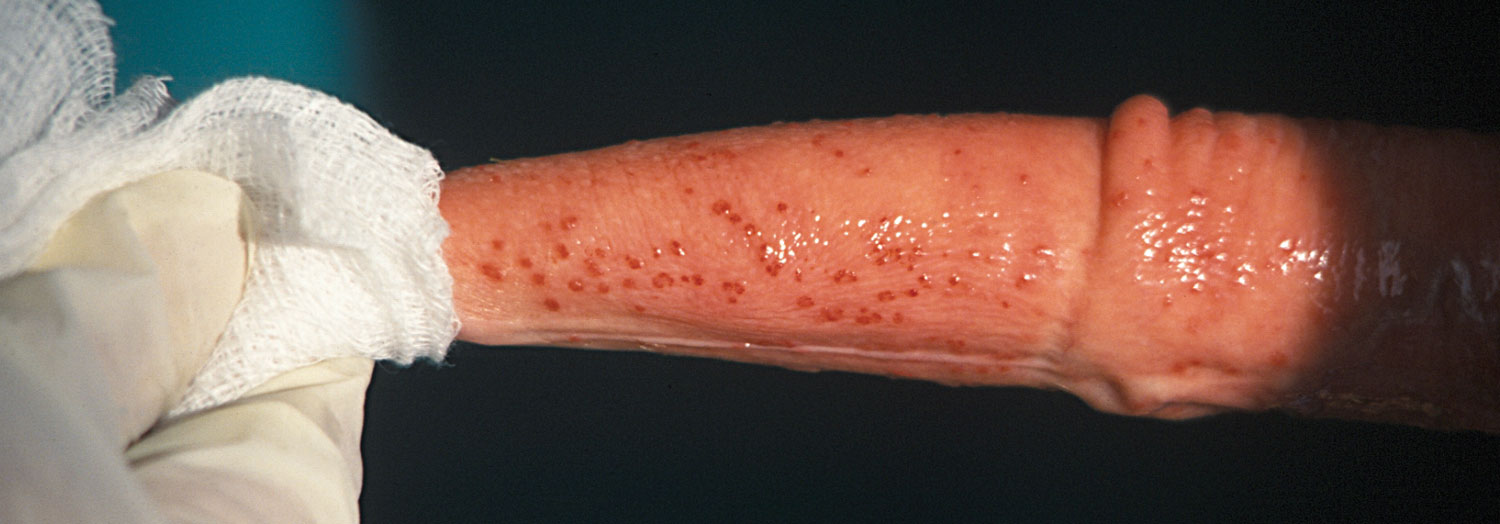
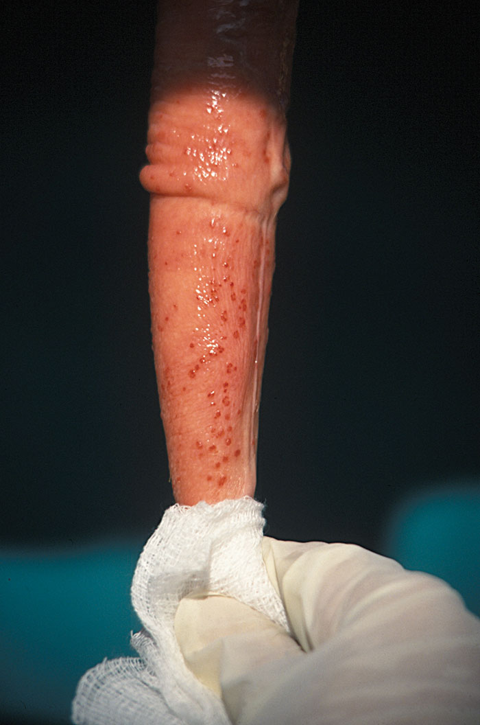
A Ureaplasma sp. was first isolated from cases of nongonococcal urethritis in humans.80 Ureaplasma diversum has since been isolated from normal cattle as well as cattle with granular vulvovaginitis, endometritis, salpingitis, seminal vesiculitis and granular balanoposthitis (Figure 206.1), and from aborted foetuses.3, 7, 8, 9, 17, 23, 51, 55, 56, 57, 65, 69, 74, 76, 79 Infection during pregnancy can also result in the birth of weak calves.60, 74 Three serogroups of U. diversum, designated A, B and C, are recognized, all of which have been isolated from the genitalia of both cows and bulls.49
Organisms assigned to the class Mollicutes generally possess few biochemical characteristics that can be used for species identification. Serological tests have come to play an essential and dominant role in this respect.66, 90, 93, 96 Tests involving membrane antigens are used almost exclusively for laboratory identification. They include growth inhibition, metabolic inhibition and immuno-fluorescence techniques. The latter in particular provide rapid and reliable identification of Mycoplasma spp. under a variety of conditions.4, 89, 93
Epidemiology
In a large survey conducted in South Africa using samples from cattle submitted for Tritrichomonas foetus and Campylobacter spp. isolation and the diagnosis of the cause of abortions, mycoplasmas were found in 14,6 per cent of females, 43 per cent of semen samples, 25 per cent of preputial washes, 3,3 per cent of foetuses and 15 per cent of placentas. 89 Mycoplasma bovigenitalium was the most common isolate.
Diseases caused by Mycoplasma and Ureaplasma spp. are principally associated with intensive animal husbandry systems, indicating the multifactorial nature of these diseases.21, 30
Coitus is probably the most important mechanism for transmitting these organisms.89 The prepuce and urethral orifice appears to be the most common site of M. bovigenitalium and U. diversum infection,25, 56, 76 and venereal transmission of these organisms has been confirmed.49, 69 Since a high proportion of semen and preputial samples are contaminated with Mycoplasma and Ureaplasma spp.,5, 25, 26, 41, 42, 57, 69, 70, 78, 89 artificial insemination probably also constitutes an important means of spread.
Mechanical transmission following close contact between infected and susceptible animals, and fomites, such as instruments used for vaginal examinations and semen collection may also be significant in this respect. Transmission may occur by the respiratory route, followed by systemic dissemination to the genital tract.8, 49
Pathogenesis
The pathogenesis of genital mycoplasmosis is poorly defined. Genital infections are easily produced by depositing organisms directly into the genital tract of either the male or the female. However, disease appears to be multifactorial, as simple infection does not invariably induce lesion development. Strain differences exist with regard to their ability to cause disease and their affinity for different organs in the cow and the bull and in experimental models.6, 49 The susceptibility of different animals can also be expected to play a role, and ovarian cyclicity appears to increase the susceptibility of heifers to vaginal lesions.71
It is unclear how mycoplasmas spread from a site of local infection in the lower reproductive tract to the upper reproductive tract to cause disease. This applies particularly to bulls, in which the most common habitat of Mycoplasma spp. is the lower urogenital tract, including the prepuce, glans penis and the distal urethra, from whence their dissemination to the upper genital tract may occur. Localization of Mycoplasma spp. in these regions produces no clinical signs of disease but may incite a local immune response,67 despite which local infections may persist for more than a year. Seminal vesiculitis in bulls is generally followed by dissemination of infection to the epididymides.49
Inconsistent results have been obtained in experiments in which M. bovigenitalium has been introduced into the genital tract of cows.1, 6, 39, 73 This could be due to a variation in the pathogenicity of different strains.2 Additionally, a combination of one or more strains in the same animal may enhance pathogenicity.89
The respiratory tract is considered to be the predilection site for M. bovis.24 Fertility can be severely affected when M. bovis is inoculated into the genital system of the female, as was shown in an experiment in which several heifers were inseminated with semen contaminated with M. bovis.38 Abnormalities in the duration of the subsequent oestrus cycle and a vaginal discharge were evident. None of the infected animals conceived, even after repeated insemination, compared to a conception rate of 83 per cent in the control group.
Male infertility or poor conception rates may be the consequence of the effect of mycoplasmas on spermatozoa.50 There is an association between the presence of M. bovigenitalium in secretions of the vesicular glands of clinically normal bulls, and diminished sperm motility.35 Inhibition of motility appears to be the result of adherence of the organisms principally to the acrosome, but also to the midpiece and tail of spermatozoa.68 Infection also reduces the post-thawing motility of frozen sperm.35 The organism may affect the ultrastructure of spermatozoa.49 However, in bulls with no detectable pathology of the testes and epididymides, sperm morphology is unaffected by the presence of mycoplasmas in the ejaculate.84 Mycoplasma bovis and Ureaplasma diversum have been shown to inhibit sperm penetration of the oocyte, possibly by a direct binding to sulphoglycolipid receptors on the sperm membrane.21, 59, 76
The adverse effect on conception rates in cattle50 may be due to effects other than the effect on semen. The effect of a mild to severe endometritis, possible inhibition of ciliary action in the oviducts, the disturbance of prostaglandin synthesis by the endometrium, and membrane damage of oviductal cells as manifested by elevated calmodulin levels should also be considered.47, 76, 83, 85 None of the Mycoplasma spp. which areknownto localize in the uterus, however, affected oviductal cell ciliary motility.85 Ureaplasma diversum has no apparent effect on the bovine embryo, despite adhering to the outer surface of the zona pellucida.11, 12, 76 Uterine infection is generally short-lived, but organisms persist for longer periods in the vagina and cervix.49, 55
The pathogenesis of abortion due to mycoplasmal infections is unknown. The organism may be introduced at breeding and persist to cause abortion, or it may spread to the uterus by a haematogenous or ascending route.60 It appears that abortion may be the consequence both of a foetal mycoplasmaemia and a concomitant placentitis.86
Infection does elicit an immune response but serum antibody concentrations are low and inconsistent and reinfection is frequent.49, 52 Vaginal U. diversum infection results in a specific humoral immune response consisting primarily of IgA in the cervicovaginal mucous.52 The results obtained in one study indicated an increased incidence of infection in young cows as compared with older animals.58 The prevalence of U. diversum in vaginal swabs from heifers was found to be unrelated to their blood urea nitrogen concentration.77
Clinical signs
Genital Mycoplasma spp. and U. diversum infections in cows usually present as a condition characterized by infertility and aberrant oestrus cycles.
A zero pregnancy rate is possible in infected herds.72 Late returns to service are caused by embryonal and foetal death. Infertility is often more severe in cases which are associated with artificial insemination because the organisms are placed directly into the uterus.17, 53
Granular vulvovaginitis is another common manifestation of disease. Acute cases of this syndrome are characterized by an intermittent mucopurulent vaginal discharge, swelling and hyperaemia of the vulva, and the presence of many pale, red or brownish-grey raised ‘granules’ 1 to 2mm in diameter on the vestibular and caudal vaginal mucosa. Pustules 2 to 5 mm in diameter form on the vulva of some affected animals. The discharge may become copious and, in some cases, it is visible on the skin and hair in the vicinity of the vulva three to six days after coitus. As the infection subsides, the discharge becomes less and may be noticeable only as a cloudiness of the bullstring at the following oestrus.48 Lesions usually resolve within 30 days, but the condition often recurs at subsequent oestrus periods. An analogous condition referred to as granular balanoposthitis is seen in bulls. Atypical forms of vulvovaginitis have been described, possibly due to the combined effects of other pathogens.29
Abortions may follow experimental exposure of pregnant cows to mollicutes, although only M. bovis and U. diversum have consistently caused abortion.37, 49, 86 Cows usually abort 11 to 18 days after infection, and the organism may be recovered from foetal and placental tissues as well as from the uterine contents. Clinical signs in the experimental cases were slight and were accompanied only by a moderate fever at the time of the mycoplasmaemia. Of the mollicutes, U. diversum is the most important cause of abortions after natural exposure. It is associated with a syndrome of sporadic abortions at any stage of gestation, stillbirths, and neonatal pneumonia, weakness and death.74 Placentas are often retained following abortion.60
Heifers may develop lameness due to arthritis, with swelling of the affected joints, anorexia and cachexia following experimental infection. Intra-uterine infection with M. bovis can result in the birth of weak calves suffering from congenital limb abnormalities.33
Pathology
Colonization of the genital tract of cattle by most Mycoplasma or Ureaplasma spp. in natural infections is normally not associated with lesions.25 Granular vulvovaginitis, however, is a common result of experimentally induced infections of M. bovigenitalium and Ureaplasma spp.18, 19 Varying degrees of endometritis, salpingitis and salpingoperitonitis follow the intra-uterine instillation of M. bovis cultures.36 A moderate amount of purulent uterine exudate is present during the first two weeks. Extensive gross lesions consisting of severe, generalized, suppurative endometritis, salpingitis and peri-oophoritis, and bursal adhesions have also been reported in long-standing cases.38, 40 Local infection may be associated with a mucopurulent exudate in the vagina, cervix and uterus.75 The exudate in the uterus may become inspissated.8
Placentitis is a constant feature of abortion caused by U. diversum. The intercotyledonary chorioallantois is thickened, opaque, and whitish to brownish, and histopathological examination reveals a necrotic, fibrosing placentitis with mononuclear cell infiltration and vasculitis. The amnion is thickened and red, with fibrosis, oedema and inflammation on histopathological examination.74 The aborted foetuses manifest a diffuse pneumonia and peribronchiolar lymphoid hyperplasia.60, 74
Lesions in the vesicular glands and epididymides in bulls experimentally infected with M. bovigenitalium are characterized by the presence of mild to moderate focal accumulations of lymphocytes and macrophages.67 Intravesicular injection of M. bovis results in a seminal vesiculitis and persistent shedding of the organisms in vesicular gland secretions for up to eight-and-a-half months.54 Attempts to reproduce vesiculitis by other routes have not been successful.
Similarly, infecting bulls intravesicularly with Ureaplasma organisms results in an enlargement of the vesicular glands, discernable on rectal palpation within 24 hours of inoculation. Moderate to severe, diffuse, chronic vesiculitis with moderate interstitial fibrosis and epithelial degeneration have been reported to occur four to eight weeks after infection.95
Diagnosis
Specimens from cows and heifers with histories of repeated breeding, conception failure, abortion in the early stages of pregnancy and/or the presence of mucopurulent discharges, should be examined for mollicutes. Methods for the collection of vaginal mucus, cervical specimens and preputial washings are available.89 Swabs of vulvar exudate should be made if a granular vulvovaginitis is being investigated. Cervical mucus should be collected in sterile pipettes in cases where lesions in the uterus and oviducts are suspected. Where infertility due to a mycoplasmal infection is suspected, uterine sampling may reveal the organism, but many samples taken at the time of return to service can be expected to be negative.19
Specimens from bulls should include preputial washings as well as vesicular gland fluid if upper genital tract involvement is suspected. Mycoplasmas are not isolated consistently from the semen of bulls with vesiculitis caused by these organisms, nor from clinically normal carriers.7 At least three collections are necessary to determine the presence of infection.
Samples should be packed on ice and delivered to the laboratory on the day of collection. Alternatively, the use of specific transport media and immediate freezing, preferably in liquid nitrogen or dry ice, have been advocated.76, 96
Mollicutes are fastidious in their growth requirements, and specifically formulated media are essential for their isolation. For diagnostic purposes, the use of a combination of different media is recommended. This enhances the frequency of primary isolation and success of laboratory maintenance. The use of different media, namely Hayflick’s agar and broth,37 Chalquest’s agar and broth,14 and Morgan’s agar and broth,61 have been recommended.89
Detection of M. bovis from semen and preputial lavage samples using a capture enzyme immunoassay has proven to be highly sensitive and specific.66 A polymerase chain reaction (PCR) assay has also been developed for U. diversum, and has demonstrated superior sensitivity to culture methods.13
Due to the common occurrence of asymptomatic infections with these organisms, positive isolations should always be interpreted in the light of the presence or absence of clinical or other evidence of disease.27
Serology is of no value in the diagnosis of genital mycoplasmosis. 24
Differential diagnosis
The two most important diseases that should be differentiated from genital mycoplasmosis are infectious pustular vulvovaginitis and the disease known as ‘epivag’ (see Infectious bovine rhinotracheitis/infectious pustular vulvovaginitis and infectious pustular balanoposthitis and Epivag, respectively). While the exact aetiology of seminal vesiculitis is not known, other infectious agents, including Chlamydophila spp., should be eliminated. Special laboratory procedures are necessary to differentiate these conditions.
Control
Intra-uterine instillation of 1 g of oxytetracycline 24 hours after breeding has been shown to improve pregnancy rates in animals infected with U. diversum.16, 17 In one study, the feeding of 350 mg chlortetracycline per head per day for 30 days also improved pregnancy rates in an infected herd when compared with untreated controls, although it did not cause a significant reduction in infection rate in the heifers or bulls,71 while in another the systemic and topical treatment with oxytetracycline of heifers suffering from a combined infection with multiple Mycoplasma spp. plus other viral and bacterial pathogens with oxytetracycline resulted in rapid resolution of lesions.29 The only reported treatment which has shown any merit in infected bulls is tylosin administered parenterally at 10 mg/kg for five days.34
A high proportion of semen samples collected from healthy bulls at artificial insemination centres are contaminated with mollicutes.7, 28, 46, 78, 89 Contamination with M. bovigenitalium, M. bovis and U. diversum should be a cause for concern, and routine screening and decontamination of semen is therefore desirable to avoid the spread of these organisms.3, 91 Antibiotics, such as penicillin and streptomycin, which are normally added during the processing of such semen do not eliminate mollicutes. Diluents containing lincomycin, spectinomycin, minocycline, gentamycin or tylosin have shown varying degrees of efficacy in controlling Mycoplasma and Ureaplasma spp. in semen.7, 81, 90, 92, 94 No drug is fully bacteriocidal, and combinations are advocated in order to ensure efficacy against a range of pathogens.48, 81 Photosensitive agents have been tested for the disinfection of semen, but have not been successful to date.20 Egg yolk, a component of the cryprotective extenders used in the preservation of bovine semen, is frequently contaminated with mycoplasmas, and the use of diluents which do not contain egg yolk may be preferable for this reason.10
Sanitary sleeves placed over the insemination pipette which prevent infection from being carried forward from the vagina are advocated in known infected herds. Other measures aimed at improving general hygiene, such as regular replacement of bedding and sanitary measures during semen collection and examinations of animals, are also indicated. Due to the association of these micro-organisms with intensification, a decrease in stocking density can be expected to cut down on transmission of organisms.
Vaccination with experimental adjuvanted U. diversum whole cell or cell wall vaccines have resulted in significantly elevated IgG levels in serum and cervicovaginal mucus, but not in protection from the development of lesions.62, 63 No vaccines are available in South Africa.
The risks of the transmission of mycoplasmas and other pathogens via transferred embryos has been reviewed.87 Ureaplasmas adherent to the zona pellucida of in vivo-produced embryos are not removed during 10 sequential washes.11, 12 Treatment with antibiotics is effective in inactivating the organisms in infected embryos.87 Despite the large number of in vivo-derived embryos which have been transferred, no cases of disease transmission by this method have been documented.87
References
- afshar, a., stuart, p. & huch, r.a., 1966. Granular vulvovaginitis of cattle associated with Mycoplasma bovigenitalium. The Veterinary Record, 78, 512–519.
- al-aubaidi, j.m., mcentee, k., lein, d.h. & roberts, s.j., 1972. Bovine seminal vesiculitis and epididymitis caused by Mycoplasma bovigenitalium. Cornell Veterinarian, 62, 581–596.
- almquist, j.o. & zaugg, n.l., 1974. Fertility of bovine semen in milk diluent containing combinations of penicillin-neomycin and lincomycin-spectinomycin. Journal of Dairy Science, 57, 1211–1213.
- baas, e.j. & jasper, d.e., 1972. Agar block technique for identification of mycoplasmas by use of fluorescent antibody. Applied Microbiology, 23, 1097–1100.
- ball, h.j., armstrong, d., mccaughey, w.j. & kennedy, s., 1987. Experimental intrauterine inoculation of cows at oestrus with Mycoplasma canadense. The Veterinary Record, 119, 370.
- ball, h.j., armstrong, d., kennedy, s. & mccaughey, w.j., 1987. Experimental intrauterine inoculation of cows at oestrus with a bovine ureaplasma strain. Irish Veterinary Journal, 41, 371–372.
- ball, h.j., logan, e.f. & orr, w., 1987. Isolation of mycoplasmas from bovine semen in Northern Ireland. The Veterinary Record, 120, 322–324.
- ball, h.j., mccaughey, w.j., mackie, d.p. & pearson, g.r., 1981. Experimental genital infection of heifers with ureaplasmas. Research in Veterinary Science, 30, 312–317.
- boughton, e., sheilagh, a.n. & gayford, p.j.r., 1983. Mycoplasma canadense from bovine fetuses. The Veterinary Record, 112, 87.
- bousseau, s., brillard, j.p., guienne, b.m.l., guerin, b., camus, a. & lechat, m., 1998. Comparison of bacteriological qualities of various egg yolk sources and the in vitro and in vivo fertilizing potential of bovine semen frozen in egg yolk or lecithin based diluents. Theriogenology, 50, 699–706.
- britton, a.p., ruhnke, h.l., miller, r.b. & johnson, w.h., 1988. Adherence of Ureaplasma diversum to the bovine zona pellucida. Theriogenology, 29, 229.
- britton, a.p., ruhnke, h.l., miller, r.b., johnson, w.h., leslie, k.e. & rosendal, s., 1987. In vitro exposure of bovine morulae to Ureaplasma diversum. Canadian Journal of Veterinary Research, 51, 198–203.
- cardoso, m.v., blanchard, a., ferris, s., verlengia, r., timenetsky, j. & da cunha, r.a.f., 2000. Detection of Ureaplasma diversum in cattle using a newly developed PCR-based detection assay. Veterinary Microbiology, 72, 241–250.
- chalquest, r.r., 1962. Cultivation of the infectious synovitis-type pleuropneumonia-like organisms. Avian Diseases, 6, 36–43.
- cottew, g.s., 1970. Mycoplasmas isolated from cattle in Australia. Australian Veterinary Journal, 46, 378–381.
- doig, p.a., 1981. Bovine genital mycoplasmosis. Canadian Veterinary Journal, 22, 339–343.
- doig, p.a., ruhnke, h.l., mckay, a.l. & palmer, n.c., 1979. Bovine granular vulvitis associated with Ureaplasma infection. The Canadian Veterinary Journal, 20, 89–94.
- doig, p.a., ruhnke, h.l. & palmer, n.c., 1980. Experimental bovine genital ureaplasmosis 1. Granular vulvitis following vulvar inoculation. Canadian Journal of Comparative Medicine, 44, 252–258.
- doig, p.a., ruhnke, h.l. & palmer, n.c., 1980. Experimental vulvitis, endometritis and salpingitis following uterine inoculation. Canadian Journal of Comparative Medicine, 44, 259–266.
- eaglesome, m.d., bielanski, a., hare, w.c.d. & ruhnke, h.l., 1994. Studies on inactivation of pathogenic microorganisms in culture media and in bovine semen by photosensitive agents. Veterinary Microbiology, 38, 277–284.
- eaglesome, m.d. & garcia, m.m., 1990. The effect of Mycoplasma bovis on fertilization processes in vitro with bull spermatozoa and zona-free hamster oocytes. Veterinary Microbiology, 21, 329–337.
- erno, h., plastridge, w.n. & tourtelloute, m.e., 1967. Mycoplasma isolation of prepuce and semen of bulls. Acta Veterinaria Scandinavia, 8, 123–135.
- farstad, w., krogenaes, a. & friis, n.f., 1996. An outbreak of transmissible granular vulvovaginitis in cattle cased by Ureaplasma diversum. Norsk Veterinaertidskrift, 108, 159–164.
- feenstra, a., madsen, e.b., friis, n.f., meyling, a. & ahrens, p., 1991. A field study of Mycoplasma bovis infection in cattle. Journal of Veterinary Medicine, 38, 195–202.
- fish, n.a., rosendal, s. & miller, r.b., 1985. The distribution of mycoplasmas and ureaplasmas in the genital tract of normal artificial insemination bulls. Canadian Veterinary Journal, 26, 13–15.
- friis, n.f. & blom, e., 1983. Isolation of Mycoplasma canadense from bull semen. Acta Veterinaria Scandinavia, 24, 315–317.
- gaines, j.d., 1989. Investigating the role of infectious diseases and toxins in the subfertile dairy herd. Veterinary Medicine, 84, 1195–1199.
- garcia, m.m., truscott, r.b., mclaren, j., stewart, r.b. & kingscote, b., 1986. Absence of Mycoplasma bovis in unprocessed frozen bull semen from Canadian artificial insemination centres. The Veterinary Record, 119, 11–12.
- gilbert, r.o. & oettle, e.e., 1990. An outbreak of granulomatous vulvitis in feedlot heifers. Journal of the South African Veterinary Association, 61, 41–43.
- gourlay, r.n., 1981. Mycoplasmosis in cattle, sheep and goats. Israel Journal of Medical Science, 17, 531–536.
- gourlay, r.n. & howard, c.j., 1979. In: The Mycoplasmas, Vol. II, London and New York: Academic Press.
- gourlay, r.n., howard, c.j., thomas, l.h. & stott, e.j., 1976. Experimentally produced calf pneumonia. Research in Veterinary Science, 20, 167–173.
- grunert, e., bolting, d., stockhofe, n. & pickel, m., 1992. Mycoplasma bovis infection in newborn calves. Tierärztliche Umschau, 47, 99–100, 102.
- gummow, b., staley, g.p. & gouws, j.j., 1992. The diagnosis and treatment of bovine genital ureaplasmosis: A case study. Journal of the South African Veterinary Association, 63, 128–131.
- hall, c.e. & mcentee, k., 1981. Reduced post-thawing survival of sperm in bulls with mycoplasmal vesiculitis. Cornell Veterinarian, 71, 111–112.
- hartman, h.a., tourteloutte, m.e., nielsen, s.w. & plastridge, w.n., 1964. Experimental bovine uterine mycoplasmosis. Research in Veterinary Science, 5, 303–310.
- hayflick, l., 1965. Tissue cultures and mycoplasmas. Texas Reports on Biology and Medicine, 1, 23, 285–303.
- hirth, r.s., nielsen, s.w. & plastridge, w.n., 1966. Bovine salpingo-oophoritis produced with semen containing Mycoplasma. Pathologia Veterinaria, 3, 616–632.
- hirth, r.s., nielsen, s.w. & tourtelloute, m.e., 1970. Characterization and comparative genital tract pathogenicity of bovine mycoplasmas. Infection and Immunity, 2, 101–104.
- hoare, m., 1969. A survey of the incidence of Mycoplasma infection in the oviducts of dairy cows. The Veterinary Record, 85, 351–355.
- hodges, r.t. & holland, j.t.s., 1980. The recovery of ureaplasmas from the semen and prepuce of bulls. New Zealand Veterinary Journal, 28, 89–90.
- holzmann, a., thiemann, g., flatcher, j. & prilhofer, k., 1982. Changes in semen of steers with positive Mycoplasma evidence. Zentralblatt für Veterinärmedizin, 29, 710–715.
- horvath, g., stipkovits, l., varga, z., zoldag, l. & meszaros, j., 1983. Infection of cows by Mycoplasma bovis. Archiv für Experimentelle Veterinärmedizin, 37, 401–403.
- howard, c.j., 1984. Animal ureaplasmas: Their ecological niche and role in disease. Israel Journal of Medical Science, 20, 954–957.
- jasper, d.e., 1977. Mycoplasma and mycoplasma mastitis. Journal of the American Veterinary Medical Association, 170, 1167–1172.
- jurmanova, k., weznik, z., cerna, j. & mazurova, j., 1983. Demonstration and role of mycoplasmas and ureaplasmas in bull semen and the control of Mycoplasma infections in bulls. Archiv für Experimentelle Veterinärmedizin, 37, 421–428.
- kapoor, p.k., mahajan, s.k., garg, d.n. & singh, y., 1993. Pathogenic effects of mollicutes from bovines with reproductive disorders in hamster tracheal ring organ culture. Indian Veterinary Journal, 70, 393–396.
- kirchoff, h., 1982. Die Bedeutung der Mykoplasmen in den Genital-organen von Rind, Pferd und Schwein. Berliner und Münchener Tierärztliche Wochenschrift, 95, 121–125.
- kirkbride, c.a., 1987. Mycoplasma, Ureaplasma and Acholeplasma infections of bovine genitalia. Veterinary Clinics of North America, Food Animal Practice, 3, 575–591.
- kissie, b., juhasz, s. & stipkovits, l., 1985. Effect of Mycoplasma contamination of bull semen on fertilization. Acta Veterinaria Hungarica, 33, 107–117.
- kreplin, c.m.a., 1988. A study of infertility and the immune response associated with Ureaplasma diversum infection in cattle. Dissertation Abstracts International, 49, 1567-B.
- kreplin, c.m.a., ruhnke, h.l., miller, r.b. & doig, p.a., 1987. The effect of intrauterine inoculation with Ureaplasma diversum on bovine fertility. Canadian Journal of Veterinary Research, 51, 440–443.
- kuhn, m.j. & hopkins, s.m., 1983. A clinical review of bovine ureaplasmosis. Iowa State Veterinarian, 45, 20–24.
- la faunce, n.a. & mcentee, k., 1982. Experimental Mycoplasma bovis seminal vesiculitis in the bull. Cornell Veterinarian, 72, 150–167.
- langford, e.v., 1975. Mycoplasma species recovered from the reproductive tracts of Western Canadian cows. Canadian Journal of Comparative Medicine, 39, 133–137.
- langford, e.v., 1975. Mycoplasma recovered from bovine male and female genitalia and aborted foeti. Proceedings of the 18th Annual Meeting of Veterinary Laboratory Diagnosticians, San Diego, California.
- le grand, d., poumarat, f. & martel, j.l., 1995. Infectious genital disease caused by Ureaplasma diversum: Investigations in cattle in France. Veterinary Research, 26, 11–20.
- leon, b.a., campos, e. bolanos, h. & caballero, m., 1995. Risk factors for Ureaplasma diversum infection in cattle in a tropical environment. Revista de Biologia Tropical, 43, 21–25.
- lingwood, c.a., quinn, p.a., wilansky, s., nutikka, a., ruhnke, h.l. & miller, r.b., 1990. Common sulfoglycolipid receptor for mycoplasmas involved in animal and human infertility. Biology of Reproduction, 4 3, 694–697.
- miller, r.b., ruhnke, h.l., doig, p.a., poitras, b.j. & palmer, n.c., 1983. The effect of Ureaplasma diversum inoculated into the amniotic cavity in cows. Theriogenology, 20, 367–374.
- morgan, j.h. & lewis, j., 1977. A medium for the primary isolation of T-mycoplasmas from cattle. Medical Laboratory Sciences, 34, 177–178.
- mulira, g.l. & saunders, j.r., 1994. Immune response of heifers to vaginal submucosal or subcutaneous vaccination and intravaginal challenge with Ureaplasma diversum. Canadian Journal of Veterinary Research, 58, 109–113.
- mulira, g.l. & saunders, j.r., 1994. Humoral and secretory antibodies to Ureaplasma diversum in heifers following subcutaneous vaccination and vaginal infection. Canadian Journal of Veterinary Research, 58, 104–108.
- mulira, g.a., saunders, j.r. & barth, a.d., 1992. Isolation of Ureaplasma diversum and mycoplasmas from genital tracts of beef and dairy cattle in Saskatchewan. Canadian Veterinary Journal, 33, 46–49.
- mulira, g.a., saunders, j.r. & kariuki, d.p., 1989. Isolation of Ureaplasma diversum from bovine granular vulvitis in Kenya. Bulletin of Animal Health and Production in Africa, 37, 347–350.
- nielsen, k.h., stewart, r.b., garcia, m.m. & eaglesome, m.d., 1987. Enzyme immunoassay for detection of Mycoplasma bovis antigens in bull semen and preputial washings. The Veterinary Record, 120, 596– 598.
- panangala, v.s., hall, c.e., caveney, n.t., lein, d.h. & winter, a.j., 1982. Mycoplasma bovigenitalium in upper genital tract of bulls: Spontaneous and induced infections. Cornell Veterinarian, 72, 292–303.
- panangala, v.s., winter, a.j., wijesinha, a. & foote, r.h., 1981. Decreased motility of bull spermatozoa caused by Mycoplasma bovigenitalium. American Journal of Veterinary Research, 42, 2090–2093.
- pilaszek, j. & truszczynski, m., 1988. Affinity of microorganisms of the genus Ureaplasma to the reproductive organs of cattle. Comparative Immunology, Microbiology and Infectious Diseases, 11, 177–180.
- rae, a.g., 1982. Isolation of mycoplasmas from bovine semen. The Veterinary Record, 109, 463.
- rae, d.o., chenoweth, p.j., brown, m.b., genho, p.c., moore, s.a. & jacobsen, k.e., 1993. Reproductive performance of beef heifers: Effects of vulvo-vaginitis, Ureaplasma diversum and prebreeding antibiotic administration. Theriogenology, 40, 497–508.
- reid, s.w., madill, d.g. & vreugdenhil, a.h., 1989. Ureaplasma vulvovaginitis and infertility in eight southern Ontario dairy herds. Canadian Veterinary Journal, 30, 255.
- roberts, d.h., 1968. Immunological response in cows to Mycoplasma bovigenitalium strain 1836. Journal of Hygiene Cambridge, 66, 585–593.
- ruhnke, h.l., palmer, n.c., doig, p.a. & miller, r.b., 1984. Bovine abortion and neonate death associated with Ureaplasma diversum. Theriogenology, 21, 295–301.
- saed, o.m. & al-aubaidi, j.m., 1983. Infertility in heifers caused by a pathogenic strain of Mycoplasma bovigenitalium. Cornell Veterinarian, 73, 125–130.
- sanderson, m.w. & chenoweth, p.j., 1999. The role of Ureaplasma diversum in bovine reproduction. Compendium on Continuing Education for the Practicing Veterinarian, 21, S98–S101.
- sanderson, m.w., chenoweth, p.j., yeary, t. & nietfield, j.c., 1999. Prevalence of Ureaplasma diversum in beef replacement heifers and the relationship to blood urea nitrogen level. Proceedings of the Annual Conference of the Society for Theriogenology, Nashville Tennessee, 22–24 September 1999, 5.
- schutte, a.p., 1975. Bees mycoplasmose en onvrugbaarheid. Symposium: Reproductive Disorders and Fertility of Ruminants, South African Veterinary Association, July 1975.
- schweighardt, h., kaltenbock, b., lauerman, e. & pechan, p., 1985. Ureaplasmen als Verusacher van granularen Vulvovaginitiden beim Rind. Wiener Tierärztliche Monatschrift, 72, 209–214.
- shepard, m.c., 1954. The recovery of pleuropneumonia-like organisms from Negro men with and without non-gonococcal urethritis. American Journal of Syphilis Gonorrhoea and Venereal Diseases, 38, 113–124.
- shin, s.j., lein, d.h., patten, v.h. & ruhnke, h.l., 1988. A new antibiotic combination for frozen bovine semen. 1. Control of Mycoplasmas, Ureaplasmas, Campylobacter fetus subsp. venerealis and Haemophilus somnus. Theriogenology, 29, 577–591.
- singh, y., garg, d.n., kapoor, p.k. & mahajan, s.k., 1991. Experimental pathogenicity of mollicutes from bovines with reproductive disorders in rabbit fallopian tube organ culture. Indian Journal of Experimental Biology, 29, 773–777.
- smits, b., rosendal, s., ruhnke, h.l., plante, c., o’brien, p.j. & miller, r.b., 1994. Effects of Ureaplasma diversum on bovine oviductal explants: Quantitative measurement using a calmodulin assay. Canadian Journal of Veterinary Research, 58, 144–121.
- sprecher, d.j., coe, p.h. & walker, r.d., 1999. Relationships among seminal culture, seminal white blood cells, and the percentage of primary sperm abnormalities in bulls evaluated prior to the breeding season. Theriogenology. 51, 1197–1206.
- stalheim, o.h.v. & gallaghar, j.e., 1975. Effects of Mycoplasma spp., Trichomonas fetus and Campylobacter fetus on ciliary activity of bovine uterine tube organ cultures. American Journal of Veterinary Research, 36, 1077–1080.
- stalheim, o.h.v. & proctor, s.j., 1976. Experimentally induced bovine abortion with Mycoplasma agalactiae subsp. bovis. American Journal of Veterinary Research, 37, 879–883.
- stringfellow, d.a. & givens, m.d., 2000. Preventing disease transmission through the transfer of in-vivo-derived bovine embryos. Livestock Production Science, 62, 237–251.
- tourtellotte, m.e. & lein, d.h., 1976. Infertility of cattle caused by mycoplasmas. Health and Laboratory Science, 13, 152–157.
- trichard, c.j.v. & jacobsz, e.p., 1985. Mycoplasmas recovered from bovine genitalia, aborted foetuses and placentas in the Republic of South Africa. Onderstepoort Journal of Veterinary Research, 52, 105–110.
- truscott, r.b., 1981. A comparison of the in vitro activity of two antibiotics against bovine ureaplasmas. Canadian Journal of Comparative Medicine, 45, 113–115.
- truscott, r.b. & abreo, c., 1977. Antibiotics for elimination of mycoplasmas and ureaplasma from bovine semen. Journal of Dairy Science, 60, 954–960.
- truscott, r.b. & ruhnke, h.l., 1984. The effect of antibiotics against bovine mycoplasmas and ureaplasmas. Canadian Journal of Comparative Medicine, 48, 171–174.
- tully, j.g. & razin, s., 1983. Methods in Mycoplasmology, Vol. I & II. New York: Academic Press.
- visser, i.j.r., ter laak, e.a. & jansen, h.b., 1999. Failure of antibiotics gentamycin, tylosin, lincomycin and spectinomycin to eliminate Mycoplasma bovis in artificially infected frozen bovine semen. Theriogenology, 51, 689–697.
- waelchli-suter, r.o., doig, p.a., ruhnke, h.l., palmer, n.c. & barker, c.a.v., 1982. Experimental genital ureaplasmosis in the bull. Schweizer Archiv für Tierheilkunde, 124, 273–295.
- wittowski, g., roth, b. & kirchhoff, h., 1984. Nachweis von Mykoplasmen und Ureaplasmen im Genitaltrakt des Rindes. Berliner un Münchener Tierärztliche Wochenschrift, 97, 189–190, 193–197.
- yaeger, m. & holler, l.d., 1997. Bacterial causes of bovine infertility and abortion. In: youngquist, r.s., (ed.). Current Therapy and Large Animal Theriogenology. Philadelphia: W.B. Saunders. 364–372.


