- Infectious Diseases of Livestock
- Part 3
- DISEASE COMPLEXES / UNKNOWN AETIOLOGY: Epivag
- GENERAL INTRODUCTION: SPIROCHAETES
- Swine dysentery
- Borrelia theileri infection
- Borrelia suilla infection
- Lyme disease in livestock
- Leptospirosis
- GENERAL INTRODUCTION: AEROBIC ⁄ MICRO-AEROPHILIC, MOTILE, HELICAL ⁄ VIBROID GRAM-NEGATIVE BACTERIA
- Genital campylobacteriosis in cattle
- Proliferative enteropathies of pigs
- Campylobacter jejuni infection
- GENERAL INTRODUCTION: GRAM-NEGATIVE AEROBIC OR CAPNOPHILIC RODS AND COCCI
- Moraxella spp. infections
- Bordetella bronchiseptica infections
- Pseudomonas spp. infections
- Glanders
- Melioidosis
- Brucella spp. infections
- Bovine brucellosis
- Brucella ovis infection
- Brucella melitensis infection
- Brucella suis infection
- Brucella infections in terrestrial wildlife
- GENERAL INTRODUCTION: FACULTATIVELY ANAEROBIC GRAM NEGATIVE RODS
- Klebsiella spp. infections
- Escherichia coli infections
- Salmonella spp. infections
- Bovine salmonellosis
- Ovine and caprine salmonellosis
- Porcine salmonellosis
- Equine salmonellosis
- Yersinia spp. infections
- Haemophilus and Histophilus spp. infections
- Haemophilus parasuis infection
- Histophilus somni disease complex in cattle
- Actinobacillus spp. infections
- Actinobacillus equuli infections
- Gram-negative pleomorphic infections: Actinobacillus seminis, Histophilus ovis and Histophilus somni
- Porcine pleuropneumonia
- Actinobacillus suis infections
- Pasteurella and Mannheimia spp. infections
- Pneumonic mannheimiosis and pasteurellosis of cattle
- Haemorrhagic septicaemia
- Pasteurellosis in sheep and goats
- Porcine pasteurellosis
- Progressive atrophic rhinitis
- GENERAL INTRODUCTION: ANAEROBIC GRAM-NEGATIVE, IRREGULAR RODS
- Fusobacterium necrophorum, Dichelobacter (Bacteroides) nodosus and Bacteroides spp. infections
- GENERAL INTRODUCTION: GRAM-POSITIVE COCCI
- Staphylococcus spp. infections
- Staphylococcus aureus infections
- Exudative epidermitis
- Other Staphylococcus spp. infections
- Streptococcus spp. infections
- Strangles
- Streptococcus suis infections
- Streptococcus porcinus infections
- Other Streptococcus spp. infections
- GENERAL INTRODUCTION: ENDOSPORE-FORMING GRAM-POSITIVE RODS AND COCCI
- Anthrax
- Clostridium perfringens group infections
- Clostridium perfringens type A infections
- Clostridium perfringens type B infections
- Clostridium perfringens type C infections
- Clostridium perfringens type D infections
- Malignant oedema⁄gas gangrene group of Clostridium spp.
- Clostridium chauvoei infections
- Clostridium novyi infections
- Clostridium septicum infections
- Other clostridial infections
- Tetanus
- Botulism
- GENERAL INTRODUCTION: REGULAR, NON-SPORING, GRAM-POSITIVE RODS
- Listeriosis
- Erysipelothrix rhusiopathiae infections
- GENERAL INTRODUCTION: IRREGULAR, NON-SPORING, GRAM-POSITIVE RODS
- Corynebacterium pseudotuberculosis infections
- Corynebacterium renale group infections
- Bolo disease
- Actinomyces bovis infections
- Trueperella pyogenes infections
- Actinobaculum suis infections
- Actinomyces hyovaginalis infections
- GENERAL INTRODUCTION: MYCOBACTERIA
- Tuberculosis
- Paratuberculosis
- GENERAL INTRODUCTION: ACTINOMYCETES
- Nocardiosis
- Rhodococcus equi infections
- Dermatophilosis
- GENERAL INTRODUCTION: MOLLICUTES
- Contagious bovine pleuropneumonia
- Contagious caprine pleuropneumonia
- Mycoplasmal pneumonia of pigs
- Mycoplasmal polyserositis and arthritis of pigs
- Mycoplasmal arthritis of pigs
- Bovine genital mycoplasmosis
- Neurotoxin-producing group of Clostridium spp.
- Contagious equine metritis
- Tyzzer's disease
- MYCOTIC AND ALGAL DISEASES: Mycoses
- MYCOTIC AND ALGAL DISEASES: Pneumocystosis
- MYCOTIC AND ALGAL DISEASES: Protothecosis and other algal diseases
- DISEASE COMPLEXES / UNKNOWN AETIOLOGY: Epivag
- DISEASE COMPLEXES / UNKNOWN AETIOLOGY: Ulcerative balanoposthitis and vulvovaginitis of sheep
- DISEASE COMPLEXES / UNKNOWN AETIOLOGY: Ill thrift
- Eperythrozoonosis
- Bovine haemobartonellosis
DISEASE COMPLEXES / UNKNOWN AETIOLOGY: Epivag
This content is distributed under the following licence: Attribution-NonCommercial CC BY-NC  View Creative Commons Licence details here
Epivag
A P SCHUTTE
Introduction
Epivag is an insidious, infectious venereal disease of cattle that is characterized in cows by an anterior vaginitis with a thick, tenacious, mucopurulent discharge from the vulva, and in bulls by chronic epididymitis and seminal vesiculitis that results in fibrosis and induration of the epididymis, seminal vesicles and testes. The fertility of affected cows is usually impaired, but abortion is not a feature of the disease. In bulls the disease usually results in reduced fertility, and complete impotency in some. Because the lesions that develop in the genital organs of both sexes are basically those of an epididymitis or vaginitis, the name ‘epivag’ was coined for the disease.4
The condition appears to be confined to Africa. It was first described in Kenya by Daubney, Hudson and Anderson in 1938,1 and has since been diagnosed in Zimbabwe and South Africa.12 Earlier reports in South Africa indicate that the disease was most prevalent in what is now Gauteng and Free State provinces, but it has occurred to a lesser extent in KwaZulu-Natal Province. In South Africa, at present, the disease is only sporadically encountered in poorly managed dairy herds in which natural breeding is practised. Although no reports are available, the same situation most probably prevails in other countries in southern, Central and East Africa.
Aetiology and epidemiology
The aetiology of the disease has not, as yet, been elucidated, but, in view of its epidemiology, one or possibly more infectious agents are thought to be involved. The consensus of opinion is that epivag is caused by a virus, but all attempts to determine its aetiology have failed. Bovine herpesvirus 1 has been isolated from seminal plasma and vaginal discharges of affected animals.2, 5, 6, 9 However, experimental attempts to reproduce the disease by infecting cattle with it, and with bovine herpesvirus 4, have been unsuccessful.7, 8, 10, 11
Epivag spreads in herds in which the cattle are allowed to breed naturally; it does not occur in herds in which artificial insemination is practised. Affected bulls in the early or advanced stage of the disease transmit the disease venereally to susceptible heifers and cows. Similarly, infected females with mild or severe vaginitis may cause venereal transmission of the infection to susceptible bulls. It would seem that females are still capable of spreading the infection venereally to bulls several months or perhaps even years after they have apparently recovered from the disease.
According to Van Rensburg,12 the disease is more acute in some herds than in others; environmental conditions, such as climate and possibly also nutritional factors, may influence the severity of the disease.
Clinical signs and pathology
Clinical evidence of the infection in bulls may only be detectable four to ten weeks after natural service of an infected cow. The earliest lesions following infection are seminal vesiculitis and a slight palpable enlargement of one or both of the spermatic cords. Eventually the epididymes and testes also become involved. The latter lesions usually commence unilaterally, but in the later stages of the disease are often bilateral. The libido of bulls with early or advanced lesions is usually unimpaired; affected bulls may therefore continue to serve and spread the infection. In some bulls, however, the libido is reduced and they may even become completely impotent.
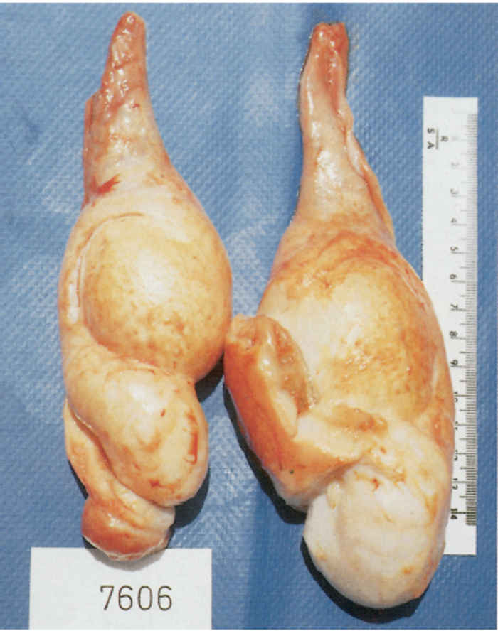
The acute lesions in the seminal vesicles are characterized by enlargement and pain on palpation. When evaluating the size of the seminal glands, cognizance should be taken of the variable size of these glands in healthy bulls. In chronically affected bulls the glands are enlarged, firm, usually not painful on palpation, and the lobulation is less distinct (Figure 212.1).
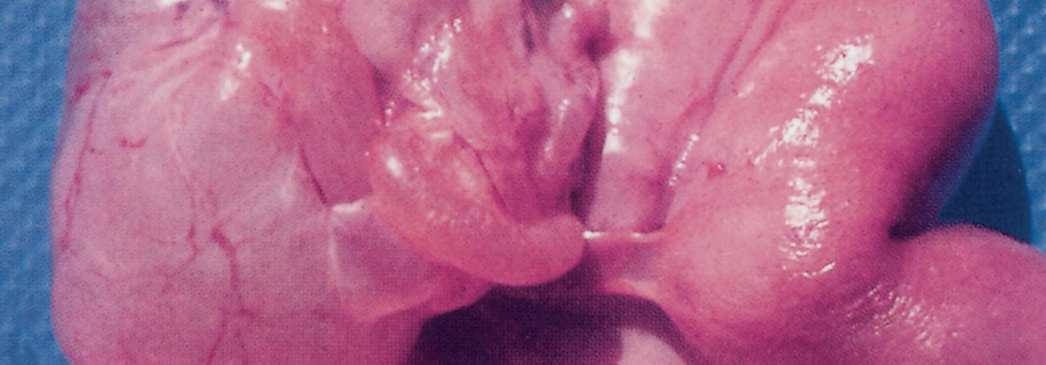
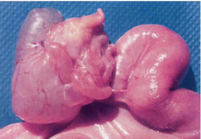
In naturally occurring acute cases, an initial diffuse swelling, probably as a result of degeneration, of the affected testis masks the changes in the epididymis. Epididymal lesions are only evident once the testicular swelling has subsided. The tail of the epididymis may be slightly or markedly enlarged, loses resilience and elasticity, and becomes a hard spherical mass with an irregularly lobulated surface due to the chronic interstitial epididymitis and consequent ductular obstructions that develop. Later in the course of the disease, similar changes are detectable in the head of the epididymis and eventually the normal structure of the epididymis is completely replaced by fibrous tissue.3 There are adhesions between the tunica vaginalis propria and tunica vaginalis communis, and the testis becomes atrophied and the interstitial tissue fibrosed (Figure 212.2). Although the quality of the semen of affected bulls is reduced, they are still, in the majority of cases, fertile for a period before sterility supervenes. Recovery in bulls is extremely rare.
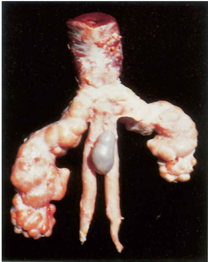
Figure 212.1 Vesicula seminalis. Note the irregular and asymmetrical enlargement of the gland as a consequence of the chronic inflammation
The course of the disease varies in heifers and cows. About seven to ten days after coitus a diffuse anterior vaginitis is generally detectable with the accumulation of a thick, tenacious, yellowish, mucopurulent, odourless exudate which adheres to the mucous membrane of the vagina. The exudate is discharged periodically for about two weeks, resulting in soiling of the tail and buttocks, giving the latter a characteristic glistening, streaky appearance. The volume of exudate varies; at times it may be copious and as much as 500 ml may accumulate in the vagina. Acutely affected female animals may conceive and produce offspring. In approximately 25 per cent of cases the inflammation extends into the cervix and uterus, which also results in the production of copious amounts of pus, as well as into the oviducts, resulting in chronic salpingitis, hydrosalpynx, peri-oophoritis, and fibrous adhesions between the Fallopian tubes and adjacent tissues (Figure 212.3). These animals are usually sterile. In most cases these changes are confined to the pelvic region but the inflammatory process may also extend into the abdominal cavity, resulting in widespread fibrous peritonitis and the development of adhesions between abdominal organs.3
Diagnosis and differential diagnosis
As the aetiology of epivag is not known, a diagnosis is based entirely on the clinical signs: presence of genital lesions (seminal vesiculitis and epididymal and testicular lesions in bulls, and the thick, tenacious, yellow, mucopurulent, odourless vaginal discharge that involves a number of females), and a history of prolonged intercalving periods and sterility in some animals in herds where natural service is practised.
Bovine herpesvirus 1 causes infectious pustular vulvovaginitis or infectious pustular balanoposthitis, but does not result in seminal vesiculitis and epididymitis in bulls, and the lesions in the vagina of cows are usually small, multifocal, vesicular or erosive, while a copious exudate, such as that often present in epivag, is not a feature of the disease. Trichomonal infection in cows can be differentiated from epivag by the difference in the nature and volume of the vaginal discharge; in trichomoniasis, the uterine content and vaginal discharge are less tenacious and voluminous than in epivag and the fluid, although also odourless, is greyish-white and contains trichomonads in great numbers.
Seminal vesiculitis may also occur in young virgin bulls on a high protein and carbohydrate diet. The aetiology of this syndrome has not been elucidated but it is speculated that hormonal influences and possibly certain infections (Mycoplasma spp.) may play a role. Infection of the seminal vesicles by pyogenic bacteria (Trueperella pyogenes, Streptococcus spp.), Brucella abortus, Mycobacterium bovis, Chlamydophila (Chlamydia) sp. and Mycoplasma spp. may also occasionally cause vesiculitis.
Control
The use of artificial insemination in a herd is considered to be the best method of control for epivag, but the semen should originate from certified disease-free bulls, the hygiene of the insemination technique should be of prescribed standard, and the management system applied by the farmer should be suitable for performing insemination. It must be emphasized that those cows which have been infected should be allowed a period of sexual rest before insemination, during which time oestrous cycles should be regular.
References
- DAUBNEY, R., HUDSON, J.R. & ANDERSON, J., 1938. Preliminary description of a form of sterility in cattle associated with vaginitis in female stock and with chronic changes in the epididymis and the testicles of bulls. East African Agricultural Journal, 3, 31–34.
- HELLIG, H., 1965. Investigation into natural outbreaks of infectious pustular vulvo-vaginitis in cattle in South Africa. Journal of the South African Veterinary Medical Association, 36, 219–226.
- HENNING, M.W., 1956. Infectious bovine infertility. In: Animal Diseases in South Africa. 3rd edn. Pretoria: Central News Agency Ltd.
- HUDSON, J.R., 1949. A specific venereal disease of cattle characterized by epididymitis in bulls and vaginitis in cows and heifers. Proceedings of the Fourteenth International Veterinary Congress, London. Vol. II. pp. 487–491.
- KAMINJOLO, J.S., NYAGA, P.N., ONNUSE, J.K. & MUTIGA, E.R., 1975. Infectious bovine rhinotracheitis/infectious pustular vulvo-vaginitis viral isolates from cattle with epididymitis and vaginitis. American Journal of Veterinary Research, 36, 123–125.
- MARÉ, L.J. & VAN RENSBURG, S.J., 1961. The isolation of viruses associated with infertility in cattle: A preliminary report. Journal of the South African Veterinary Medical Association, 32, 201–210.
- MCINTOSH, B.M., HAIG, D.A. & ALEXANDER, R.A., 1952. Isolation of viruses associated with epididymitis and vaginitis in cattle. Journal of the South African Veterinary Medical Association, 23, 165–166.
- SCHUTTE, A.P, 1993. Taurus, Private Bag X5, Irene, South Africa. Unpublished data.
- SWANEPOEL, R. & CHRISTIE, S., 1972. A survey of infectious bovine rhinotracheitis in Rhodesian cattle. Rhodesian Veterinary Journal, 3, 20–25.
- THEODORIDIS, A., 1978. Preliminary characterization of viruses isolated from cases of epididymitis and vaginitis in cattle. Onderstepoort Journal of Veterinary Research, 45, 187–195.
- THEODORIDIS, A., 1985. Studies on bovine herpesviruses. Part I. Isolation and characterization of viruses isolated from the genital tract of cattle. Onderstepoort Journal of Veterinary Research, 52, 239–254.
- VAN RENSBURG, S.W.J., 1953. Bovine sterility caused by infectious diseases in South Africa. The British Veterinary Journal, 109, 226–233.


