- Infectious Diseases of Livestock
- Part 3
- Erysipelothrix rhusiopathiae infections
- GENERAL INTRODUCTION: SPIROCHAETES
- Swine dysentery
- Borrelia theileri infection
- Borrelia suilla infection
- Lyme disease in livestock
- Leptospirosis
- GENERAL INTRODUCTION: AEROBIC ⁄ MICRO-AEROPHILIC, MOTILE, HELICAL ⁄ VIBROID GRAM-NEGATIVE BACTERIA
- Genital campylobacteriosis in cattle
- Proliferative enteropathies of pigs
- Campylobacter jejuni infection
- GENERAL INTRODUCTION: GRAM-NEGATIVE AEROBIC OR CAPNOPHILIC RODS AND COCCI
- Moraxella spp. infections
- Bordetella bronchiseptica infections
- Pseudomonas spp. infections
- Glanders
- Melioidosis
- Brucella spp. infections
- Bovine brucellosis
- Brucella ovis infection
- Brucella melitensis infection
- Brucella suis infection
- Brucella infections in terrestrial wildlife
- GENERAL INTRODUCTION: FACULTATIVELY ANAEROBIC GRAM NEGATIVE RODS
- Klebsiella spp. infections
- Escherichia coli infections
- Salmonella spp. infections
- Bovine salmonellosis
- Ovine and caprine salmonellosis
- Porcine salmonellosis
- Equine salmonellosis
- Yersinia spp. infections
- Haemophilus and Histophilus spp. infections
- Haemophilus parasuis infection
- Histophilus somni disease complex in cattle
- Actinobacillus spp. infections
- Actinobacillus equuli infections
- Gram-negative pleomorphic infections: Actinobacillus seminis, Histophilus ovis and Histophilus somni
- Porcine pleuropneumonia
- Actinobacillus suis infections
- Pasteurella and Mannheimia spp. infections
- Pneumonic mannheimiosis and pasteurellosis of cattle
- Haemorrhagic septicaemia
- Pasteurellosis in sheep and goats
- Porcine pasteurellosis
- Progressive atrophic rhinitis
- GENERAL INTRODUCTION: ANAEROBIC GRAM-NEGATIVE, IRREGULAR RODS
- Fusobacterium necrophorum, Dichelobacter (Bacteroides) nodosus and Bacteroides spp. infections
- GENERAL INTRODUCTION: GRAM-POSITIVE COCCI
- Staphylococcus spp. infections
- Staphylococcus aureus infections
- Exudative epidermitis
- Other Staphylococcus spp. infections
- Streptococcus spp. infections
- Strangles
- Streptococcus suis infections
- Streptococcus porcinus infections
- Other Streptococcus spp. infections
- GENERAL INTRODUCTION: ENDOSPORE-FORMING GRAM-POSITIVE RODS AND COCCI
- Anthrax
- Clostridium perfringens group infections
- Clostridium perfringens type A infections
- Clostridium perfringens type B infections
- Clostridium perfringens type C infections
- Clostridium perfringens type D infections
- Malignant oedema⁄gas gangrene group of Clostridium spp.
- Clostridium chauvoei infections
- Clostridium novyi infections
- Clostridium septicum infections
- Other clostridial infections
- Tetanus
- Botulism
- GENERAL INTRODUCTION: REGULAR, NON-SPORING, GRAM-POSITIVE RODS
- Listeriosis
- Erysipelothrix rhusiopathiae infections
- GENERAL INTRODUCTION: IRREGULAR, NON-SPORING, GRAM-POSITIVE RODS
- Corynebacterium pseudotuberculosis infections
- Corynebacterium renale group infections
- Bolo disease
- Actinomyces bovis infections
- Trueperella pyogenes infections
- Actinobaculum suis infections
- Actinomyces hyovaginalis infections
- GENERAL INTRODUCTION: MYCOBACTERIA
- Tuberculosis
- Paratuberculosis
- GENERAL INTRODUCTION: ACTINOMYCETES
- Nocardiosis
- Rhodococcus equi infections
- Dermatophilosis
- GENERAL INTRODUCTION: MOLLICUTES
- Contagious bovine pleuropneumonia
- Contagious caprine pleuropneumonia
- Mycoplasmal pneumonia of pigs
- Mycoplasmal polyserositis and arthritis of pigs
- Mycoplasmal arthritis of pigs
- Bovine genital mycoplasmosis
- Neurotoxin-producing group of Clostridium spp.
- Contagious equine metritis
- Tyzzer's disease
- MYCOTIC AND ALGAL DISEASES: Mycoses
- MYCOTIC AND ALGAL DISEASES: Pneumocystosis
- MYCOTIC AND ALGAL DISEASES: Protothecosis and other algal diseases
- DISEASE COMPLEXES / UNKNOWN AETIOLOGY: Epivag
- DISEASE COMPLEXES / UNKNOWN AETIOLOGY: Ulcerative balanoposthitis and vulvovaginitis of sheep
- DISEASE COMPLEXES / UNKNOWN AETIOLOGY: Ill thrift
- Eperythrozoonosis
- Bovine haemobartonellosis
Erysipelothrix rhusiopathiae infections
This content is distributed under the following licence: Attribution-NonCommercial CC BY-NC  View Creative Commons Licence details here
Erysipelothrix rhusiopathiae infections
M-L PENRITH AND B T SPENCER
Erysipelas in pigs
Synonyms: Diamond skin disease, swine erysipelas, vleksiekte (Afrik.), rouget (Fr.), mal rubra (Port.)
Introduction
Erysipelas is a peracute, acute, subacute or chronic infectious disease of pigs caused by Erysipelothrix rhusiopathiae. The peracute or acute forms manifest as an often fatal septicaemia. The common names ‘diamond skin disease’ or ‘vleksiekte’ (Afrik.) are derived from the subacute form, which is characterized by large, roughly diamond-shaped, slightly raised, well-demarcated, purplish-red patches on the skin. Pigs suffering from the chronic form of the disease are usually unthrifty and may have vegetative valvular endocarditis and/or chronic polyarthritis.
A bacillus that was probably E. rhusiopathiae was isolated by Koch in 1878 from the blood of mice suffering from septicaemia.39 The organism was associated with the disease known as ‘rouget’ in pigs in 1882/83, which was accurately described for the first time by Löffler in 1886.39
Outbreaks of erysipelas occur only sporadically in South Africa, probably because most of the breeding stock are vaccinated regularly.
In pigs, erysipelas can result in reduced production due to mortality, unthriftiness and increased condemnations at slaughter.
Aetiology
Erysipelothrix rhusiopathiae is usually regarded as the only species in the genus, although, based on differing N-acetyl-beta- glucosaminidase activity, a new species, E. tonsillae, has been proposed.28, 31 The taxonomic position of the genus in relation to other bacterial genera has not yet been resolved.14
The organisms are facultatively anaerobic, non-motile, non-sporulating, usually slender, straight or slightly curved rods 0,2 to 0,4 μm in width and 0,5 to 2,5 μm in length. They may, however, be filamentous and 4 to 60 μm or more in length.14, 39 They are Gram-positive, stain well with the various aniline dyes,39 ferment glucose and lactose weakly, and produce hydrogen sulphide in triple sugar iron agar.14, 23 They grow readily on most of the standard laboratory media, but cultivation is enhanced by the addition of glucose, and, to a lesser extent, blood and serum to the medium in a slightly alkaline pH. Most strains grow better if isolated primarily in a reduced oxygen environment containing 5 to 10 per cent carbon dioxide.39 Smooth (S-form) colonies are round, convex, slimy, translucent, about 0,3 to 1,5 mm in diameter, and have entire or undulating edges. These colonies are formed within 24 to 48 hours, and are surrounded by a narrow rim of incomplete haemolysis (α-haemolysin) when grown on a blood agar medium.14, 32 Rough (R-form) colonies are generally opaque, flatter and larger than S-form colonies,14, 23 and have irregular edges, and a surface which appears matt. Serovars 1 and 2 generally produce S-form colonies on blood agar, while less virulent serovars produce R-form colonies. However, morphological distinction between the two forms is not always accurate and intermediate forms exist; S-form colonies may change to intermediate or R-form colonies, and vice versa.14, 39
Of the 25 serovars that have been identified, serovars 1 and 2 are those most frequently isolated from cases of erysipelas; the others are relatively rare.7, 12, 14, 23, 39
Erysipelothrix rhusiopathiae is remarkably resistant for a non-sporulating organism. It retains its infectivity in putrefied meat for more than four months, in pickled and salted bacon for several weeks, in well-smoked hams for more than three months, in drinking water for five days, and in sewage for up to 35 days.11, 14, 38 The survival of the organism is favoured by environmental factors such as low temperature, alkaline pH and abundant organic matter.14 Survival in soil under any conditions does not exceed 35 days.39
There is a high correlation between pigs and mice of susceptibility to artificially induced infection with highly or less virulent strains, while mixed results are obtained in infections with strains of intermediate virulence.5 White mice are highly susceptible to infection with E. rhusiopathiae, and for this reason they are used in a test known as the mouse protection test, which is used for the identification of new isolates of E. rhusiopathiae. Almost all strains of E. rhusiopathiae tested by the tube or slide technique are coagulase-positive. This technique is useful for differentiating between E. rhusiopathiae, Listeria spp. and Corynebacterium spp. 33
Epidemiology
Erysipelothrix rhusiopathiae is a cosmopolitan pathogen. Erysipelas can occur in pigs kept under a variety of conditions and, unlike most of the currently important diseases of pigs, is not associated with intensification of production. Sporadic outbreaks in pigs occur periodically in South Africa, 9 and the disease was recently reported in Kenya.36 Other susceptible species are humans, cattle, sheep, horses, white mice, pigeons, turkeys and several other species of birds,11, 14, 34 but the disease is probably only important in pigs, sheep and turkeys.39
Pigs of all ages may contract the disease. It has been suggested that pigs aged between two months and one year, and pregnant sows, are most susceptible,15, 20 while, according to Wood,39 pigs younger than three months and older than three years are least susceptible. However, recent reports of field outbreaks in the USA and Kenya involved pigs of 10 to 18 months old and pigs ranging from about six months to adult, respectively.1, 36 In a herd, a large number of pigs may be affected by the acute septicaemic disease within a short time, or only occasional sporadic cases may occur.
Most infections are acquired from ingestion of food, soil and faeces contaminated with E. rhusiopathiae.32, 39 Transmission may also occur via bites of infected flies or by contamination of skin wounds.39
Under natural conditions, E. rhusiopathiae is readily spread by diseased or subclinically infected carrier pigs. All pigs should be considered potential carriers,19, 34 as the bacterium may be recovered from the tonsils, gastrointestinal tract, gall bladder or faeces of healthy, immune pigs.34, 39 Recrudescence of infection may occur when these pigs are stressed, with subsequent spread of infection to other pigs.34 Other possible sources of the organism are swill, slaughter-house offal, contaminated water and soil, and stable manure.17, 32, 39 The organism can be spread by slurry to cultivated lands,27, 39 where it can survive for two to three weeks.20 Wild birds and rodents have also been incriminated as possible carriers of infection, as they may harbour the bacterium in their gastrointestinal tract.11, 14 Erysipelothrix rhusiopathiae has been isolated from the surfaces of fish, shellfish and fish boxes.14, 32, 39
Sudden changes in the weather, such as excessively hot or humid weather conditions, overfeeding (particularly of protein), poor hygiene, handling, transportation, and high population densities may predispose to outbreaks of erysipelas. 26, 34
Pathogenesis
Following ingestion of the organisms, infection occurs via the tonsils and gut-associated lymphoid tissue. In many pigs, the bacteria remain localized in the tonsillar tissues, but some pigs develop bacteraemia that may result in frank disease. The severity of the disease depends on the size of the infective dose, the virulence of the serovar involved, and the susceptibility of the pig.8 Peracute, acute or subacute disease usually develops in highly susceptible pigs, while those with some degree of immunity develop chronic disease as a result of localization of bacteria in the joints and on the heart valves.8, 34 Subacute and chronic disease may also be observed in pigs that recover from the acute, septicaemic form of the disease. Death during septicaemic disease is generally due to septic shock and the effects of disseminated intravascular coagulation.
The sharply circumscribed cutaneous lesions in animals suffering from the subacute form of the disease, which is also known as diamond skin disease, result from vasculitis and thrombosis of the arterioles in the dermis. The proliferative changes in the joints of the chronic cases are a sequel to antibody directed against the antigen in the joints, with the formation of immune complexes, activation of complement and the presence of neutrophils.35 Following these reactions, lysosomal enzymes liberated by neutrophils damage the cartilage and other tissues in the joints. Localization of bacteria on heart valves results in a chronic valvular endocarditis, with consequent development of valvular insufficiency and chronic congestive heart failure.20 Boars may be temporarily infertile after recovery from acute erysipelas, presumably as a result of the adverse effect of fever on spermatogenesis.32
Erysipelothrix rhusiopathiae may persist indefinitely in clinically healthy pigs, in pigs that have recovered from the disease, or in pigs suffering from chronic arthritis.32 However, isolation of organisms from chronic lesions of arthritis and endocarditis may be difficult.
The factors responsible for the virulence of E. rhusiopathiae are not fully understood. Hyaluronidase was considered to play a role but its consistent production by pathogenic strains has not been demonstrated.39 Neuraminidase produced by E. rhusiopathiae does, however, appear to be a virulence factor. Virulent strains appear to produce neuraminidase in greater quantities during their logarithmic growth phases than avirulent strains, or strains of low virulence. It is postulated that the large amounts of neurominidase produced during the septicaemic stage is a major factor in the ‘multiple mechanisms mediating the widespread vascular damage, thrombosis and the shocklike coagulopathy resulting in diapedesis’.39
Clinical signs
The incubation period varies from one to seven days. Pigs affected by the peracute form of the disease die within 24 hours, usually without any noticeable clinical signs. In acutely affected pigs the onset of disease is sudden and several animals in a herd are usually affected simultaneously. The initial signs include high fever, anorexia, thirst, somnolence, vomition, and conjunctivitis. Discoloration of the skin usually appears within one day of illness; dark-red or purplish patches develop in the skin of the ears, snout, neck, ventral parts of the thorax, abdomen, groin, perineum, and axillae.15, 32, 34, 39 These erythematous areas are generally not sharply circumscribed, and they may coalesce and affect large parts of the body.2 In the pigmented breeds it is not possible to visualize this skin discoloration. Affected pigs often develop weakness of the hindquarters and walk with a stiff gait. At first the faeces are hard and may be streaked with blood or mucus, but diarrhoea may subsequently develop. Difficult breathing may also be noticed. In acutely affected animals, the course of disease is two to four days before death supervenes.11 The prognosis in pigs suffering from the acute septicaemic disease is usually unfavourable; the mortality rate varies from 50 to 100 per cent. In some animals recovery is only partial, as they may develop the subacute or chronic disease after a lapse of time.
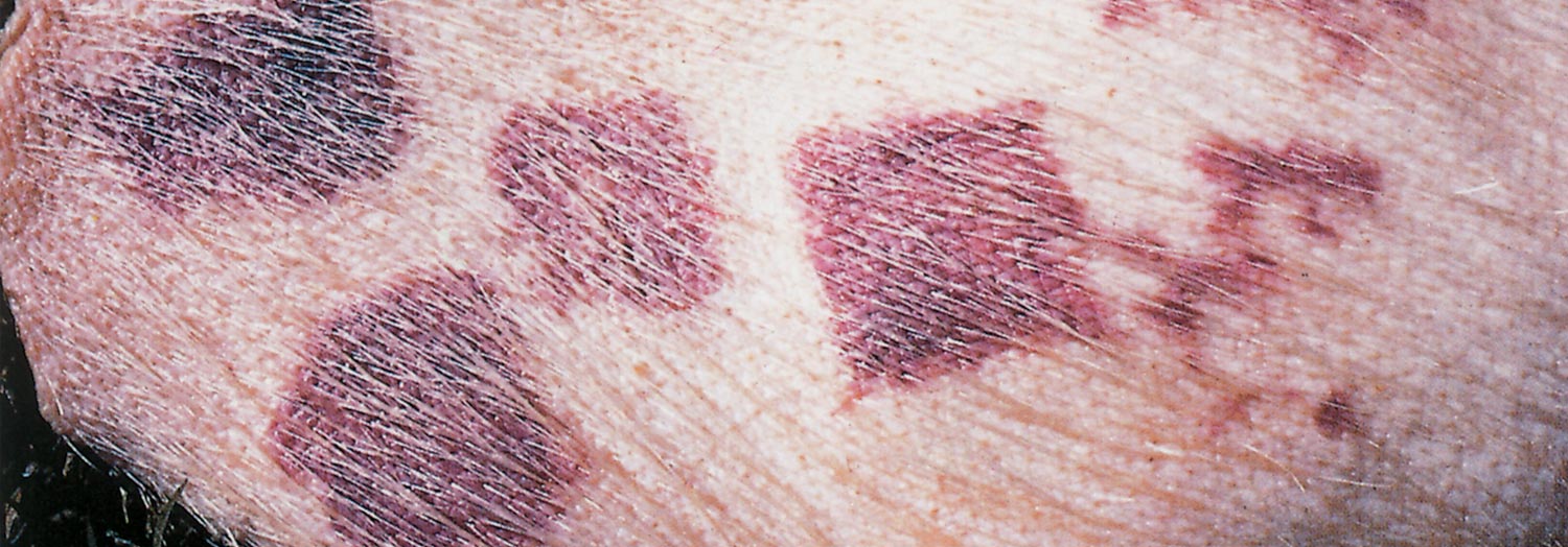
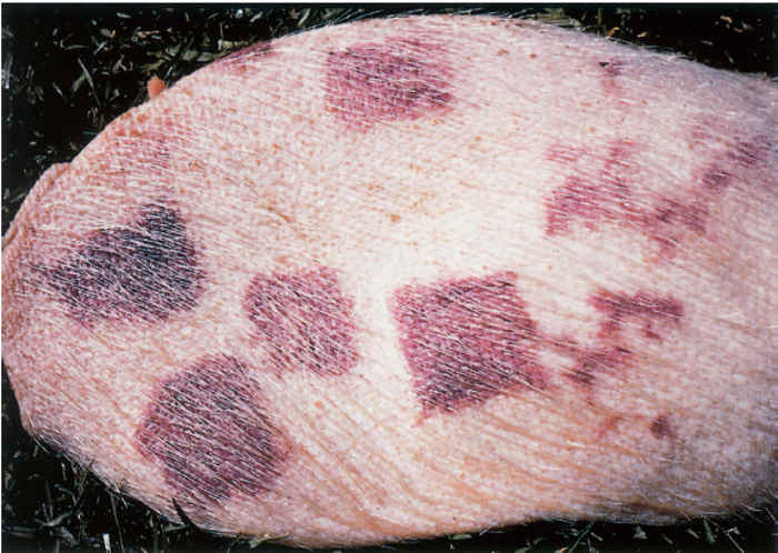
In the milder, subacute disease, initial signs include fever, malaise, anorexia, reluctance to stand, and bristling of the hairs where the skin lesions are going to develop. Within three to four days after the appearance of these signs, numerous well-circumscribed, purplish-red, intensely hyperaemic and slightly raised patches, with a diamond, rectangular or rhomboid shape, develop in the skin (Figure 188.1). In mild cases the patches have pale centres and disappear after a few days. Desquamation of the epidermis of the affected skin ensues, and the animal recovers in seven to ten days.11 In severely affected pigs vesicles develop in the raised patches which may rupture to form crusts. In some animals the affected skin becomes black, hard and necrotic. These areas may slough, leaving large, raw ulcers. The ears and tail may also be shed as a result of dry gangrene. The mortality rate is usually not higher than 2 per cent and is often the result of secondary infections. Pregnant sows may abort or give birth to dead piglets.20, 32
The chronic form of the disease is characterized by arthritis and/or vegetative valvular endocarditis. It may follow mild or inapparent systemic infection, or it may develop as a sequel to acute or subacute disease. Alternatively, it may develop in vaccinated pigs subjected to natural exposure. The hock, stifle and elbow joints are most commonly affected, but the carpus and tarsus may also be involved. The affected joints are often visibly swollen and painful, and the animal walks with a stiff gait, on its toes and with its back arched. Affected pigs generally prefer to lie down, lose their appetites, and become unthrifty and stunted. In some, the joints are not noticeably swollen, but the animal is nevertheless stiff, weak in the hindquarters and unthrifty.
Chronic arthritis occurs most often in animals older than ten weeks of age and may persist for several years. Most pigs with cardiac involvement do not reveal any noteworthy signs, but some show fatigue, listlessness, loss of appetite, and signs of congestive heart failure, manifested by resting on the sternum and elbows, or sitting on their haunches.11 Pulmonary involvement as a sequel to the cardiac lesions is characterized by a slight cough, and dyspnoea, particularly when the animals move, is common. Cardiac and respiratory involvement occasionally results in cyanosis of mucous membranes, congested patches in the skin of the ears and neck, oedema of the extremities, and paresis of the hindquarters. The prognosis is unfavourable. Affected pigs remain unthrifty and may pine for weeks; death is often sudden as a result of heart failure.
Pathology
The lesions vary with the severity and duration of the disease. 20, 39 In peracute cases, the animal may die before lesions develop.
Pigs that die of acute disease may exhibit purple flushing of the skin, particularly of the abdomen and extremities; haemorrhages in various organs and on subcutaneous, serosal and mucosal surfaces; enlargement and congestion of the spleen as well as generalized organ congestion; and infarction of the gastric mucosa.20, 39 These lesions are characteristic of septicaemia. Microscopically, diffuse endothelial swelling with the development of fibrinoid vasculitis and hyaline thrombi may be observed.20
Pigs that survive for more than two days may exhibit more typical lesions of diamond skin disease, in addition to septicaemic lesions. The typical diamond-, rectangular- or rhomboidal-shaped skin lesions develop in subacute cases in two to four days.20, 39 They are slightly raised and can therefore be palpated even when not clearly visible in darkskinned pigs. They may be uniformly red to purplish, or only the margins and the centre may be discoloured.20 Red or partly discoloured lesions may resolve or progress to the more severe, dark or purple lesions that undergo necrosis, and may coalesce, in which case large areas of dermal necrosis develop.20 Pinpoint haemorrhages may be evident on the capsular and cortical surfaces of the kidneys, with more extensive medullary haemorrhage.20 Microscopical lesions in the skin are associated with arteritis characterized by intramural infiltration of neutrophils and cellular thrombi.20 Cutaneous vasculitis in pigs is usually indicative of erysipelas.40 Bacterial colonies are usually present in the dermis as well as in the kidneys, which are characterized microscopically by haemorrhage and varying degrees of glomerulonephritis, with fibrinoid necrosis of the glomerular tufts.
Acute arthritis is characterized by hyperaemia of the synovial membranes and increased volume and viscosity of joint fluid. Microscopically, synovial arterioles may exhibit fibrinoid vasculitis and cellular thrombosis.20 Lesions in other organs are non-specific and may include hepatic leukocytosis, interstitial pneumonia, degeneration of skeletal muscles, choroiditis with heavy infiltration of neutrophils, and cerebral leukocytosis and perivasculitis.20
Chronic erysipelas is characterized by arthritis and/or endocarditis. As the joint lesions become chronic, villous hyperplasia of the synovial membrane occurs, with proliferations or ‘tags’ that project into the articular space. When proliferating tissue extends as a pannus over the articular cartilage, the articular surface may be destroyed, with detachment of the cartilage, periostitis, lymphoid proliferation and eventual ankylosis of the joint.11, 20, 22, 39 Diskospondylitis is occasionally present.
Vegetative valvular endocarditis occurs most commonly on the mitral valves. Vegetations may be very large. Depending on the age of the thrombi, bacteria may be evident near the surface or deeper into the lesion, but they are generally radially aligned.20 Septic emboli and infarcts may be present in the kidneys and spleen.
Diagnosis
A presumptive diagnosis of erysipelas may be made from the clinical signs and the lesions. Bacteriological culture of the organism from tissue specimens is necessary to confirm the diagnosis.
In the septicaemic disease, rod-shaped organisms are found in smears made from the blood, spleen, liver and kidneys. In chronic heart and joint lesions, as well as in cultures, long, thread-like filamentous forms of the bacteria tend to develop.
The organisms are readily cultured from the blood and specimens of internal organs of septicaemic animals and from the dermis of diamond-shaped skin lesions. Isolation from chronic lesions from affected joints and valves of the heart may be difficult.
Serological tests, including plate, tube and microtitration agglutination, ELISA, gel-diffusion precipitation, passive haemagglutination, haemagglutination-inhibition, complement fixation and indirect immunofluorescence tests have been used, but are not diagnostically conclusive for acute infection and at best can be used as herd tests for detecting chronic infections.6, 39
Differential diagnosis
Clinical signs and lesions of acute erysipelas may in some instances be difficult to differentiate from similar signs that may be present in pigs suffering from African swine fever, classical swine fever (hog cholera), and bacterial septicaemias, particularly salmonellosis, Streptococcus spp. infections, and pasteurellosis. The enlarged spleen may be suggestive of the septicaemic form of anthrax, which occurs rarely in pigs. Swine fevers, in particular African swine fever, generally cause much higher mortality, and do not respond to antimicrobial treatment. The majority of Salmonella Choleraesuis infections are associated with hepatic necrosis and microgranulomas in addition to septicaemic lesions, and longer-standing cases may develop diphtheritic enteritis or pneumonia. Pneumonic lesions are present in most cases of pasteurellosis. None of the conditions mentioned cause the skin lesions typical of erysipelas. The possibility of septicaemic anthrax can be excluded by examination of a blood smear. Microbiological culture of specimens of various internal organs of acutely ill animals usually reveals the presence of the causative agent, but in older infections it may be necessary to culture the organisms from the joints.39
Arthritis caused by E. rhusiopathiae must be differentiated from that caused by organisms such as Mycoplasma hyosinoviae, M. hyorhinis, Trueperella pyogenes, Haemophilus spp., Streptococcus spp. and Staphylococcus spp. as well as non-infectious osteochondrosis.
Apart from E. rhusiopathiae, chronic vegetative endocarditis in pigs may be caused by Streptococcus and Pasteurella spp.
Control
Penicillin is the drug of choice, and response to the administration of a single long-acting preparation at 10 000 to 20 000 units/kg body weight is often rapid and dramatic in acutely and subacutely affected pigs.39 In-feed medication with oxytetracyclines at levels of up to 200 ppm is ineffective in cases of subacute erysipelas.26, 29, 30
When administered during the early stages of the disease, antiserum will often bring about a rapid recovery within a very short time, but will provide subsequent protection for only about two weeks.23, 39 However, administration of antiserum may exacerbate pre-existing arthritis caused by E. rhusiopathiae.24 Hyperimmune serum is not available in southern Africa.
Serovar 2 is the most immunogenic strain39 and is used in the majority of vaccines. In South Africa, a bacterin is used to immunize pigs in breeding herds and grower pigs. In breeding herds, the gilts and young boars should be immunized twice, a month apart, at selection or purchase, and thereafter once every six months. Depending on the management practice, this booster vaccination should either be administered at a specific time to the whole herd, or the sows can be vaccinated, for example, when their litters are weaned. Boars are vaccinated twice a year. Weaners and growers are not usually vaccinated on farms. On farms where the disease is present in grower pigs, vaccination is recommended when the pigs are handled for the first time after weaning. Stress should be limited as far as possible.
Bacterins provide protection against the septicaemic disease but would appear to enhance the susceptibility of pigs to arthritis.4 Combination vaccines may be used to control erysipelas.16
Erysipelothrix rhusiopathiae infection in other animal species and humans
Post-dipping lameness due to subacute to chronic dermatitis and cellulitis in lambs and adult sheep as a result of Erysipelothrix infection occurs occasionally following dipping of especially recently shorn sheep in contaminated dip fluid, or when they graze on pastures that have been irrigated with pig faeces.13 Lesions occur often about the fetlocks but may extend to the metacarpus or metatarsus. 10, 11, 34, 37
Usually only a few animals in a flock are affected, but the morbidity may be as high as 50 per cent.20 Sheep become infected through wounds acquired as a result of castration, docking, shearing or dipping. The lesions may remain localized at the point of entry, but bacteraemia, followed by polyarthritis, may occur. Death rarely ensues. Affected animals show fever, severe lameness and rapid wasting. Incision through the affected areas reveals congestion and a serous to serofibrinous exudate. In the chronic stage, the joints reveal periarticular fibrosis and periosteal and perichondrial osteophyte formation.3, 13, 20
The presence of E. rhusiopathiae in dipping fluid can be eliminated by the addition of 10 per cent zinc sulphate together with a detergent to aid its penetration. Delaying dipping the sheep until a week after shearing is effective in the prevention of post-dipping lameness. Modern acaracides are more labile than those previously used, with the result that the dip wash has to be replaced more frequently, and this reduces contamination.11
Other disease conditions that have been attributed to E. rhusiopathiae include endocarditis and arthritis in horses,21, 25 while arthritis of the tibiotarsal, stifle and carpal joints in calves as a result of E. rhusiopathiae infection has been reported.18
Disease in humans, referred to as erysipeloid, is usually the result of a wound infection. It is a mild disease generally confined to the hands of persons handling infected pork or fish or virulent cultures, or performing necropsies on infected animals.34 The localized skin lesions are usually reddish- purple and swollen and may gradually extend to involve the entire hand, but seldom spread beyond the wrist. Sometimes weals and vesicles are formed on the fingers or hand. Swelling and tenderness of neighbouring joints, usually the elbow or carpal joint, and of the regional lymph nodes are other clinical signs which may occur. Septicaemia is rare, but arthritis and endocarditis have been reported.14
References
- AMASS, S.E. & SCHOLZ, D.A., 1998. Acute nonfatal erysipelas in sows in a commercial farrow-to-finish operation. Journal of the American Veterinary Medical Association, 212, 708–709.
- BASTIANELLO, S.S. & SPENCER, B.T., 1984. A report of swine erysipelas in a litter of piglets. Journal of the South African Veterinary Association, 55, 195–198.
- BATH, G.F., 1991. Faculty of Veterinary Science, University of Pretoria. Personal communication.
- BÖHM, K.H., BOLLWAHN, W., FISCHER, J., MUMME, J. & EHARD, H., 1988. Vaccination against erysipelas in breeding farms. Tenth International Pig Veterinary Society Congress, Rio de Janeiro, August 1988.
- EAMENS, G.J., 1988. Pathogenicity of field isolates of Erysipelothrix rhusiopathiae in mice, rats and pigs. Australian Veterinary Journal, 65, 280–284.
- EAMENS, G.J., CHIN, J.C. & NICHOLLS, P.J., 1989. Comparison of inoculation regimes for the experimental production of swine erysipelas arthritis. II. Serological findings in a gel diffusion precipitin test and enzyme-linked immunosorbent assay. Australian Veterinary Journal, 66, 216–220.
- EAMENS, G.J., TURNER, M.J. & CATT, R.E., 1988. Serotypes of Erysipelothrix rhusiopathiae in Australian pigs, small ruminants, poultry and captive wild birds and animals. Australian Veterinary Journal, 65, 249–252.
- GYLES, C.L., 1986. Erysipelothrix. In: gyles, c.l. & thoen, c.o., (eds). Pathogenesis of Bacterial Infection in Animals. Ames, Iowa: Iowa State University Press.
- HAIG, D.A. & ADELAAR, T.E., 1944. A case of swine erysipelas in the Union of South Africa. Onderstepoort Journal of Veterinary Research, 20, 57–59.
- HARBOUR, H.E. & KERSHAW, G.F., 1949. Erysipelothrix rhusiopathiae infection in sheep. The Veterinary Record, 61, 37–38.
- HENNING, M.W., 1956. Animal Diseases in South Africa, 3rd edn. Pretoria: Central News Agency Ltd.
- HUNTER, P., 1989. Veterinary Research Institute, Onderstepoort, South Africa. Unpublished data.
- HUNTER, P. & SPENCER, B.T., 1987. Veterinary Research Institute, Onderstepoort. Unpublished data.
- JONES, D., 1986. Genus Erysipelothrix Rosenbach 1909, 367AL. In: sneath, p.h.a., mair, n.s., sharpe, m.e. & holt, j.g., (eds). Bergey’s Manual of Systemic Bacteriology, Vol. 2. Baltimore: Williams & Wilkins.
- LOVEDAY, R.K., 1962. Acute swine erysipelas in suckling pigs. Journal of the South African Veterinary Medical Association, 33, 3–5.
- MCCARTHY, D.H., PORTER, D.B., DOUGLASS, J. & SLUSSER, C.A., 1986. Preventing atrophic rhinitis, erysipelas and pasteurellosis in pigs. Veterinary Medicine, 81, 1169–1170, 1172–1174.
- MOLIN, G., SODERLIND, O., URSING, J., NORRUNG, V., TERNSTROM, A. & LOWENHIELM, C., 1989. Occurrence of Erysipelothrix rhusiopathiae on pork and in pig slurry, and the distribution of specific antibodies in abattoir workers. Journal of Applied Bacteriology, 67, 347–352.
- MOULTON, J.E., RHODE, E.R. & WHEAT, J.D., 1953. Erysipelas arthritis in calves. Journal of the American Veterinary Medical Association, 123, 335–340.
- OKOLO, M.I.O., 1986. Isolation of Erysipelothrix rhusiopathiae from apparently healthy pigs reared under intensive and free range systems of management. Microbios, 47, 29–35.
- palmer, n., 1993. Bones and joints. In: jubb, k.v., kennedy, p.c. & palmer, n., 1985 (eds). Pathology of Domestic Animals, 4th edn. Vol. I, New York: Academic Press.
- PATERSON, J.S. & HEATLEY, T.G., 1938. A case of infection of the horse with Erysipelothrix rhusiopathiae (swine erysipelas). Veterinary Journal, 94, 33–34.
- RIEKA, P., COSTA, L., VENDRELL, J., ESPUNA, E. & CASADEVALL, P., 1984. Studies on a field case of arthritis in weaned pigs caused by E. rhusiopathiae. Proceedings of the International Pig Veterinary Society, Ghent, 27–31 August 1984.
- SCANLAN, C.M., 1988. Introduction to Veterinary Bacteriology, Ames, Iowa: Iowa State University Press.
- SCHOENING, W.H., GREY, C.G. & OSTEEN, O.L., 1942. Swine erysipelas. United States Department of Agriculture, Yearbook of Agriculture, 686–694.
- SEAHORN, T.L., BRUMBAUGH, G.W., CARTER, G.K. & WOOD, R.L., 1989. Erysipelothrix rhusiopathiae bacteremia in a horse. Cornell Veterinarian, 79, 151–156.
- SPENCER, B.T., 1992. P.O. Box 32518, Glenstantia 0010. Personal observations.
- SPENCER, B.T. & HUNTER, P., 1991. P.O. Box 32518, Glenstantia 0010. Unpublished data.
- TAKAHASHI, T., NAGAMINE, N., KUIMA, M., SUZUKI, S., TAKAGI, M., TAMURA, Y., NAKAMURA, M., MURAMATSU, M. & SAWADA, T., 1996. Serovars of Erysipelothrix strains isolated from pigs affected with erysipelas in Japan. Journal of Veterinary Medical Science, 58, 587–589.
- TAKAHASHI, T., SAWADA, T., OHMAE, K., TAKAGI, M., MURAMATSU, M., TERAKADO, N., SETO, K., MARUYAMA, T. & KANSAKI, M., 1986. Serotypes and antibiotic resistance of Erysipelothrix rhusiopathiae strains isolated from pigs affected with chronic swine erysipelas. Japan Agricultural Research Quarterly, 19, 287–294.
- TAKAHASHI, T., SAWADA, T., OHMAE, K., TERAKADO, N., MURAMATSU, M., SETO, K., MURUYAMA, T. & KANZAKI, M., 1983. Antibiotic resistance of Erysipelothrix rhusiopathiae isolated from pigs with chronic swine erysipelas. Antimicrobial Agents and Chemotherapy, 25, 385–386.
- TAKAHASHI, T., TAMURA, T., SAWADA, T., SUZUKI, S., MURAMATSU, M., FUJISAWA, T., BENNO, Y. & MITSUOKA, T., 1989. Enzymatic profiles of Erysopelothrix rhusiopathiae and Erysipelothrix tonsillae. Research in Veterinary Science, 47, 275–276.
- TAYLOR, D.J., 1989. Erysipelas. In: Pig Diseases. 5th edn. Cambridge: Burlington Press.
- TESH, M.J. & WOOD, R.L., 1988. Detection of coagulase activity in Erysipelothrix rhusiopathiae. Journal of Clinical Microbiology, 26, 1058–1060.
- TIMONEY, J.F., GILLESPIE, J.H., SCOTT, F.W. & BARLOUGH, J.E., 1988. Hagan and Brunner’s Microbiology and Infectious Diseases of Domestic Animals. 8th edn. Cornell University, USA: Cornell University Press.
- TIZARD, I., 1987. Veterinary Immunology: An Introduction. 3rd edn. London: W.B. Saunders Co.
- WABACHA, J.K., GITAU, G.K., NDUHIU, J.M., THAIYA, A.G., MBITHI, P.M.F. & MUNYUA, S.J.M., 1998. An outbreak of urticarial form of swine erysipelas in a medium-scale piggery in Kiambu District, Kenya. Journal of the South African Veterinary Association, 69, 61–63.
- WHITTEN, L.D., HARBOUR, H.E. & ALLAN, W.S., 1948. Cutaneous Erysipelothrix infection in sheep. An etiological agent in post-dipping lameness. Australian Veterinary Journal, 24, 157.
- WOOD, R.L., 1984. Swine erysipelas—a review of prevalence and research. Journal of the American Veterinary Medical Association, 184, 944–949.
- WOOD, R.L., 1992. Swine erysipelas. In: leman, a.d., straw, b., mengeling, w.l., d’allaire, s., & taylor, d.j., (eds). Diseases of Swine. 7th edn. Ames: Iowa State University Press.
- YAGER, J.A. & SCOTT, D.W., 1993. The skin and appendages. In jubb, k.v., kennedy, p.c. & palmer, n., 1985 (eds). Pathology of Domestic Animals, 4th edn. Vol. I, New York: Academic Press.


