- Infectious Diseases of Livestock
- Part 3
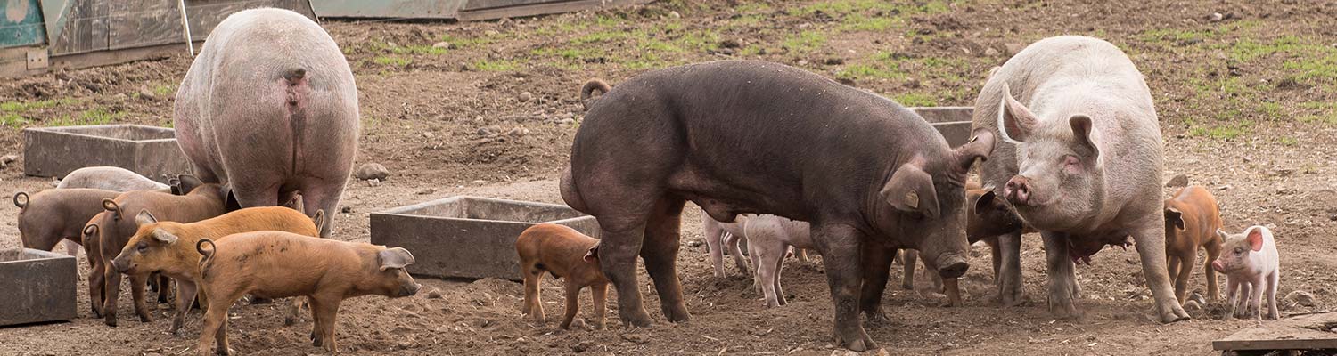
- Brucella suis infection
- GENERAL INTRODUCTION: SPIROCHAETES
- Swine dysentery
- Borrelia theileri infection
- Borrelia suilla infection
- Lyme disease in livestock
- Leptospirosis
- GENERAL INTRODUCTION: AEROBIC ⁄ MICRO-AEROPHILIC, MOTILE, HELICAL ⁄ VIBROID GRAM-NEGATIVE BACTERIA
- Genital campylobacteriosis in cattle
- Proliferative enteropathies of pigs
- Campylobacter jejuni infection
- GENERAL INTRODUCTION: GRAM-NEGATIVE AEROBIC OR CAPNOPHILIC RODS AND COCCI
- Moraxella spp. infections
- Bordetella bronchiseptica infections
- Pseudomonas spp. infections
- Glanders
- Melioidosis
- Brucella spp. infections
- Bovine brucellosis
- Brucella ovis infection
- Brucella melitensis infection
- Brucella suis infection
- Brucella infections in terrestrial wildlife
- GENERAL INTRODUCTION: FACULTATIVELY ANAEROBIC GRAM NEGATIVE RODS
- Klebsiella spp. infections
- Escherichia coli infections
- Salmonella spp. infections
- Bovine salmonellosis
- Ovine and caprine salmonellosis
- Porcine salmonellosis
- Equine salmonellosis
- Yersinia spp. infections
- Haemophilus and Histophilus spp. infections
- Haemophilus parasuis infection
- Histophilus somni disease complex in cattle
- Actinobacillus spp. infections
- Actinobacillus equuli infections
- Gram-negative pleomorphic infections: Actinobacillus seminis, Histophilus ovis and Histophilus somni
- Porcine pleuropneumonia
- Actinobacillus suis infections
- Pasteurella and Mannheimia spp. infections
- Pneumonic mannheimiosis and pasteurellosis of cattle
- Haemorrhagic septicaemia
- Pasteurellosis in sheep and goats
- Porcine pasteurellosis
- Progressive atrophic rhinitis
- GENERAL INTRODUCTION: ANAEROBIC GRAM-NEGATIVE, IRREGULAR RODS
- Fusobacterium necrophorum, Dichelobacter (Bacteroides) nodosus and Bacteroides spp. infections
- GENERAL INTRODUCTION: GRAM-POSITIVE COCCI
- Staphylococcus spp. infections
- Staphylococcus aureus infections
- Exudative epidermitis
- Other Staphylococcus spp. infections
- Streptococcus spp. infections
- Strangles
- Streptococcus suis infections
- Streptococcus porcinus infections
- Other Streptococcus spp. infections
- GENERAL INTRODUCTION: ENDOSPORE-FORMING GRAM-POSITIVE RODS AND COCCI
- Anthrax
- Clostridium perfringens group infections
- Clostridium perfringens type A infections
- Clostridium perfringens type B infections
- Clostridium perfringens type C infections
- Clostridium perfringens type D infections
- Malignant oedema⁄gas gangrene group of Clostridium spp.
- Clostridium chauvoei infections
- Clostridium novyi infections
- Clostridium septicum infections
- Other clostridial infections
- Tetanus
- Botulism
- GENERAL INTRODUCTION: REGULAR, NON-SPORING, GRAM-POSITIVE RODS
- Listeriosis
- Erysipelothrix rhusiopathiae infections
- GENERAL INTRODUCTION: IRREGULAR, NON-SPORING, GRAM-POSITIVE RODS
- Corynebacterium pseudotuberculosis infections
- Corynebacterium renale group infections
- Bolo disease
- Actinomyces bovis infections
- Trueperella pyogenes infections
- Actinobaculum suis infections
- Actinomyces hyovaginalis infections
- GENERAL INTRODUCTION: MYCOBACTERIA
- Tuberculosis
- Paratuberculosis
- GENERAL INTRODUCTION: ACTINOMYCETES
- Nocardiosis
- Rhodococcus equi infections
- Dermatophilosis
- GENERAL INTRODUCTION: MOLLICUTES
- Contagious bovine pleuropneumonia
- Contagious caprine pleuropneumonia
- Mycoplasmal pneumonia of pigs
- Mycoplasmal polyserositis and arthritis of pigs
- Mycoplasmal arthritis of pigs
- Bovine genital mycoplasmosis
- Neurotoxin-producing group of Clostridium spp.
- Contagious equine metritis
- Tyzzer's disease
- MYCOTIC AND ALGAL DISEASES: Mycoses
- MYCOTIC AND ALGAL DISEASES: Pneumocystosis
- MYCOTIC AND ALGAL DISEASES: Protothecosis and other algal diseases
- DISEASE COMPLEXES / UNKNOWN AETIOLOGY: Epivag
- DISEASE COMPLEXES / UNKNOWN AETIOLOGY: Ulcerative balanoposthitis and vulvovaginitis of sheep
- DISEASE COMPLEXES / UNKNOWN AETIOLOGY: Ill thrift
- Eperythrozoonosis
- Bovine haemobartonellosis
Brucella suis infection
This content is distributed under the following licence: Attribution-NonCommercial CC BY-NC  View Creative Commons Licence details here
NJ Maclachlan and M-L Penrith (Editors). J Godfroid and JM Blasco, Brucella suis infection, 2018.
Brucella suis infection
Previous authors: J GODFROID, C O THOEN AND R D ANGUS
Current authors:
J X L GODFROID - Professor of Microbiology, DVM, MSc, PhD, University of Tromsø - the Arctic University of Norway, Hansine Hansens veg 18, Tromsø, 9019, Norway
J M BLASCO - Emeritus Researcher, DVM, PhD, Cita/Ia2/University Zaragoza Avenue, Montañana 930, Zaragoza, 50011, Spain
Introduction
Brucellosis in pigs is caused primarily by biovar 1, 2 or 3 of the five biovars of Brucella suis. Clinical signs in sows are abortion, birth of stillborn piglets, perinatal mortality, mastitis, arthritis and infertility. Orchitis and sterility may occur in infected boars. In endemic situations B. suis infections may be subclinical and farmers may be unaware that their herds are infected. Brucella suis biovars 1 and 3 are important zoonoses.
Porcine brucellosis was first described by Traum in Indiana, USA, in 1914.44 The disease has a worldwide distribution , and despite the paucity of data its prevalence is considered to be low.4 Swine brucellosis appears to be endemic in most parts of South America and Southeast Asia, and is also believed to be widespread across sub-Saharan Africa.29, 31, 36 Although reported in Mozambique, it has not yet been diagnosed in South Africa.6 In the USA and Australia the infection (caused by B. suis biovar 1) is mainly restricted to feral pigs,37, 45 while the wild boar (Sus scrofa) and the European hare (Lepus europaeus) are the wild reservoirs of swine brucellosis caused by B. suis biovar 2 in Continental Europe (except Finland, Norway, and Sweden).15, 23, 26
Aetiology and epidemiology
Brucella suis organisms are small Gram negative coccobacillioccurring naturally in the smooth (S) form and whose colonies cannot be differentiated macroscopically from isolates of other S Brucella species.1-3 There are five recognized biovars of B. suis (named from biovar 1 to biovar 5). Biovars 1, 2 and 3 are main causes of brucellosis in swine.2, 7, 8 Biovar 4 has been isolated from reindeer (Rangifer tarandus and its various subspecies) in the Arctic including Russia, Alaska and Canada where it is considered to be endemic in semi-domesticated reindeer. Brucella suis has also been isolated sporadically in moose (Alces alces), American bison (Bison bison), musk oxen (Ovibos moschatus), arctic foxes (Alopex lagopus) and wolves (Canis lupus) in the Arctic and constitutes a serious zoonotic problem.18 Brucella suis biovar 5 has been isolated exclusively from wild rodents in the former USSR.36 Very rarely brucellosis in pigs is caused by B. abortus and B. melitensis.36
Brucella suis biovars 1, 2 and 3 can be isolated in suitable culture media and identified by simple bacteriological and molecular tests (see below).
Brucella suis biovars 1 and 3 are mainly pathogenic for pigs and humans but have also been reported to infect cattle sporadically.6, 16 These two biovars have a worldwide distribution. Epidemics of brucellosis in humans have been reported in the USA among packing-house workers where the usual source was infected pigs19 and also in Australia among those involved in the killing and slaughter of feral pigs.37 Human brucellosis caused by B. suis biovars 1 and 3 is thus almost entirely occupational1, 22 except in countries such as Brazil and Colombia in South America, where B. suis has become established in cattle, and brucellosis in humans caused by biovars 1 and 3 is emerging as an increasingly serious public health problem as a result of the consumption of unpasteurized milk because of infection of the bovine udder.6, 29
Brucella suis biovar 2 differs from B. suis biovars 1 and 3 in that it is restricted to Continental Europe and Northern Africa and infects wild boar and the European hare, which are the wild reservoirs.15, 23, 26 Otherwise, swine brucellosis due to B. suis biovar 2 closely resembles the disease caused by B. suis biovars 1 and 3.1 Remarkably, B. suis biovar 2 is very rarely a human pathogen and has only been reported twice as the cause of human brucellosis in immune-compromised patients.30 Brucella suis biovar 2 can sporadically infect cattle.20 However, B. suis biovar 2 is not likely to be transmitted between cattle because they are spillover and not preferential hosts for B. suis biovar 2, and are thus not likely to sustain the infection.20
In modern industrial pig farming, infection is generally introduced into a herd by the purchase of infected animals. Boars may be the main source of infection and transmission occurs chiefly via the semen at mating. Indeed, persistent granulomatous lesions in the testes and accessory sex organs, from which bacteria can be shed in the semen for months or years, may be present in boars even though they appear healthy. Hence the potential exists for spreading the disease through artificial insemination.5, 36, 41 Other routes of spread are transplacental and the ingestion of aborted materials, infected milk or contaminated feedstuff.1 The feeding of contaminated kitchen waste containing uncooked pork may also be important.36 Transmission of B. suis biovar 2 from wild boar to pigs in open air breeding systems or backyard farms is thought to be mainly through the venereal route. Infected hares can be involved in B. suis biovar 2 outbreaks in domestic pigs26 through swill feeding with offal from hunted infected hares, or ingestion of dead hares.
In suitable environmental conditions. B. suis can survive for several months. Animal premises may thus remain contaminated for prolonged periods. Direct sunlight considerably reduces the survival time. Brucella suis resists drying and freezing, and also survives for considerable periods in cold climates in aborted foetuses and manure. However, environmental bacterial persistence is accepted to be of low epidemiological importance since direct or close animal to animal contact is required for transmission. Maintenance of B. suis in a population requires continued infection of susceptible hosts.36
The course of the clinical disease is usually acute when B. suis is introduced for first time into a non-infected herd of pigs. Infection spreads rapidly in a herd and more than 50 per cent of the animals may become infected in a relatively short period of time, manifesting mainly as a high rate of abortions and infertility. Infection rates of up to 70-80 per cent are not uncommon in the early stages of outbreaks, resulting in severe economic losses. If no control measures are implemented, some infected individuals will either show a chronic form of the disease with or without excretion of B. suis or more infrequently free themselves of the infection (self-cure), while others may remain uninfected. The latter are at risk and may subsequently become infected because of the presence of B. suis in the herd. This sequence of events explains why a herd may be infected for years but for long periods may be considered to be free of clinical disease.36
Pathogenesis, clinical signs and pathology
The pathogenesis of B. suis infection in pigs has not been elucidated fully. Brucellosis in pigs is usually a chronic infection that persists for life. The infected animals develop inflammatory responses in lymphoid tissues and organs rich in reticulo-endothelial cells, like the spleen.Hosts mostly become infected through mucous membranes. Through licking, sniffing or ingesting aborted foetuses and foetal membranes, or ingesting contaminated milk, B. suis comes into contact with the oral and nasal mucosae, tonsils, and gastrointestinal or reproductive mucosae. After entry, the pathogenesis of B. suis is believed to be similar to infection of preferential hosts by other brucellae. Brucella suis localize initially in lymphoreticular tissues draining the site of infection followed by a bacteraemia that may persist for up to 90 days or more.1 During and subsequent to the bacteraemia, localization of B. suis occurs in various tissues including the male and female genital organs, skeletal system, joints, mammary glands, lymph nodes, spleen, liver, kidneys and urinary bladder.24, 42 Localization of B. suis in these organs or tissues accounts for the main clinical signs of the disease, and the main routes of excretion. Infection with B. suis may also be sublinical.1
Like Brucella infections in other animal species, the onset of infection depends on the virulence and dose of the infecting strain as well as the susceptibility of the host as determined by innate and acquired immunity mechanisms. It has been reported that the virulence of B. suis biovars 1,2 and 3 are similar in pigs, but no experimental evidence is available. The minimal infective dose has not been established properly but it has been reported that doses ranging between 105-106 CFU induce infection in most animals.36
The clinical signs of brucellosis in pigs, when present, vary but are similar to those in cattle, sheep and goats. Reproductive failure characterized by abortion, stillbirth and infertility in sows and orchitis, epididymitis and infertility in boars are the main clinical features of B. suis infection. Abortion may occur at any stage of pregnancy.1 Early abortion may be missed, and one may think that there is an infertility problem in the herd. Sows usually do not abort more than once following infection but may become infertile thereafter. Some piglets infected in utero may die within a few hours of birth, the mortality rate often being very high, but others survive and retain the infection into adulthood.41 Sows may suffer from mastitis. In boars, there may be no clinical signs or there may be swelling of the testes, which subsequently undergo atrophy.1 Mortality in older pigs is usually low but chronic disease causes economic losses because of the forced culling of boars and sows as a result of infertility caused by lesions in the genital tract or as a result of lameness caused by arthritis, bursitis and tendovaginitis.1
The majority of visible lesions in the genital organs consist of progressively enlarging granulomas that may become calcified. In the uterus and Fallopian tube, they are generally 2 to 5 mm in diameter and have a miliary distribution. In addition to this miliary granulomatous metritis, mucoid endometritis may be present in which numerous Brucella organisms are present in the exudate in the lumen of the uterus. In boars, it is often possible to isolate the organisms in the absence of gross pathology. However, orchitis and infection of the accessory sex glands are common.36 Orchitis caused by B. suis results in multiple abscesses accompanied occasionally by fibrinopurulent or haemorrhagic periorchitis. Testes may appear enlarged or atrophic. Abscesses in the testes or epididymis have a caseous core surrounded by epithelioid macrophages with a connective tissue capsule. Infection of accessory glands can be associated with vesicular gland hypertrophy, and microabscesses in vesicular glands, prostate, or bulbourethral glands. Calcified foci may also be seen in the testes and accessory sexual glands and organs, particularly in the epididymis and seminal vesicles. Testicular atrophy and a variable degree of enlargement of the epididymis are characteristics of the chronic phase of the disease.36 The inflammatory reaction, which may be present in one or more joints, is fibrinopurulent and may cause lameness. If the vertebrae (often the lumbar region) are involved, granulomatous osteomyelitis develops that can destroy adjacent intervertebral discs. Such affected animals often manifest with posterior paresis and sometimes paralysis. No specific lesions occur in aborted foetuses.
Rangifer brucellosis in reindeer (Rangifer tarandus tarandus) and caribou (Rangifer tarandus groenlandicus) is caused by B. suis biovar 4 throughout the Arctic region, Siberia, Canada and Alaska and constitutes a serious zoonotic problem.18 When clinical signs are present in these animals, abortions and metritis are seen in females and orchitis occurs in males. In both sexes, abscess formation in joints, bursitis and lameness are observed in some cases. Transmission to humans may be by direct contact, or through the consumption of raw milk or other inadequately heated products from infected animals.18
Diagnosis
Brucella suis infection in pigs may be present in the absence of typical clinical signs or gross pathology, which is not pathognomonic. However, in endemic regions, organs (particularly the genital organs) showing granulomatous lesions during routine post-mortem inspection, can be suggestive of infection.
Direct tests
A presumptive diagnosis can be made by microscopic examination of Stamp’s stained smears from vaginal swabs, placentas and/or aborted foetuses, and semen. However, this test lacks sensitivity and specificity, and the isolation of B. suis on appropriate culture media allows the only accurate diagnosis. Vaginal secretions (swabs) from aborted sows, milk, semen, foetal membranes and samples from aborted foetuses (stomach contents, spleen and lung) are the samples of choice in live pigs. At necropsy the spleen and lymph nodes from the head, mammary, and genital tract are the samples of choice. Brucella suis can also be isolated from the testis, epididymis, vesicular glands, prostate and bulbo-urethral glands of boars.2, 27, 36
Brucella suis grows well at 37°C on general multipurpose culture media (i.e. blood agar) without the addition of blood or serum. However, due to the frequent presence of commensal bacteria and fungi in field samples, the use of selective media is recommended.4 Brucella suis can grow in air but the highest sensitivity of the culture is obtained in a 5 to 10 per cent CO2 atmosphere.1 Farrell’s is the most widely used selective medium for B. suis but some of the antibiotics contained in this medium can be inhibitory for some brucellae including some B. suis strains.10 Therefore, the simultaneous use of both Farrell’s and the CITA’s media is recommended for the isolation of smooth brucellae, including B. suis.10 Colonies generally appear after 3 to 4 days, but cultures should not be discarded as negative until after 8 days of incubation. Brucella suis colonies are morphologically indistinguishable from other smooth brucellae and can be presumptively identified by positive reactions in the oxidase and urease tests and by agglutination with monospecific antisera, since the three most relevant biovars (1, 2 and 3) agglutinate with the anti-A but not the anti-M monospecific antiserum.4 With DNA from the isolated colonies, several PCR procedures allow proper identification of B. suis.4 Other additional microbiological tests used for the identification of the different biovars in other Brucella species are not straightforward for the differentiation of B. suis biovars, but two PCR tests are suitable for that purpose.21, 27
The diagnosis of B. suis directly from field samples has been also attempted using several PCR-based protocols.4 However, due to the presence of DNA inhibitors in animal tissues and field samples, the sensitivity of these methods is significantly lower than that of classical microbiological culture.36
Indirect tests
Virtually all serological tests used for the diagnosis of brucellosis in ruminants have been applied for diagnostic purposes for brucellosis in pigs.4, 15, 36, 38, 43 However, it is widely accepted that none of the conventional serological tests used in domestic ruminants are fully reliable for diagnosing pig brucellosis when applied to individual animals. Therefore, it is recommended that serological tests be interpreted on a herd rather than on an individual animal basis.4, 9, 15, 36
The serological tests for brucellosis in pigs are performed using the same cell wall-associated B. abortus smooth lipopolysaccharide (S-LPS) antigen used for the diagnosis of B. abortus and B. melitensis infections in domestic ruminants (see B. melitensis and B. abortus). The O side-chain polysaccharide (O-PS; composed of repeated residues of 4-formamindo-4, 6-dideoxymanose linked in α-1,2 or α-1,3 conformation) is the major S-LPS epitope involved in serological tests for brucellosis. Unfortunately, the O-PS is very similar to the O side-chain polysaccharides of other bacteria such as Yersinia enterocolitica O:9, Escherichia coli O:157, and others.4, 15 Yersinia enterocolitica O:9 infection in pigs is quite widespread and, accordingly, this represents a major source of false positive serological reactions.15 This is the most likely explanation for reports of positive responses on brucellosis tests in pigs in the absence of epidemiologic or bacteriologic evidence of infection.4, 15
Serological tests can be performed either at herd or individual level. It is important to note that such tests are adequate for the detection of an infected herd but have limitations in detecting infected individual pigs.4, 9
It is difficult to assess the actual sensitivity and specificity of the various serological tests that are available because different antibody isotypes are detected by the different tests and the relative quantity of these isotypes in a pig’s serum varies during the course of a B. suis infection.4 Hence, a test that is unable to detect an early infection - e.g. the complement fixation test (CFT) - might give a positive result in the case of an active-chronic infection.11, 17 Because pig serum may sometimes contain nonspecific antibodies, thought to be IgM, the specificity of the standard agglutination test (SAT) is reduced, and is not be recommended for the diagnosis of swine brucellosis.4, 15 In addition, pig complement interacts with guinea-pig complement to produce anti complementary activity that reduces the sensitivity of the CFT. In fact, the diagnostic sensitivity of the CFT is significantly lower than that of the rose Bengal test (RBT) and other S-LPS tests.12, 36 The S-LPS tests with the highest diagnostic sensitivities for diagnosing swine brucellosis are the RBT and some commercial indirect enzyme-linked immunosorbent assays (i-ELISA).12, 36 The competitive ELISA (c-ELISA) and the Fluorescence Polarization Assay (FPA)34 have a lower diagnostic performance than both the RBT and the i-ELISA.12, 33 It must be noted, however, that B. suis may be isolated from seronegative pigs,17 which has important implications in eradication programmes. The above results are in close agreement with the conclusions from the only systematic review of the literature on the subject.15
It has been shown that natural and experimentally induced Y. enterocolitica O:9 infections produce false positive serological reactions (FPSR) in S-LPS brucellosis tests that are virtually indistinguishable from those caused by brucellosis.15, 46 Accordingly, none of the above S-LPS tests provide 100 per cent specificity when testing a brucellosis-free pig population affected by the FPSR.12, 36 This problem is particularly important and frequent in brucellosis-free countries and interferes with the international trade of live pigs. Several serological tests have been proposed to solve this FPSR problem in pigs, including both c-ELISA and i-ELISA with S-LPS, as well as an i-ELISA using an extract of rough (R) brucellae.32, 35 The results of these investigations show that serial and cumbersome testing schedules would be necessary to increase the specificity of the diagnosis, and moreover, none of them has been proven to be fully specific to differentiate brucellosis from FPSR.4, 15, 36
Soluble cytosolic proteins are presumed to be specific from the Brucella genus, thus being potential candidates to resolve the FPSR problem.4 When used in serological tests, these antigens do not demonstrate suitable diagnostic performance in pigs.12 Lymphocyte transformation tests with these proteins have been studied to measure cell-mediated responses of infected pigs.25 Preliminary results suggest that there is good correlation of the results of these tests with those obtained by bacteriological isolation of B. suis from tissues. None of these lymphocyte tests are in common use and proven effective to resolve the FPSR problem.
Allergic skin tests have been widely used in Eastern Europe, the former USSR and China for the diagnosis of brucellosis in pig herds.4 As the antigens used for the allergic tests are the Brucella cytosolic proteins, no cross-reactions with Y. enterocolitica O:9 are expected, and thus the diagnostic specificity is very high. However, the sensitivity of the allergic tests is similar to that of the serological tests in that they are not reliable for the diagnosis of the disease in individual pigs but they are very valuable when interpreted at herd level.11, 36 A commercially produced allergen is available for the allergic skin test has been shown to be very useful in the context of FPSR in cattle.39 Studies in Spain showed the suitability of this allergic skin test in pigs when used at a herd level to exclude brucellosis in the presence of FPSR due to Y. enterocolitica O:9.11
Control
Swine brucellosis is usually not a disease that occurs on intensive pig farms with modern indoor facilities. It is critical to prevent B. suis-infected animals from entering these intensive systems, particularly nucleus breeding herds at the top of the pyramid and artificial insemination centres. Thus, strict biosafety measures must be implemented in these intensive farming systems as well as where domestic pigs are reared in outdoor breeding systems or small back yard farms to avoid contact with feral pigs or wild reservoirs. Where there is a risk of introducing swine brucellosis, regular serological surveillance should be performed on a herd basis, using the skin test as a confirmatory test at herd level whenever positive serological reactions are detected.11
Adequate fencing, although expensive, to avoid contact with wild reservoir hosts of B. suis is important to prevent infection of free-range pig farms.
Eradication of B. suis from a herd is based solely on the depopulation of all animals in the infected holdings. However, culling of large herds kept in in- or outdoor farming systems may not be economically feasible or impracticable.
An alternative control strategy could be testing with culling of infected animals. However, the current serological tests are not suitable to apply at an individual level and should be used at a herd level. It is therefore not possible to eradicate the disease from an infected herd by means of serological test-and-removal programmes.14, 17, 28, 36
Alternatively, test and culling strategies based on the use of a combination of serological tests and the skin test in breeding stock can be used to eradicate the disease in a herd4, 14 as some infected animals that are negative serologically, react positively to the skin test, and vice versa. Where practicable, it is thus useful to perform a combination of both serological and skin tests.14
When total depopulation is not feasible and eradication not possible, antibiotic treatment, alone or in combination with test-and-culling, can be applied to control the disease, minimizing the clinical effects and economic impact. Although further work is necessary a combined regimen of oxytetracycline and tildipirosin can be a cost-effective practical alternative to control swine brucellosis where depopulation is not feasible.13 In addition to antibiotic treatment, selective culling (i.e. intensifying the slaughter of sows that aborted or that are infertile), and testing and slaughter infected pigs on the basis of brucellin skin test results) could significantly reduce the prevalence of disease in a herd.14
Replacement stock should originate from herds known to be officially free of brucellosis. According to the Office International des Epizooties (OIE), for international and other trade purposes the disease status of the herd and that of the area in which the herd is situated are of more importance than the testing of individual animals.4
No vaccine is effective in preventing brucellosis in swine. Although the Chinese B. suis strain 2 vaccine given orally has been reported to be effective,47 data on its safety and efficacy are not available. Moreover, experimental and field data showed vaccination with the B. abortus RB51 vaccine does not protect against swine brucellosis.40
References
- ALTON, G. G., 1990. Brucella suis. In: NEILSEN, K. & DUNCAN, J.R., (eds.). Animal Brucellosis. Boca Raton, FL: CRC Press. 411–422.
- ALTON, G. G., JONES, L. M., ANGUS, R. D. & VERGER, J. M., 1988. Techniques for the Brucellosis Laboratory. lst edn. Paris: Institut National de la Recherche Agronomique.
- ANON, 1986. Joint FAO/WHO Expert Committee on Brucellosis, 1986. 6th report. Geneva: World Health Organization.
- ANON, 2018. Manual of Standards for Diagnostic Tests and Vaccines. Paris: Office International des Epizooties. http://www.oie.int/en/standard-setting/terrestrial-manual/access-online/ (Accessed on line, October 08, 2018).
- BLOOD, D. C., HENDERSON, J. A. & RADOSTITS, O. M., 1983. Veterinary Medicine. 6th edn. London: Baillière Tindall.
- CORBEL, M. J., 1997. Brucellosis: an overview. Emerging Infectious Diseases, 3, 213-221.
- CORBEL, M. J. & BRINLEY MORGAN, W. J., 1984. Genus Brucella Meyer and Shen 1920. In: KRIEG, N.R. & HOLT, J.G., (eds). Bergey’s Manual of Systematic Bacteriology. Baltimore, London: Williams & Wilkins.
- CORBEL, M. J., GILL, K. P. W., THOMAS, E. L. & HENDRY, D., 1983. Methods for the Identification of Brucella. Booklet No. 85, Alnwick NE66 2PF, United Kingdom: MAFF Publications.
- CRICHTON, R. & MEDVECZKY, N. E., 1987. The identity, distribution and epizootiological significance of Brucella isolates in Australia, 1981 to 1985. Australian Veterinary Journal, 64, 48–52.
- DE MIGUEL, M. J., MARÍN, C. M., MUÑOZ, P. M., DIESTE, L., GRILLÓ, M. J. & BLASCO, J. M., 2011. Development of a selective culture medium for primary isolation of the main Brucella species. Journal of Clinical Microbiology, 49, 1458-63.
- DIESTE-PÉREZ, L., BLASCO, J. M. D. E., MIGUEL, M. J., MARÍN, C. M., BARBERÁN, M., CONDE-ÁLVAREZ, R., MORIYÓN, I. & MUÑOZ, P. M., 2014. Performance of skin tests with allergens from B. melitensis B115 and rough B. abortus mutants for diagnosing swine brucellosis. Veterinary Microbiology, 168, 161-168.
- DIESTE-PÉREZ, L., BLASCO, J. M. D. E., MIGUEL, M. J., MORIYÓN, I. & MUÑOZ, P. M., 2015. Diagnostic performance of serological tests for swine brucellosis in the presence of false positive serological reactions. Journal of Microbiological Methods, 111, 57-63.
- DIESTE-PÉREZ, L., FRAILE, L. D. E., MIGUEL, M. J., BARBERÁN, M., BLASCO, J. M. & MUÑOZ, P. M., 2015. Studies on a suitable antibiotic therapy for treating swine brucellosis. Journal of Veterinary Pharmacology and Therapeutics, 38, 357-364.
- DIESTE-PÉREZ, L., FRANKENA, K., BLASCO, J. M., MUÑOZ, P. M. & DE JONG, M. C., 2016. Efficacy of antibiotic treatment and test-based culling strategies for eradicating brucellosis in commercial swine herds. Preventive veterinary medicine, 126, 105-110.
- EUROPEAN FOOD SAFETY, A., 2009. Scientific Opinion of the Panel on Animal Health and Welfare (AHAW) on a request from the Commission on porcine brucellosis (Brucella suis). The European Food Safety Authority Journal, 1144, 1-112.
- EWALT, D. R., PAYEUR, J. P., RHYAN, J. C. & GEER, P. L., 1997. Brucella suis biovar 1 in naturally infected cattle: A bacteriological, serological and histological study. Journal of Veterinary Diagnostic Investigation, 9, 417-420.
- FERRIS, R. A., SCHOENBAUM, M. A. & CRAWFORD, R. P., 1995. Comparison of serological tests and bacteriologic cultures for detection of brucellosis in swine from naturally infected herds. Journal of the American Veterinary Medical Association, 207, 1332-1333.
- FORBES, L. B., 1991. Isolates of Brucella suis biovar 4 from animals and humans in Canada, 1982–1990. Canadian Veterinary Journal, 32, 686-688.
- FOX, M. D. & KAUFMANN, A. F., 1977. Brucellosis in the United States 1965–74. Journal of Infectious Diseases, 136, 312-316.
- FRETIN, D., MORI, M., CZAPLICKI, G., QUINET, C., MAQUET, B., GODFROID, J. & SAEGERMAN, C., 2013. Unexpected Brucella suis biovar 2 Infection in a dairy cow, Belgium. Emerging Infectious Diseases, 19, 2053-2054.
- FRETIN, D., WHATMORE, A. M., AL DAHOUK, S., NEUBAUER, H., GARIN-BASTUJI, B., ALBERT, D., VAN HESSCHE, M., MENART, M., GODFROID, J., WALRAVENS, K. & WATTIAU, P., 2008. Brucella suis identification and biovar typing by real-time PCR. Veterinary Microbiology, 131, 376-385.
- GIBBS, E. P. J., 1997. The public health risks associated with wild and feral swine. Revue Scientifique et Technique OIE, 16, 594-598.
- GODFROID, J., MICHEL, P., UYTTERHAEGEN, L., DE SMED, T. C., RASSENEUR, F., BOELAERT, F., SAEGERMAN, C. & PATIGNY, X., 1994. Brucellose enzootique à Brucella suis biotype 2 chez le sanglier (Sus scrofa) en Belgique. Annales de Médecine Vétérinaire, 138, 263-268.
- JUBB, K. V. F., KENNEDY, P. C. & PALMER, N., 1993. Pathology of Domestic Animals. 4th edn. San Diego, California. Academic Press, Inc.
- KANEENE, J. M., ANDERSON, R. K., JOHNSON, D. W., ANGUS, R. D., MUCOPLAT, C. C., PIETZ, D. E., VANDERWAGON, L. C. & SLOANE, E. E., 1978. Cell-mediated immune responses in swine from a herd infected with Brucella suis. American Journal of Veterinary Research, 39, 1607-1611.
- KREIZINGER, Z., FOSTER, J. T., RÓNAI, Z., SULYOK, K. M., WEHMANN, E., JÁNOSI, S. & GYURANECZ, M., 2014. Genetic relatedness of Brucella suis biovar 2 isolates from hares, wild boars and domestic pigs. Veterinary Microbiology, 172, 492-498.
- LÓPEZ-GOÑI, I., GARCÍA-YOLDI, D., MARÍN, C. M. D. E., MIGUEL, M. J., BARQUERO-CALVO, E., GUZMÁN-VERRI, C., ALBERT, D. & GARIN-BASTUJI, B., 2011. New Bruce-ladder multiplex PCR assay for the biovar typing of Brucella suis and the discrimination of Brucella suis and Brucella canis. Veterinary Microbiology, 154, 152-155.
- LORD, V. R., CHERWONOGRODZKY, J. W., MARCANO, M. J. & MELENDEZ, G. E., 1997. Serological and bacteriological study of swine brucellosis. Journal of Clinical Microbiology, 35, 295-297.
- LUCERO, N. E., AYALA, S. M., ESCOBAR, G. I. & JACOB, N. R., 2008. Brucella isolated in humans and animals in Latin America from 1968 to 2006. Epidemiology & Infection, 136, 496-503.
- MAILLES, A., OGIELSKA, M., KEMICHE, F., GARIN-BASTUJI, B., BRIEU, N., BURNUSUS, Z., CREUWELS, A., DANJEAN, M. P., GUIET, P., NASSER, V., TOURRAND, B., VALOUR, F., MAURIN, M., O'CALLAGHAN, D., MICK, V., VAILLANT, V. J. A. Y. M., LAVIGNE, J. P. & DE VALK, H., 2017. Brucella suis biovar 2 infection in humans in France: emerging infection or better recognition? Epidemiology & Infection, 145, 2711-2716.
- MCDERMOTT, J. J. & ARIMI, S. M., 2002. Brucellosis in sub-Saharan Africa: epidemiology, control and impact. Veterinary Microbiology, 90, 111-134.
- MCGIVEN, J. A., NICOLA, A., COMMANDER, N. J., DUNCOMBE, L., TAYLOR, A. V., VILLARI, S., DAINTY, A., THIRLWALL, R., BOUZELMAT, N., PERRETT, L. L., BREW, S. D. & STACK, J. A., 2012. An evaluation of the capability of existing and novel serodiagnostic methods for porcine brucellosis to reduce false positive serological reactions. Veterinary Microbiology, 160, 378-386.
- MUÑOZ, P. M., BLASCO, J. M., ENGEL, B. D. E., MIGUEL, M. J., MARÍN, C. M., DIESTE, L. & MAINAR-JAIME, R. C., 2012. Assessment of performance of selected serological tests for diagnosing brucellosis in pigs. Veterinary Immunology and Immunopathology, 146, 150-158.
- NIELSEN, K., GALL, D., SMITH, P., VIGLIOCCO, A., PEREZ, B., SAMARTINO, L., NICOLETTI, P., DAJER, A., ELZER, P. & ENRIGHT, F., 1999. Validation of the fluorescence polarization assay as a serological test for the presumptive diagnosis of porcine brucellosis. Veterinary Microbiology, 68, 245-53.
- NIELSEN, K., SMITH, P., YU, W., NICOLETTI, P., JUNGERSEN, G., STACK, J. & GODFROID, J., 2006. Serological discrimination by indirect enzyme immunoassay between the antibody response to Brucella sp. and Yersinia enterocolitica O:9 in cattle and pigs. Veterinary Immunology and Immunopathology, 109, 69-78.
- OLSEN, S., GARIN-BASTUJI, B., BLASCO, J. M., NICOLA, A. & SAMARTINO, L., 2012. Brucellosis. In: ZIMMERMAN, J., KARRIKER, L., RAMIREZ, A., SCHWARTZ, K. & STEVENSON, G., (eds), Diseases of Swine, 10th edn. John Wiley & Sons, Inc. 697-708.
- ROBSON, J. M., HARRISON, M. W., WOOD, R. N., TILSE, M. H., MCKAY, A. B. & BRODRIBB, T. R., 1993. Brucellosis: Re-emergence and changing epidemiology in Queensland. The Medical Journal of Australia, 159, 153-158.
- ROGERS, R. J., COOK, D. R., KETTERER, P. J., BALOOCK, F. C., BLACKALL, P. J. & STEWART, R. W., 1989. An evaluation of three serological tests for antibody to Brucella suis in pigs. Australian Veterinary Journal, 66, 77-80.
- SAEGERMAN, C., VO, T. K., DE WAELE, L., GILSON, D., BASTIN, A., DUBRAY, G., FLANAGAN, P., LIMET, J. N., LETESSON, J. J. & GODFROID, J., 1999. Diagnosis of bovine brucellosis by skin test: Conditions for the test and evaluation of its performance. The Veterinary Record, 145, 214-218.
- STOFFREGEN, W. C., OLSEN, S. C. & BRICKER, B. J., 2006. Parenteral vaccination of domestic pigs with Brucella abortus strain RB51. American Journal of Veterinary Research, 67, 1802-1808.
- TAYLOR, D. J., 1983. Pig Diseases. Cambridge: The Burlington Press.
- THOEN, C. O., ENRIGHT, F. & CHEVILLE, N. C., 1993. In: GYLES, C.L. & THOEN, C.O., (eds). Pathogenesis of Bacterial Infections in Animals. 2nd edn. Ames Iowa: Iowa State University Press.
- THOEN, C. O., HOPKINS, M. P., ARMBRUST, A. L., ANGUS, R. D. & PIETZ, D. E., 1980. Development of an enzyme-linked immunosorbent assay for detecting antibodies in sera of Brucella suis infected swine. Canadian Journal of Comparative Medicine, 44, 294-298.
- TRAUM, J., 1914. Report of the Chief of the Bureau of Animal Industry, U.S. Department of Agriculture, Washington D.C. 30.
- VAN DER LEEK, M. L., BECKER, H. N., HUMPHREY, P., ADAMS, C. L., BELDEN, R. C., FRANKENBERGER, W. B. & NICOLETTI, P. L., 1993. Prevalence of Brucella sp. antibodies in feral swine in Florida. Journal of Wildlife Diseases, 29, 410-415.
- WRATHALL, A. E., BROUGHTON, E. S., GILL, K. P. W. & GOLDSMITH, G. P., 1993. Serological reactions to Brucella species in British pigs. The Veterinary Record, 132, 449-454.
- XIE, X. I. N., 1986. Orally administrable brucellosis vaccine: Brucella suis strain 2 vaccine. Vaccine, 4, 212–216.


