- Infectious Diseases of Livestock
- Part 3
- DISEASE COMPLEXES / UNKNOWN AETIOLOGY: Ulcerative balanoposthitis and vulvovaginitis of sheep
- GENERAL INTRODUCTION: SPIROCHAETES
- Swine dysentery
- Borrelia theileri infection
- Borrelia suilla infection
- Lyme disease in livestock
- Leptospirosis
- GENERAL INTRODUCTION: AEROBIC ⁄ MICRO-AEROPHILIC, MOTILE, HELICAL ⁄ VIBROID GRAM-NEGATIVE BACTERIA
- Genital campylobacteriosis in cattle
- Proliferative enteropathies of pigs
- Campylobacter jejuni infection
- GENERAL INTRODUCTION: GRAM-NEGATIVE AEROBIC OR CAPNOPHILIC RODS AND COCCI
- Moraxella spp. infections
- Bordetella bronchiseptica infections
- Pseudomonas spp. infections
- Glanders
- Melioidosis
- Brucella spp. infections
- Bovine brucellosis
- Brucella ovis infection
- Brucella melitensis infection
- Brucella suis infection
- Brucella infections in terrestrial wildlife
- GENERAL INTRODUCTION: FACULTATIVELY ANAEROBIC GRAM NEGATIVE RODS
- Klebsiella spp. infections
- Escherichia coli infections
- Salmonella spp. infections
- Bovine salmonellosis
- Ovine and caprine salmonellosis
- Porcine salmonellosis
- Equine salmonellosis
- Yersinia spp. infections
- Haemophilus and Histophilus spp. infections
- Haemophilus parasuis infection
- Histophilus somni disease complex in cattle
- Actinobacillus spp. infections
- Actinobacillus equuli infections
- Gram-negative pleomorphic infections: Actinobacillus seminis, Histophilus ovis and Histophilus somni
- Porcine pleuropneumonia
- Actinobacillus suis infections
- Pasteurella and Mannheimia spp. infections
- Pneumonic mannheimiosis and pasteurellosis of cattle
- Haemorrhagic septicaemia
- Pasteurellosis in sheep and goats
- Porcine pasteurellosis
- Progressive atrophic rhinitis
- GENERAL INTRODUCTION: ANAEROBIC GRAM-NEGATIVE, IRREGULAR RODS
- Fusobacterium necrophorum, Dichelobacter (Bacteroides) nodosus and Bacteroides spp. infections
- GENERAL INTRODUCTION: GRAM-POSITIVE COCCI
- Staphylococcus spp. infections
- Staphylococcus aureus infections
- Exudative epidermitis
- Other Staphylococcus spp. infections
- Streptococcus spp. infections
- Strangles
- Streptococcus suis infections
- Streptococcus porcinus infections
- Other Streptococcus spp. infections
- GENERAL INTRODUCTION: ENDOSPORE-FORMING GRAM-POSITIVE RODS AND COCCI
- Anthrax
- Clostridium perfringens group infections
- Clostridium perfringens type A infections
- Clostridium perfringens type B infections
- Clostridium perfringens type C infections
- Clostridium perfringens type D infections
- Malignant oedema⁄gas gangrene group of Clostridium spp.
- Clostridium chauvoei infections
- Clostridium novyi infections
- Clostridium septicum infections
- Other clostridial infections
- Tetanus
- Botulism
- GENERAL INTRODUCTION: REGULAR, NON-SPORING, GRAM-POSITIVE RODS
- Listeriosis
- Erysipelothrix rhusiopathiae infections
- GENERAL INTRODUCTION: IRREGULAR, NON-SPORING, GRAM-POSITIVE RODS
- Corynebacterium pseudotuberculosis infections
- Corynebacterium renale group infections
- Bolo disease
- Actinomyces bovis infections
- Trueperella pyogenes infections
- Actinobaculum suis infections
- Actinomyces hyovaginalis infections
- GENERAL INTRODUCTION: MYCOBACTERIA
- Tuberculosis
- Paratuberculosis
- GENERAL INTRODUCTION: ACTINOMYCETES
- Nocardiosis
- Rhodococcus equi infections
- Dermatophilosis
- GENERAL INTRODUCTION: MOLLICUTES
- Contagious bovine pleuropneumonia
- Contagious caprine pleuropneumonia
- Mycoplasmal pneumonia of pigs
- Mycoplasmal polyserositis and arthritis of pigs
- Mycoplasmal arthritis of pigs
- Bovine genital mycoplasmosis
- Neurotoxin-producing group of Clostridium spp.
- Contagious equine metritis
- Tyzzer's disease
- MYCOTIC AND ALGAL DISEASES: Mycoses
- MYCOTIC AND ALGAL DISEASES: Pneumocystosis
- MYCOTIC AND ALGAL DISEASES: Protothecosis and other algal diseases
- DISEASE COMPLEXES / UNKNOWN AETIOLOGY: Epivag
- DISEASE COMPLEXES / UNKNOWN AETIOLOGY: Ulcerative balanoposthitis and vulvovaginitis of sheep
- DISEASE COMPLEXES / UNKNOWN AETIOLOGY: Ill thrift
- Eperythrozoonosis
- Bovine haemobartonellosis
DISEASE COMPLEXES / UNKNOWN AETIOLOGY: Ulcerative balanoposthitis and vulvovaginitis of sheep
This content is distributed under the following licence: Attribution-NonCommercial CC BY-NC  View Creative Commons Licence details here
Ulcerative balanoposthitis and vulvovaginitis of sheep
M VAN VUUREN AND C J V TRICHARD*
Introduction
Ulcerative balanoposthitis and vulvovaginitis of sheep is a disease characterized by erosions and ulcers of the external genitalia of rams and ewes. A similar syndrome has been recorded in goats. The condition is transmitted venereally. In South Africa, it occurs most frequently in Dorper sheep; 2 to 4 per cent of the national flock in South Africa is estimated to be affected at any one time. Other breeds are seldomly affected.
This disease was first encountered in South Africa in the Calvinia district in 197613 and during early 1979 in a semiintensive, mutton production unit in the north-eastern areas of the Free State Province. Although the ulcerative balanoposthitis and vulvovaginitis syndrome has not yet been recorded in other countries in southern Africa, vulvovaginitis seems to be a fairly common condition in goats in Nigeria.9 Similar disease syndromes, apparently with various causes, have been encountered in several countries. In some outbreaks in theUKrams and ewes were affected, 17 while in others in Australia, England, Canada, the USA and India, ewes have shown manifestations of vulvovaginitis, granular vaginitis, vulvitis or vaginitis.1, 10, 14, 19, 22, 33, 34
Aetiology
In South Africa the aetiology of ulcerative balanoposthitis and vulvovaginitis has recently been shown to be a combined infection with Mycoplasma mycoides mycoides large colony (LC) and Trueperella pyogenes.20, 38
Although the bacterium isolated most consistently from field cases is T. pyogenes, several other bacterial species isolated to a lesser extent include Corynebacterium spp., Erysipelothrix rhusiopathiae, Rhodococcus equi and Streptococcus spp.20 The latter organisms, however, have consistently failed to induce disease following intrapreputial instillation. This was true even when preputial massage or the penile mucosa being rubbed with sterile gauze caused mild trauma.
Apart from M. mycoides mycoides LC that is found in the majority of affected sheep, a number of other mollicutes, namely Mycoplasma bovigenitalium, M. arginini, M. mycoides capri, Mycoplasma sp. Group 7, M. agalactiae, M. capricolum, M. mycoides capri, unidentified Mycoplasma spp. and Ureaplasma spp., have been isolated from specimens taken from the penis and vulva, particularly from vesicular lesions.20, 30, 35, 38 Although not as pronounced as in natural cases, granular-vesicular vaginitis or balanitis was caused by the application of combinations of some of the mollicute cultures to the mucous membrane. Circumstantial evidence therefore suggests that either a single mollicute, or a combination of species, is responsible for the condition. Prominent lesions have also been induced following experimental infection with several Ureaplasma spp.1, 18, 35
Ulcerative lesions of the vulva, penis and prepuce, associated with Mycoplasma and Ureaplasma spp., have been described in several countries worldwide.17 Mycoplasma and Ureaplasma organisms have been isolated together, or separately, from rams and ewes and the diseases associated with this group of organisms include vulvovaginitis,10 vulvovaginitis and granular vulvitis,14 vulvitis,1 and ulcerative balanoposthitis and vulvovaginitis.38
Although Acholeplasma spp. have been isolated from animals suffering from vulvovaginitis,19 their potential role as disease causing agents is still uncertain.
Since the first report of ovine urogenital disease associated with ureaplasma,14 there have been several similar reports from other parts of the world.17, 25, 28 Ureaplasma spp. were also implicated in an outbreak of vulvitis in Northern Ireland, and vulvitis and vaginitis with ulceration of the vestibule were evident in some cases following experimental transmission.1
Several workers who believed ulcerative balanophosthitis and vulvovaginitis is infectious, but failed to associate the disease with mycoplasmas or other bacteria, have suggested that a virus could be the cause. No viruses or Chlamydophila spp. have been isolated from affected animals in South Africa, but in Australia a caprine herpesvirus has been isolated from lesions in goats with ulcerative vulvitis and ulcerative balanoposthitis.33
Although viruses have consistently not been isolated concurrently with the disease,numerouspathogenic bacteria have been isolated from the lesions.12, 20 The role of secondary bacteria in the progression of the mycoplasmal and ureaplasmal infections is supported by reports that clinical lesions improve after broad-spectrum antibacterial treatment.1
Epidemiology
Ulcerative balanoposthitis and vulvovaginitis in sheep has been recorded on a large number of farms in the North West, Limpopo and Eastern Cape provinces, and southern parts of the Free State Province in South Africa. 18, 35, 39 In all outbreaks rams and ewes were severely affected but the disease was particularly prevalent in rams. Young sheep less than 30 months of age are 2,5 times more likely to be affected than adult sheep.20
In South Africa the condition has mostly been observed in the Dorper and Karakul breeds and to a much lesser extent in Merino, South African Mutton Merino, Ile de’France rams, and Angora goats.13
There is little doubt that natural transmission of ulcerative balanoposthitis and vulvovaginitis in South Africa is venereal.13, 35, 36, 39 Some ewes may remain infected for up to a year after the initial clinical signs.4, 27
Inapparently infected carrier rams and ewes would seem to be important in the transmission of infection within and between flocks1, 2, 4, 5, 14, 16, 17, 21, 22, 26, 27, 36, 39 as sudden acute outbreaks occur shortly after their introduction into susceptible flocks.17, 39 Latent infection in ewes has resulted in sudden outbreaks of vulvovaginitis in late pregnancy or after lambing.1, 5, 14, 22 Mollicutes have been isolated from the vaginas of apparently healthy ewes which either exhibited normal oestrus cycles, were pregnant, or had recently lambed or mated.21, 25, 26, 27, 29, 35, 36
Ulcerative balanoposthitis and vulvovaginitis has been reported in sheep before the mating season,15 during the breeding season15, 17 and before lambing.10, 17 An outbreak of the disease can affect up to 50 per cent of a flock.17 A reduction of lambing percentage in affected flocks has been reported.6 Young lambs of either sex may become infected in the prepubertal stage4, 5, 14, 25 and act as inapparent carriers or reservoirs of infection. It is also possible for transmission of the disease to young animals to occur during the perinatal period,4, 5, 25 in utero, or during parturition.5 Ram-to-ram spread during homosexual activities and mechanical ram-to-ewe contact during mating are important in the transmission of the disease. Trauma and secondary bacterial infection by organisms such as T. pyogenes appear to be predisposing factors of the disease. For example, wet wool surrounding the vulva and frequent mating in an uncontrolled breeding programme cause excessive friction and laceration of the mucous membranes of the male and female genitalia.
The mechanical spread of infection of the penis and prepuce from one ram to another by handlers during the collection of semen has been recorded.11 Rotational mating of ewes promotes venereal transmission in flocks, as will the re-introduction of rams which have apparently been successfully treated.
Clinical signs and pathology
Outbreaks of acute ulcerative balanoposthitis and vulvovaginitis in sheep flocks generally follow five to six days after the introduction of infected animals, particularly rams.41 Reluctance or complete refusal to serve are the first signs in rams. They may also be depressed and sometimes stand aside and assume an arched back stance. If an affected ram does mate, blood may ooze from the preputial opening. Preputial haemorrhage may also occur after urination, which is often accompanied by straining. The skin and hair or wool of the ventral aspect of the abdomen, particularly around the prepuce, are often blood-stained. Swelling of the preputial opening and of the sheath may occur in some cases.
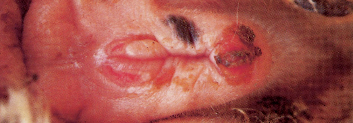
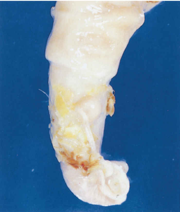
Often the penis can only be manually extruded with difficulty, if at all. The mucous membrane of the penis is severely inflamed, covered by a fibrinous or mucopurulent exudate (Figure 213.1), tear and bleed easily and contain small, scattered papulo-vesicular lesions which quickly develop into erosions or even ulcers. In some cases the erosions are extensive and ulcers may cover most of the glans penis. Ulcers of variable size may also occur at the preputial orifice, often completely surrounding it. If scabs are present, their removal exposes raw, bleeding surfaces covered by variable amounts of necrotic material. In severe cases, fibrosis and preputial adhesions result in phimosis. Paraphimosis, which is occasionally seen, culminates in extensive trauma and soiling of the penis. While clinical signs are developing, ewes which have been served will be noticed to have blood-stained hindquarters, particularly around the vulva and on the tail.
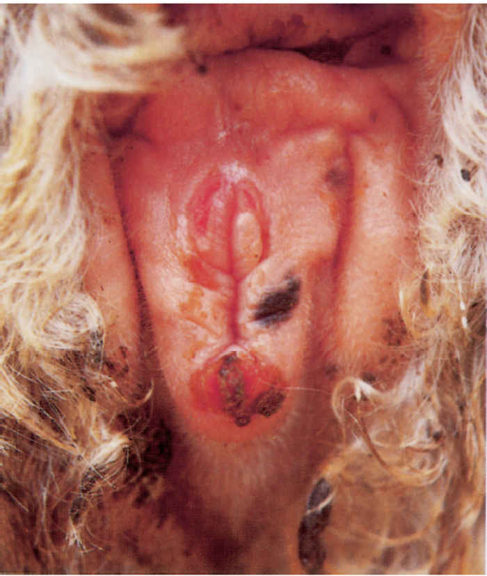
In Dorper ewes which have their tails docked very short, thus exposing their external genitalia, the vulval lips are swollen, oedematous and congested, and a blood-stained fluid or reddish, stringy mucus oozes from the external orifice. Frequent wagging of the tail-butt and urination, sometimes accompanied by straining, are observed in some cases. In acute cases the mucosa of the vagina is swollen and hyperaemic, with numerous greyish-white papulo-vesicular lesions scattered over the caudal region.1, 14, 17 Small erosions and papular lesions, which bleed easily when handled such as during vaginoscopic examination, may be present throughout the vaginal mucosa. Larger scab-covered ulcers are usually found on the vulval lips at the mucocutaneous junction, and particularly at the ventral tip of the vulva (Figure 213.2).
In ewes light microscopical lesions of the vagina and vulva include diffuse infiltration by mild to moderate numbers of predominantly lymphocytes and isolated plasma cells particularly noticeable at the muco-epidermal junction.38
Diagnosis and differential diagnosis
The diagnosis of ulcerative balanoposthitis and vulvovaginitis is at present based on clinical signs and lesions and certain epidemiological features. A fairly accurate diagnosis can be made if the typical acute lesions suddenly appear after four to six days of sexual activity.
Ulcerative balanoposthitis and vulvovaginitis should be differentiated from enzootic balanoposthitis (sheath rot) in rams and wethers, and vulvitis in ewes which have been fed on protein-rich diets. This latter disease syndrome follows a protracted course. The primary lesion in rams and wethers is a small scab on the skin dorsal to the preputium. The lesion gradually enlarges and extends into the preputial orifice to eventually involve the penis. In ewes the vulval lips are involved. This syndrome invariably occurs independently of mating. Corynebacterium spp. can usually be cultured from the lesions.7, 8, 32
Ulcerative to proliferative vulvitis or posthitis are manifestations of ulcerative dermatosis and contagious ecthyma (orf) and usually follow the appearance of similar lesions on the lips, face and legs of prepubertal lambs. Neither condition is associated with mating and the causative poxviruses can be isolated from the lesions.
Control
Systemic or local treatment with antimicrobial drugs of the classes macrolides, fluoroquinolones, amphenicols and tetracyclines are usually effective in acute cases.3, 20
However, clinical signs in treated animals which have apparently recovered recur soon after their re-introduction to the ewe flock. This suggests either that treatment is only effective against the secondary bacterial infections, or that effective immunity does not follow recovery.37
At present, good management practices are among the few measures that can be taken to control the disease. Only clinically healthy breeding animals should be purchased. Before the commencement of each breeding season, rams should be mated to groups of ten non-infected ewes. If no lesions appear within eight days of service, the rams may be used on the breeding flock with very little risk. Culling of infected rams is recommended.
First-time breeding rams and ewes should be managed as a unit and they should not be allowed to come into contact with older, previously bred sheep. This system should be permanently maintained, and implies that young rams should be housed separately from the older rams. Although artificial insemination is ideal for the control of the problem, the technique is impractical on most large and extensive farming units.
After the initial introduction of infective stock into a flock, the disease seems to stabilize after two or three breeding seasons, after which it is of little practical importance.
References
- BALL, H.J. & MCCAUGHEY, W.J., 1982. Experimental production of vulvitis in ewes with a Ureaplasma isolate. The Veterinary Record, 110, 581.
- BALL, H.J. & MCCAUGHEY, W.J., 1984. Investigations into the elimination of ureaplasmas from the uro-genital tract of ewes. British Veterinary Journal, 140, 292–299.
- BALL, H.J. & MCCAUGHEY, W.J., 1985. Experimental intramuscular inoculation of tiamulin and oxytetracycline in the elimination of Ureaplasma from sheep. The Veterinary Record, 117, 640–641.
- BALL, H.J., MCCAUGHEY, W.J. & IRWIN, D., 1984. Persistence of Ureaplasma genital infection in naturally-infected ewes. British Veterinary Journal, 140, 347–353.
- BALL, H.J., MCCAUGHEY, W.J., KENNEDY, S. & MCLOUGHLIN, M., 1985. Experimental intrauterine inoculation of pregnant ewes with Ureaplasma. Veterinary Research Communications, 9, 35–43.
- BATH, G. & DE WET, J., 2000. Pizzle Disease. Sheep and Goat Diseases. Cape Town: Tafelberg Publishers Limited. pp. 74–76.
- BARAJAS ROJAS, J.A. & BIBERSTEIN, E.L., 1974. Diphtheroid agent of ovine posthitis: Its relationship to Corynebacterium renale. Journal of Comparative Pathology, 84, 301–307.
- BROOK, A.H., SOUTHCOTT, W.H. & STACY, B.D., 1966. Etiology of ovine posthitis: Relationship between urine and a causal organism. Australian Veterinary Journal, 42, 9–12.
- CHIMA, J.C., ERNO, H. & OJO, M.O., 1986. Characterization and identification of caprine genital mycoplasmas. Acta Veterinaria Scandinavica, 27, 531–539.
- COTTEW, G.S., LLOYD, L.C., PARSONSON, I.M. & HORE, D.E., 1974. Isolation of a Mycoplasma from vulvovaginitis in sheep. Australian Veterinary Journal, 50, 576–577.
- CROUSE, W.J., 1989. State Veterinarian, Beaufort West, South Africa. Unpublished data.
- DEAS, D.W., 1983. Ulcerative balanitis and ulcerative vulvitis. In: martin, w.b., (ed.). Diseases of sheep. Oxford: Blackwell Scientific Publications. pp. 144–146.
- DE WET, J.A.L., 1989. Regional Veterinary Laboratory, Bloemfontein, South Africa. Unpublished data.
- DOIG, P.A. & RUHNKE, H.L., 1977. Isolation of Ureaplasma from sheep with granular vulvitis. The Veterinary Record, 100, 179–180.
- GUMMOW, B. & STALEY. G.P., 2000. A retrospective survey of ulcerative balanoposthitis and vulvovaginitis in South African Dorper sheep. Livestock Health and Production, 12, 31–36.
- JONES, G.E. & RAE, R.G., 1979. Ureaplasmas in sheep. The Veterinary Record, 104, 466.
- JONES, G.E., RAE, A.G., HOLMES, R.G., LISTER, S.A., JONES, J.M.W., GRATER, G.S. & RICHARDS, N., 1983. Isolation of exotic mycoplasmas from sheep in England. The Veterinary Record, 113, 540.
- JORDAAN, P., 1989. State Veterinarian, Upington, South Africa. Unpublished data.
- KAPOOR, S.G., SINGH, P.P. & PATHAK, R.C., 1984. Prevalence of Mycoplasma/Acholeplasma in the genital tract of sheep. Indian Journal of Animal Sciences, 54, 653–656.
- KIDANEMARIAM, A., 2003. Identification and characterization of the primary infectious agents associated with ovine ulcerative balanoposthitis and vulvovaginitis in South Africa. MSc thesis. University of Pretoria, South Africa.
- LIVINGSTONE, C.W. JR. & GAUER, B.B., 1975. Isolation of T-strain Mycoplasma from sheep and goats in Texas. American Journal of Veterinary Research, 36, 313–314.
- LIVINGSTONE, C.W. & GAUER, B.B., 1982. Effect of venereal transmission of ovine Ureaplasma on reproductive efficiency of ewes. American Journal of Veterinary Research, 43, 1190–1193.
- LIVINGSTONE, C.W. JR. & GAUER, B.B., 1983. Occurrence of Mycoplasma sp. (2D) in Texas sheep flocks. American Journal of Veterinary Research, 44, 868–869.
- MACKIE, D.P. & BALL, H.J., 1984. Pathogenicity of ovine ureaplasmas for the bovine mammary gland. The Veterinary Record, 114, 271–272.
- MCCAUGHEY, W.J. & BALL, H.J., 1981. Distribution of Ureaplasma in the urogenital tract of ewes. The Veterinary Record, 109, 472.
- MCCAUGHEY, W.J. & BALL, H.J., 1982. Experimental production of vulvitis in ewes with Ureaplasma isolates. The Veterinary Record, 110, 581.
- MCCAUGHEY, W.J. & BALL, H.J., 1983. Ureaplasmas in ewes. The Veterinary Record, 112, 441.
- MCCAUGHEY, W.J., BALL, H.J. & IRWIN, D., 1979. Ureaplasma species isolated from ewes in Northern Ireland. The Veterinary Record, 104, 397–398.
- MCCAUGHEY, W.J., BALL, H.J. & IRWIN, D., 1981. Isolation of Ureaplasma from ewes following synchronized mating. Irish Veterinary Journal, 35, 210–213.
- SCHUTTE, A.P., 1979, Taurus, Private Bag X5, Irene, South Africa. Unpublished data.
- SINGH, N., RAJYAN, B.S. & MOHANTY, G.C., 1974. Granular vulvovaginitis (GVV) in goats associated with Mycoplasma agalactiae. Cornell Veterinarian, 64, 435–442.
- SOUTHCOTT, W.H., 1962. The etiology of ovine posthitis: Transmission of the disease. Australian Veterinary Journal, 38, 441–446.
- TARIGAN, S. & WEBB, R.F., 1987. Caprine herpesvirus from balanoposthitis. Australian Veterinary Journal, 64, 321.
- TIWANA, J.S., SINGH, N. & KWATRA, M.S., 1984. Isolation of Mycoplasma and Acholeplasma from vulvovaginitis in goats. Indian Journal of Comparative Microbiology, Immunology and Infectious Diseases, 5, 17–19.
- TRICHARD, C.J.V., 1989. Onderstepoort Veterinary Institute, Pretoria, South Africa. Unpublished data.
- TRICHARD, C.J.V. & JORDAAN, P., 1989. Onderstepoort Veterinary Institute, Pretoria, South Africa. Unpublished data.
- TRICHARD, C.J.V. & VAN TONDER, E.M., 1989. Onderstepoort Veterinary Institute, Pretoria, South Africa. Unpublished data.
- TRICHARD, C.J.V., JORDAAN, P., PROZESKY, L., JACOBSZ, E.P. & HENTON, M.J., 1993. The identification of Mycoplasma mycoides mycoides LC as the aetiological agent of balanoposthitis and vulvovaginitis in sheep in South Africa. Onderstepoort Journal of Veterinary Research, 60, 29–37.
- VAN TONDER, E.M., 1989. Regional Veterinary Laboratory, Middelburg, Eastern Cape Province, South Africa. Unpublished data.
- VAN TONDER, E.M. & HENTON, M.M., 1989. Regional Veterinary Laboratory, Middelburg, Eastern Cape Province, South Africa. Unpublished data.
- VAN TONDER, E.M. & TRICHARD, C.J.V., 1989. Regional Veterinary Laboratory, Middelburg, Eastern Cape Province, South Africa. Unpublished data.


