- Infectious Diseases of Livestock
- Part 3
- Brucella melitensis infection
- GENERAL INTRODUCTION: SPIROCHAETES
- Swine dysentery
- Borrelia theileri infection
- Borrelia suilla infection
- Lyme disease in livestock
- Leptospirosis
- GENERAL INTRODUCTION: AEROBIC ⁄ MICRO-AEROPHILIC, MOTILE, HELICAL ⁄ VIBROID GRAM-NEGATIVE BACTERIA
- Genital campylobacteriosis in cattle
- Proliferative enteropathies of pigs
- Campylobacter jejuni infection
- GENERAL INTRODUCTION: GRAM-NEGATIVE AEROBIC OR CAPNOPHILIC RODS AND COCCI
- Moraxella spp. infections
- Bordetella bronchiseptica infections
- Pseudomonas spp. infections
- Glanders
- Melioidosis
- Brucella spp. infections
- Bovine brucellosis
- Brucella ovis infection
- Brucella melitensis infection
- Brucella suis infection
- Brucella infections in terrestrial wildlife
- GENERAL INTRODUCTION: FACULTATIVELY ANAEROBIC GRAM NEGATIVE RODS
- Klebsiella spp. infections
- Escherichia coli infections
- Salmonella spp. infections
- Bovine salmonellosis
- Ovine and caprine salmonellosis
- Porcine salmonellosis
- Equine salmonellosis
- Yersinia spp. infections
- Haemophilus and Histophilus spp. infections
- Haemophilus parasuis infection
- Histophilus somni disease complex in cattle
- Actinobacillus spp. infections
- Actinobacillus equuli infections
- Gram-negative pleomorphic infections: Actinobacillus seminis, Histophilus ovis and Histophilus somni
- Porcine pleuropneumonia
- Actinobacillus suis infections
- Pasteurella and Mannheimia spp. infections
- Pneumonic mannheimiosis and pasteurellosis of cattle
- Haemorrhagic septicaemia
- Pasteurellosis in sheep and goats
- Porcine pasteurellosis
- Progressive atrophic rhinitis
- GENERAL INTRODUCTION: ANAEROBIC GRAM-NEGATIVE, IRREGULAR RODS
- Fusobacterium necrophorum, Dichelobacter (Bacteroides) nodosus and Bacteroides spp. infections
- GENERAL INTRODUCTION: GRAM-POSITIVE COCCI
- Staphylococcus spp. infections
- Staphylococcus aureus infections
- Exudative epidermitis
- Other Staphylococcus spp. infections
- Streptococcus spp. infections
- Strangles
- Streptococcus suis infections
- Streptococcus porcinus infections
- Other Streptococcus spp. infections
- GENERAL INTRODUCTION: ENDOSPORE-FORMING GRAM-POSITIVE RODS AND COCCI
- Anthrax
- Clostridium perfringens group infections
- Clostridium perfringens type A infections
- Clostridium perfringens type B infections
- Clostridium perfringens type C infections
- Clostridium perfringens type D infections
- Malignant oedema⁄gas gangrene group of Clostridium spp.
- Clostridium chauvoei infections
- Clostridium novyi infections
- Clostridium septicum infections
- Other clostridial infections
- Tetanus
- Botulism
- GENERAL INTRODUCTION: REGULAR, NON-SPORING, GRAM-POSITIVE RODS
- Listeriosis
- Erysipelothrix rhusiopathiae infections
- GENERAL INTRODUCTION: IRREGULAR, NON-SPORING, GRAM-POSITIVE RODS
- Corynebacterium pseudotuberculosis infections
- Corynebacterium renale group infections
- Bolo disease
- Actinomyces bovis infections
- Trueperella pyogenes infections
- Actinobaculum suis infections
- Actinomyces hyovaginalis infections
- GENERAL INTRODUCTION: MYCOBACTERIA
- Tuberculosis
- Paratuberculosis
- GENERAL INTRODUCTION: ACTINOMYCETES
- Nocardiosis
- Rhodococcus equi infections
- Dermatophilosis
- GENERAL INTRODUCTION: MOLLICUTES
- Contagious bovine pleuropneumonia
- Contagious caprine pleuropneumonia
- Mycoplasmal pneumonia of pigs
- Mycoplasmal polyserositis and arthritis of pigs
- Mycoplasmal arthritis of pigs
- Bovine genital mycoplasmosis
- Neurotoxin-producing group of Clostridium spp.
- Contagious equine metritis
- Tyzzer's disease
- MYCOTIC AND ALGAL DISEASES: Mycoses
- MYCOTIC AND ALGAL DISEASES: Pneumocystosis
- MYCOTIC AND ALGAL DISEASES: Protothecosis and other algal diseases
- DISEASE COMPLEXES / UNKNOWN AETIOLOGY: Epivag
- DISEASE COMPLEXES / UNKNOWN AETIOLOGY: Ulcerative balanoposthitis and vulvovaginitis of sheep
- DISEASE COMPLEXES / UNKNOWN AETIOLOGY: Ill thrift
- Eperythrozoonosis
- Bovine haemobartonellosis
Brucella melitensis infection
This content is distributed under the following licence: Attribution-NonCommercial CC BY-NC  View Creative Commons Licence details here
NJ Maclachlan and M-L Penrith (Editors). J Godfroid, B Garin-Bastuji and J M Blasco, Brucella melitensis infection, 2018.
Brucella melitensis infections
Previous authors: J GODFROID, B GARIN-BASTUJI, J M BLASCO, J THOMSON AND C O THOEN
Current authors:
J GODFROID - Professor of Microbiology, DVM, MSc, PhD, University of Tromsø - the Arctic University of Norway, Hansine Hansens veg 18, Tromsø, 9019, Norway
B GARIN-BASTUJI - Senior Research Director - Scientific Adviser, European & International Affairs Department, French Agency for Food, Environmental & Occupational Health & Safety (ANSES), 14 rue Pierre et Marie Curie, Maisons-Alfort Cedex, F-94701, France
J M BLASCO - Emeritus Researcher, DVM, PhD, Cita/Ia2/University Zaragoza Avenue, Montañana 930, Zaragoza, 50011, Spain
Introduction
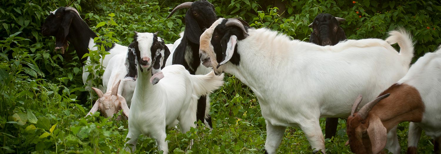
Brucella melitensis is the main causative agent of caprine and ovine brucellosis. It is also pathogenic for several other mammal species including humans.8, 12 Infected cows may abort and shed B. melitensis in their milk.74, 75 Brucellosis in small ruminants is characterized by one or more of the following: abortion, reduced milk yield and retained placenta (particularly in goats) in females; orchitis and epididymitis in males; and, rarely, arthritis in both sexes.8, 12 It is worth noting that brucellosis caused by Brucella abortus infection in small ruminantsseems to be an important problem in West Africa whereas Brucella suis infection is rare in small ruminants.
The organism responsible for Malta fever in humans was first discovered by Sir David Bruce on the island of Malta in 1887.23 It had become evident to him and his co-workers that the disease was not transmitted from person to person. Zammit, a member of the Mediterranean Fever Commission, determined in 1905 that B. melitensis was the cause of contagious abortion in goats on the island and that humans contracted the disease by the consumption of infected fresh goats’ milk and milk products.84
Aetiology and epidemiology
Brucella melitensis is morphologically and tinctorially indistinguishable from B. abortus and B. suis. Species identification can be based on lysis by phages, on biochemical tests (such as oxidase, urease and catalase) or molecular tests. (see Bovine brucellosis and Brucella suis infection).10, 14 Unlike B. abortus, growth of B. melitensis is not dependent on an atmosphere of 5 to 10 per cent CO2, although there might be some exceptions.10, 14 The identification of a Brucella species to the biovar level is currently performed by four main tests, namely CO2 dependence, production of hydrogen sulphide, dye (thionin and basic fuchsin) sensitivity, and agglutination with monospecific A and M anti-sera.10, 25, 27 The three biovars (1, 2 and 3) recognized for B. melitensis show no difference in pathogenicity. Brucella melitensis biovar 3 appears to be the biovar most frequently isolated in Mediterranean countries while biovar 1 seems to predominate in Latin America.8, 26, 55
In a Multi Locus Sequence Analysis (MLSA) study,81 B. melitensis isolates clustered into three distinct lineages that correspond to the “Americas,” “West Mediterranean,” and “East Mediterranean” lineages described earlier using Multiple Locus Variable (number of tandem repeats) Analysis (MLVA) based studies.2 Although a few isolates from the USA and Argentina were included in the “Americas” clade, more than 60 per cent of these isolates clustered with strains of African origin. African isolates included in the study originated widely across the continent (Ethiopia, Somalia, Nigeria, Tanzania, Sudan, Zimbabwe) and, as few African isolates fit into the “West Mediterranean” and “East Mediterranean” lineages, they may represent lineages that are endemic in Africa. In agreement with the abovementioned MLVA study, strains falling in the “West Mediterranean” lineage were isolated exclusively in Italy, France, the former Yugoslavia and Germany. In contrast, the “East Mediterranean” lineage, while including isolates from Greece, Turkey, Cyprus, and the Balkan States, extends geographically more widely into the Middle East and Asia (Thailand, India, Pakistan, Mongolia, and Afghanistan). While most isolates in the “Americas” and “West Mediterranean” lineages corresponded to biovar 1 (81 per cent) and biovar 3 (64 per cent) respectively, both lineages included isolates of all three biovars. This MLSA study concluded that the B. melitensis biovar concept is reliant solely on the serological reaction observed with the monospecific anti A and anti M sera and is of no epidemiological significance.81 Smooth (S) B. melitensis cultures have a tendency to undergo variation during growth, especially on subculturing, and dissociate to rough forms. These changes in colonial morphology are generally associated with changes in virulence and antigenic and immunogenic properties. Such changes may also occur during the production of the live attenuated S vaccines, particularly in the case of B. melitensis Rev. 1 vaccine,14, 15, 67 which may have serious consequences (see Control, below). Brucella melitensis wild type strains are usually resistant to benzyl penicillin and sensitive to streptomycin, whereas the Rev. 1 vaccine strain is sensitive to benzyl penicillin and resistant to streptomycin.10, 13, 14 This microbiological test and several PCR procedures (see below) can be used to differentiate field B. melitensis biovar 1 strains from Rev. 1.14
Brucella melitensis infection occurs worldwide,8, 12 and is prevalent in small ruminants particularly in the Mediterranean countries, the Middle East, some regions of Asia and Latin America.8, 18, 26, 43, 55 Infections have also been encountered in dromedary camels (Camelus dromedarius), bactrian camels (Camelus bactrianus) and cattle in contact with infected sheep and goats.1, 74, 75, 80 Northern Europe, South-East Asia, Australia, New Zealand and Canada are believed to be free of the disease.8, 12 In the USA, B. melitensis was detected in 1999 in cattle and goats in Texas after absence since the early 1970s, but the source of infection was not determined.49
The first report of the presence of B. melitensis in southern Africa was in 1953 when the organism was isolated from Karakul sheep in Namibia.72 The next report followed in 1965 when it was recovered from three outbreaks of abortion in sheep in Mpumalanga and Limpopo Provinces of South Africa.73 Brucella melitensis biovar 1 was isolated in Namibia from three humans in 1988 and in Gauteng Province, South Africa in 1990 in association with abortion in a flock of goats.70 In 1994, B. melitensis infection in a flock of goats in the Makhathini Flats area of KwaZulu-Natal Province, South Africa was diagnosed by serological tests and culture following a diagnosis of Malta fever in their owner.69 A subsequent serological survey in the Province revealed a low prevalence of brucellosis in goats and identified six foci of infection.A test-and-slaughter eradication programme was implemented in which all serologically positive cases were slaughtered. Bacteriological investigation of the tissues from a number of the slaughtered goats confirmed the presence of B. melitensis biovar 1.34 The survey seemed to indicate a slow rate of spread of the disease in goats, with fewer abortions having occurred than is the case in other countries.8, 69 Very few, if any, of the local human population in the region concerned drink goats’ milk.69 The histories of the positive goats revealed that a number of them had been purchased in nearby Swaziland, where the disease has been confirmed.34 Importantly, B. melitensis was isolated from a human patient in 2016.82 A retrospective inquiry revealed that a farm had been quarantined because of seropositive goats. Recently, B. melitensis has been isolated from cattle in South Africa.51
Outbreaks of B. melitensis infections in sheep and goats are sporadic in other countries in southern Africa such as Zambia53 and Zimbabwe.57 Until 1987, it was believed that B. melitensis did not occur in Zimbabwe, but, since then, two outbreaks in goats on commercial farms in the Gweru and Harare districts and two in goats in communal areas in Matabeleland North and Mashonaland Central have been reported.57
All breeds of goats are believed to be equally susceptible to the infection, but despite the scarcity of data, resistance is assumed to vary with breed in sheep.; Maltese sheep appear to be relatively resistant, while certain fat-tailed breeds (such as Awassi) are highly susceptible.6, 7, 12 However, it seems probable that the local Nguni sheep, a fat-tailed breed in KwaZulu-Natal, South Africa is more resistant to B. melitensis infection than other breeds.34 Although it has been documented that different breeds of sheep also differ in their serological response to the B. melitensis Rev. 1 vaccine,12-14 it cannot be excluded that this reflects the heterogeneity of Rev. 1 vaccine batches rather than a difference in resistance to B. melitensis infection.
The oral route is the main route of infection through the sucking or licking of aborted foetuses and their placentas and vaginal discharges, and the ingestion of infected milk or feedstuffs. Brucella melitensis may persist in the udder for years.8, 12, 41 Inhalation of contaminated dust is also believed to be an important route of infection.6 The herding of goats and sheep into pens or kraals at night creates an ideal environment for the spread of the infection.8, 12 As in the case of B. abortus infection in cattle, B. melitensis can be transmitted from ewes to lambs. A small proportion of lambs can be infected in utero, but the majority of B. melitensis infections are probably acquired by the ingestion of infected colostrum or milk.44 Although not proven, it is likely that a self-cure mechanism, similar to that considered to occur in calves, takes effect in most of the infected lambs. However, infections in lambs greatly increase the difficulty of eradicating this disease, as B. melitensis may persist until adulthood without inducing a detectable serological immune response.44
It is believed that billy goats and rams do not play an important role in the epidemiology of disease. However, orchitis and epididymitis are common sequelae of infection and B. melitensis is excreted in semen in a significant proportion of infected males, with a concomitant risk of transmission. It has in fact been shown that artificial insemination may spread brucellosis among animals.11
Dogs kept on farms with infected flocks may become infected and develop generalized disease and they (especially the pregnant bitches) may spread the infection.8 Surprisingly, the ecological range of B. melitensis in wildlife is more restricted than the ranges of B. abortus and B. suis.43 In fact, in a study conducted in heavily infected areas in Spain it was shown that wild ruminants were not a significant brucellosis reservoir for domestic livestock.64 Spill-over of B. melitensis from infected small ruminants and cattle to a small number of wildlife species such as chamois (Rupicapra rupicapra) and ibex (Capra ibex) in the French and Italian Alps has been documented in southern Europe.40, 42, 61
The survival of B. melitensis in the environment is similar to that of B. abortus5 (see Bovine brucellosis).
Brucella melitensis is highly pathogenic for humans, causing the disease known as Malta fever (also called Mediterranean or undulant fever)8, 12 which is rated by the World Health Organization as one of the most important zoonoses.13 Brucellosis in humans is also caused by B. abortus and B. suis infections, the resultant clinical disease being indistinguishable from the disease caused by B. melitensis(although less severe).8, 12 Brucellosis in humans is transmitted by ingesting infected unpasteurized milk and milk products and by direct contact with infected animals, animal carcasses and aborted material, and the organisms themselves in laboratories, as well as by accidental inoculation with the B. melitensis Rev. 1 vaccine stain.5, 9 Worldwide, millions of humans are at risk, especially those in developing countries where the infection in animals is not well controlled, heat treatment procedures of milk (e.g. pasteurization) are not routinely applied, and poor hygienic conditions and food habits such as the consumption of raw milk favour human infection.8, 12, 13 The survival of Brucella in milk and dairy products depends on a variety of factors. However, it is generally accepted that industrial pasteurization or prolonged boiling of milk readily kill Brucella organisms. The bacteria do not persist for periods exceeding three months in ripened fermented cheese. Milk or cream used in the preparation of cheese should be pasteurized to ensure its safety for human consumption.
It is worth noting that humans are often the first to be reported as being infected in an area into which the disease has been newly introduced.6, 75, 78 Epidemic human infections caused by B. melitensis occur in people frequently in contact with infected goat herds or goat manure, as reported in Argentina.79 Localization of B. melitensis in the udder of infected camels and cattle is also a major public health problem.1, 74, 75 In laboratories handling Brucella-infected or potentially infected material it is mandatory that conditions of high level biohazard containment are used.10, 12
Pathogenesis and clinical signs
The pathogenesis of the disease is similar to that of B. abortus infection in cattle12, 71 (see Bovine brucellosis). The first sign of the presence of the disease in a susceptible herd of goats or flock of sheep is usually an abortion storm during which a high proportion of the pregnant animals abort, generally late in gestation. In some cases, particularly in nanny goats, retention of placenta and foetal membranes occurs.8, 12, 71 Milk yield following abortion is poor, and the quality of the milk may be reduced. Genital discharges of infected nanny goats are usually copious and persist for several months following abortion or kidding. Genital discharges are less copious and less persistent in infected ewes.8, 12, 41 In males, localization of B. melitensis in the testis, epididymis and accessory sex organs is common and may result in acute orchitis and epididymitis, and subsequently in reduced fertility. Bacteria may be shed in the semen. Arthritis is occasionally observed in both sexes.8, 12, 71 During subsequent breeding seasons the number of animals in a flock that abort declines progressively and eventually abortions may cease to occur. Such flocks, however, remain infected for years.16 Persistent infection of the mammary glands with constant or intermittent shedding of the organism in the milk in succeeding lactations is frequent.8 Kids or lambs of infected females may be born weak or are asymptomatic; some of them may become persistent latent carriers of the infection.44
In areas where brucellosis is endemic the prevalence of infection with B. melitensis in some flocks may be high even in the absence of any signs of overt disease.8
Pathology
The macroscopic lesions in nanny goats and ewes that abort following infection with B. melitensis include oedema and greyish-white areas of necrosis of the placenta and the presence of a brownish-red exudate between the allantochorion and the endometrium.63
Microscopically, small granulomatous or necrotic foci are evident in lymphoid tissues, organs such as reproductive organs and associated lymph nodes, and synovial membranes. In pregnant females, there may be extensive necrosis of the chorioallantoic membranes accompanied by vasculitis and acute endometritis63 with large numbers of bacteria in necrotic villi. Desquamated trophoblast cells and a few macrophages, neutrophils and plasma cells occur in the spaces between the chorionic villi and septa.
Diagnosis
Brucella melitensis can be demonstrated by microscopic examination of smears made from vaginal discharges or swabs, placenta or abomasum of the aborted foetus and stained with Stamp’s modification of the Ziehl-Neelsen method. However, as morphologically similar micro-organisms such as Brucella ovis, Chlamydophila abortus and Coxiella burnetii can be confused with B. melitensis. The most reliable and the only unequivocal method for the diagnosis of animal brucellosis is the isolation and typing of the causative bacterium using appropriate culture media.8, 10 Vaginal excretion of B. melitensis usually persists for several weeks after abortion,8 while the mammary gland is the main target of infection in small ruminants.58 Vaginal swabs and milk are therefore the samples of choice to isolate B. melitensis from live nanny goats and ewes. Attempts should be made to culture the organism from the cotyledons, abomasal content and lungs of the foetus and, in slaughtered animals, from the mammary glands and supramammary lymph nodes in females, the seminal vesicles and testes in males, and the parotid, mandibular and retropharyngeal lymph nodes and spleen in both sexes.25
Selective media should be used for isolation of B. melitensis because contaminants are usually present in field samples that will overgrow B. melitensis colonies. Farrell’s selective medium, developed for the isolation of B. abortus from milk,37 is recommended for the isolation of B. melitensis.10 However, this medium contains nalidixic acid and bacitracin which, at the concentrations used, have inhibitory effects on some B. melitensis strains, so it should be used with the less selective Thayer-Martin modified medium (mTM).59 The CITA medium, which inhibits most contaminant microorganisms but allows simultaneously the growth of all Brucella species, is the selective medium of choice for the isolation of all Brucella spp.28 The best diagnostic sensitivity is obtained using both the Farrell’s and CITA simultaneously.28
Numerous polymerase chain reaction (PCR)-based assays have been developed for the identification of members of the genus Brucella. For epidemiological trace-back, strain-specific identification is required but, despite the considerable progress made,77, 81 no technique is available for the differentiation of the three B. melitensis biovars.22 Initially all these molecular assays were performed on purified DNA obtained from previously cultured organisms. For the identification of brucellae from field samples and food products such as milk and cheese, the removal of polymerase inhibitors is required before the implementation of PCR techniques.22 Real-time PCRs have been developed for the specific detection of B. melitensis in milk.68 However, these techniques need to be further validated for routine diagnostic purposes.
The differentiation of the B. melitensis Rev. 1 vaccine strain from wild B. melitensis is accomplished by microbiological laboratory tests such as sensitivity to penicillin and streptomycin, size of colony, and growth on media containing dyes (e.g. thionin and basic fuchsin).10, 25 Chromosomally acquired streptomycin resistance is frequently due to mutations in the rpsL gene encoding the ribosomal protein S12, and a PCR-RFLP based on a mutation in the rpsL gene has been described for the identification of the B. melitensis Rev. 1 vaccine strain.24 A multiplex PCR assay (Bruce-ladder) was developed and can differentiate in a single step all of the classical Brucella spp., including those found in marine mammals and the S19, RB51, and Rev.1 vaccine strains.54
Several serological tests are used to diagnose brucellosis in small ruminants. The outer membrane of smooth Brucella spp. strains is composed of phospholipids, proteins and lipopolysaccharide [(smooth lipopolysaccharide (S-LPS)]. Smooth Brucella strains are classified into three serotypes, i.e., A+M− (A-dominant strains, such as B. abortus biovar 1 and B. melitensis biovar 2), A−M+ (M-dominant strains, such as B. melitensis biovar 1), and A+M+ (such as B. melitensis biovar 3), using a simple slide agglutination technique with the anti-A and anti-M monospecific polyclonal antisera.10 The epitopes involved have been located on the O-polysaccharide (O-PS) moiety of the S-LPS, which represents the most exposed antigenic structure on the surface of smooth Brucella spp. Most of the serological tests, particularly those using whole-cell suspensions as antigen, such as the Rose Bengal test (RBT), the complement fixation test (CFT) and most enzyme-linked immunosorbent assays (ELISA), have been developed to detect antibodies directed against O-PS antigens.4, 10, 14
The RBT and the CFT are the most widely used tests for the serological diagnosis of ovine and caprine brucellosis.14, 19, 20, 36, 41, 60 These tests are currently the official standardized tests used in the European Union Member States,35 and are the tests prescribed by the OIE for international trade.14
One of the controversial issues concerning the serological diagnosis of B. melitensis infection in small ruminants is related to which Brucella spp. and biovars are used in the production of the antigens. The antigenic suspensions (whole cells) used in both the RBT and CFT are made with B. abortus biovar 1 (an A-dominant strain).10 This implies that, theoretically, infections due to M-dominant strains (such as B. melitensis biovar 1) could be misdiagnosed.10, 56 However, no significant difference has been found in the sensitivity of the classical RBT antigen prepared with B. abortus biovar 1 (A-dominant) in sheep populations infected with either B. melitensis biovar 1 (M- dominant) or B. melitensis biovar 3 (A-dominant),19 and the same is true in the case of the soluble S-LPS antigens used in ELISAs.4
There are small variations among the RBT and CFT used worldwide. It has been shown that the CFT and the buffered agglutination tests used in North America for the diagnosis of brucellosis in goats performed as well as the RBT and CFT used in Europe.62 For the CFT, it was also shown that there was no advantage in using antigen prepared from B. melitensis instead of antigen prepared from B. abortus for the detection of infection B. melitensis biovar 1 in goats .62
The CFT has many drawbacks such as complexity, variability of reagents, prozones, anticomplementary activity of sera, difficulty to perform with haemolysed sera and subjectivity of the interpretation. Therefore, while the sensitivity of the RBT is sufficient for the surveillance of areas free of the disease at the flock level, the RBT and CFT should be used together in infected flocks to obtain an accurate individual sensitivity level in test-and-slaughter programmes. Moreover, another important drawback of both the RBT and CFT is their low specificity when testing sera from sheep and goats that have been recently vaccinated subcutaneously with the B. melitensis Rev. 1 vaccine. However, when this vaccine is applied conjunctivally the interference problem is significantly reduced in all serological tests.14, 15, 30, 47 Despite the above drawbacks, the combination of the RBT as screening test and the CFT as a complementary test has been used successfully in many countries to eradicate B. melitensis infection in small ruminants.
The serum agglutination test is not considered to be sufficiently reliable for use in the diagnosis of infection in small ruminants.14 Indirect ELISAs have, however, been reported to be adequately sensitive for this purpose.4, 20, 29, 30, 45, 47, 60 The specificity of the indirect ELISA is quite low when testing sera from animals that have been vaccinated recently with the Rev. 1 vaccine.30, 45 Other serological techniques such as competitive ELISA16, 60 and fluorescence polarization assay66 have been developed but not properly standardized and validated for wide use in small ruminants, and do not provide any advantages over the abovementioned tests.14 The milk ring test does not give as clear-cut a reaction as it does in cattle infected with B. abortus, and it is thus not recommended as a flock or herd test in small ruminants.8, 12, 14 A technique similar to the indirect serum ELISA has also been proposed for testing individual or pooled milk samples for the diagnosis of ovine and caprine brucellosis.17, 62
An allergic brucellosis skin test for the diagnosis of B. melitensis infection has been used with variable degrees of success to detect specific delayed-type hypersensitivity (DTH) responses in sheep and goats.20, 38, 39 The S-LPS itself does not play a role in DTH reactions but its presence in the allergen could cause an inflammatory reaction that interferes with the interpretation of the test. In addition, it can cause a secondary antibody response, thus interfering with future serological testing. These problems are solved by using rough Brucella strains devoid of the S-LPS O-chain polysaccharide.10 The only skin test allergen available (known as brucellergene OCB, brucellin INRA or brucellin) is a hypertonic cytosoluble extract obtained from the rough B. melitensis B115 strain, and is commercially available.10, 20 The site and route of inoculation of the allergen do not affect the sensitivity of the test.10, 20, 39 The method considered to be the most efficient and practical is to inoculate the allergen into the lower eyelid. Readings are made after 48 to 72 hours.10, 20 The brucellosis skin test is considered to be 100 per cent specific when testing unvaccinated Brucella-free animals20, 38, 39 but this is not the case when testing animals vaccinated recently with the B. melitensis Rev. 1 vaccine, which can remain positive in this test for as long as two years or more after vaccination.38
Control
Vaccination of sheep and goats with the live attenuated B. melitensis Rev. 1 vaccine, developed by Elberg in the mid- 1950s, is the only practical and effective method for reducing the incidence of brucellosis.6-9, 12-14 Due to its ability to induce abortion and the possibility of the organism being excreted in milk, it was recommended that the B. melitensis Rev. 1 vaccine be administered subcutaneously in young replacement sheep and goats, prior to the first gestation, at three to seven months of age.12-14 However, when B. melitensis strain Rev. 1 vaccine is administered by the standard method [1 to 2 × 10⁹ colony forming units (CFU) given subcutaneously], it may induce a long-lasting serological response in classical brucellosis tests (RBT and CFT) that could interfere in the serological diagnosis of the disease itself.12-15 In contrast, when this vaccine is administered by the conjunctival route, the immunity conferred is similar to that induced by the standard method but the serological response evoked is significantly reduced, making it the method of choice when combining vaccination and eradication programmes.14, 15, 30, 39, 47
When used in a herd or flock vaccination programme, inoculation of the B. melitensis Rev. 1 vaccine greatly decreases the prevalence of brucellosis in goats and sheep and therefore in human populations.12, 13, 15, 32, 33 Once the prevalence has been reduced, control of the disease may be achieved through the implementation of a programme based on the combination of B. melitensis Rev. 1 vaccination of lambs and kids, the serological testing of adults and the culling of serologically positive animals. Once the disease has been controlled by vaccination, eradication (i.e. the elimination of B. melitensis from the target animal population) can only be accomplished by a test-and-slaughter approach in flocks where the prevalence of the disease is very low. This requires the identification of animals and flocks, the control of animal movement, and adequate veterinary services and economic resources.41 In a test-and-slaughter programme two negative CFT results six months apart in all the animals in a flock is accepted as proof of eradication of the disease.7, 14, 50, 52
The exclusive vaccination of replacement males and females can be effective in controlling the infection in countries with a low or moderate brucellosis prevalence.18 However, this exclusive vaccination of young replacement animals has failed to control brucellosis in some developed countries and is frequently not applicable in resource-limited countries because of the high prevalence of the disease and because in many regions the animals are kept under extensive management conditions that may be nomadic or semi-nomadic.8, 12-15 As a result, the development of flock immunity is very low and unvaccinated adult females remain unprotected and may spread the infection further.8, 15, 41 Therefore, vaccination of the entire flock appears to be the only feasible method for controlling B. melitensis infection in small ruminants maintained under such extensive management conditions and high prevalence.14, 41 In such difficult conditions, vaccination of both young and adult replacement animals every two years is the only technique suitable for reduction of the prevalence of the disease, and is therefore the first step towards successful control.18, 21 Vaccination of pregnant animals with a full standard dose of Rev. 1 administered subcutaneously or conjunctivally is often followed by abortion, particularly when they are vaccinated during early or mid-term pregnancy.14, 15, 18, 85 The induction of abortions when vaccinating pregnant animals means that there is no entirely safe strategy for vaccination with the B. melitensis Rev. 1 vaccine. Conjunctival vaccination is safer than subcutaneous vaccination but is not safe enough to be applied without taking the pregnancy status of the ewes or nanny goats into consideration and it should be used only under restricted conditions.46, 85 The number of abortions is, however, considerably reduced when adult female animals are vaccinated conjunctivally just before mating or duringlambing/kidding and lactation. Moreover, the vaccination of lactating animals with Rev. 1 is not followed by excretion of the vaccine strain in milk.48 Therefore, when mass vaccination is the only means of controlling the disease, a vaccination campaign should be recommended using the standard dose of B. melitensis Rev.1 administered by the conjunctival route applied when the animals are not pregnant.14, 15, 18
Reducing the dose of B. melitensis Rev. 1 vaccine has been proposed as a method of avoiding the problem of inducing abortion and, accordingly, a reduced-dose vaccination strategy has been widely used and has been reported to be a safe and effective method of controlling small ruminant brucellosis.3, 32, 33 Nevertheless, controlled field and experimental results have revealed that, due to the induction of abortion in pregnant animals and the low degree of immunity conferred, reduced-dose vaccination of Rev. 1 should not be recommended as an alternative to the full standard dose.14, 18, 85
Production of Brucella or B. melitensis Rev.1 vaccine is based on a seed-lot system. Seed cultures to be used in vaccines should originate from reference centres. They must conform to minimal standards for viability, smoothness, residual infectivity and immunogenicity.13, 15 When tested in a mouse model, differences in residual virulence and immunogenicity have been demonstrated between the organisms used in different B. melitensis Rev. 1 vaccines produced worldwide. These differences could account for the discrepancies in safety that have been obtained in mass vaccination trials in different countries.18 In this context, B. melitensis Rev. 1-like strains that caused infections in sheep followed by infections in humans have been reported in South Africa and Israel.15, 67 Laboratory and data analysis in South Africa suggested that inadequacies in the manufacturing of the vaccine used led to this accident.67 Guidelines for the production of vaccine are given in the OIE Manual of Standards for Diagnostic and Vaccines.14
The Chinese live and smooth B. suis S2 vaccine that was apparently successfully used in field experiments in China83 and Libya65 did not provide adequate protection against B. melitensis infection when tested in controlled experiments.76 The Brucella abortus RB51, a rough attenuated strain, also did not protect sheep effectively against B. melitensis.31
References
- ABOU-EISHA, A. M., 2000. Brucellosis in camels and its relation to public health. Assiut Veterinary Medical Journal, 44, 54-64.
- AL DAHOUK, S. L. E., FLÈCHE, P. L., NÖCKLER, K., JACQUES, I., GRAYON, M., SCHOLZ, H. C., TOMASO, H., VERGNAUD, G. & NEUBAUER, H., 2007. Evaluation of Brucella MLVA typing for human brucellosis. Journal of Microbiological Methods, 69, 137-45.
- AL-KHALAF, S. A., MOHAMAD, B. T. & NICOLETTI, P., 1992. Control of brucellosis in Kuwait by vaccination of cattle, sheep and goats with Brucella abortus strain 19 or Brucella melitensis strain Rev. 1. Tropical Animal Health Production, 24, 45-9.
- ALONSO-URMENETA, B., MARIN, C., ARAGON, V., BLASCO, J. M., DIAZ, R. & MORIYON, I., 1998. Evaluation of lipopolysaccharides and polysaccharides of different epitopic structures in the indirect enzyme-linked immunosorbent assay for diagnosis of brucellosis in small ruminants and cattle. Clinical and Diagnostic Laboratory Immunology, 5, 749-754.
- ALTON, G. G., 1985. The epidemiology of Brucella melitensis in sheep and goats. In: VERGER, J.M. & PLOMMET, M., (eds). Brucella melitensi. Dordrecht, Boston, Lancaster: Martinus Nijhoff Publishers.
- ALTON, G. G., 1985. Rev. 1 and H38 Brucella melitensis vaccines. In: VERGER, J.M. & PLOMMET, M., (eds). Brucella melitensis. Dordrecht, Boston, Lancaster: Martinus Nijhoff Publishers.
- ALTON, G. G., 1987. Control of Brucella melitensis infection in sheep and goats—a review. Tropical Animal Health and Production, 19, 65-74.
- ALTON, G. G., 1990. Brucella melitensis. In: NIELSEN, K. & DUNCAN, J.R., (eds), Animal brucellosis. Boca Raton, FL: CRC Press Inc. 383-409.
- ALTON, G. G. & ELBERG, S. S., 1967. Rev. 1 Brucella melitensis vaccine. A review of ten years of study. Veterinary Bulletin, 37, 793-800.
- ALTON, G. G., JONES, L. M., ANGUS, R. D. & VERGER, J. M., 1988. Techniques for the brucellosis laboratory. Paris: Institut National de la Recherche Agronomique (INRA).
- AMIN, A. S., HAMDY, M. E. & IBRAHIM, A. K., 2001. Detection of Brucella melitensis in semen using the polymerase chain reaction assay. Veterinary Microbiology, 22, 37-44.
- ANON, 1986. Joint FAO/WHO Expert Committee on Brucellosis. World Health Organization Technical Report Series 740. Geneva: WHO.
- ANON, 1998. The development of new/improved brucellosis vaccines: report of a WHO meeting. World Health Organization Communicable Disease Surveillance and Response. WHO/ZDI/98.14.
- ANON, 2018. Manual of Standards for Diagnostic Tests and Vaccines. Paris: Office International des Epizooties. http://www.oie.int/en/standard-setting/terrestrial-manual/access-online/ (Accessed on line, October 08, 2018).
- BANAI, M., 2002. Control of small ruminant brucellosis by use of Brucella melitensis Rev.1 vaccine: Laboratory aspects and field observations. Veterinary Microbiology, 90, 497-519.
- BIANCIFIORI, F., GARRIDO, F., NIELSEN, K., MOSCATI, L., DURAN, M. & GALL, D., 2000. Assessment of a monoclonal antibody-based competitive enzyme linked immunosorbent assay (cELISA) for diagnosis of brucellosis in infected and Rev. 1 vaccinated sheep and goats. New Microbiologica, 23, 399-406.
- BIANCIFIORI, F., NANNINI, D., DI MATTEO, A. & BELFIORE, P., 1996. Assessment of an indirect ELISA in milk for the diagnosis of ovine brucellosis. Comparative Immunology, Microbiology and Infectious Diseases, 19, 17-24.
- BLASCO, J. M., 1997. A review of the use of B. melitensis Rev. 1 vaccine in adult sheep and goats. Preventive Veterinary Medicine, 31, 275-281.
- BLASCO, J. M., GARIN-BASTUJI, B., MARÍN, C. M., GERBIER, G., FANLO, J., JIMÉNEZ, D. E., BAGÜÉS, M. P. & CAU, C., 1994. Efficacy of different Rose Bengal and complement fixation antigens for the diagnosis of Brucella melitensis in sheep and goats. The Veterinary Record, 134, 415-420.
- BLASCO, J. M., MARÍN, C., JIMÉNEZ, D. E., P., B. M., BARBERÁN, M., HERNÁNDEZ, A., MOLINA, L., VELASCO, J., DÍAZ, R. & MORIYÓN, I., 1994. Evaluation of allergic and serological tests for diagnosing Brucella melitensis infection in sheep. Journal of Clinical Microbiology, 32, 1835-1840.
- BLASCO, J. M. & MOLINA-FLORES, B., 2011. Control and eradication of Brucella melitensis infection in sheep and goats. Veterinary Clinics: Food Animal Practice, 27, 95-104.
- BRICKER, B. J., 2002. PCR as a diagnostic tool for brucellosis. Veterinary Microbiology, 90, 435-446.
- BRUCE, D., 1887. Note on the discovery of a micro-organism in Malta fever. Practitioner, 39, 161.
- CLOECKAERT, A., GRAYON, M. & GREPINET, O., 2002. Identification of Brucella melitensis vaccine strain Rev.1 by PCR-RFLP based on a mutation in the rpsL gene. Vaccine, 20, 2546-2550.
- CORBEL, M. J., 1985. Bacteriological procedures in the diagnosis of Brucella melitensis infection. In: VERGER, M. & PLOMMET., (eds). Brucella melitensis. Dordrecht, Boston, Lancaster: Martinus Nijhoff Publishers.
- CORBEL, M. J., 1997. Brucellosis: an overview. Emerging Infectious Diseases, 3, 213-221.
- CORBEL, M. J., GILL, P. W., THOMAS, E. L., REVISED BY CORBEL, M. J. & HENDRY, D. M. L. F. D., 1983. Methods for the Identification of Brucella. Central Veterinary Laboratory, Weybridge, Surrey KT15 3NB England.
- DE MIGUEL, M. J., MARÍN, C. M., MUÑOZ, P. M., DIESTE, L., GRILLÓ, M. J. & BLASCO, J. M., 2011. Development of a selective culture medium for primary isolation of the main Brucella species. Journal of Clinical Microbiology, 49, 1458-63.
- DELGADO, S., FERNÁNDEZ, M. & CÁRMENES, P., 1995. Evaluation of an enzyme-linked immunosorbent assay for the detection of sheep infected and vaccinated with Brucella melitensis. Journal of Veterinary Diagnostic Investigation, 7, 206-209.
- DÍAZ-APARICIO, E., MARÍN, C., ALONSO, B., ARAGÓN, V., PEREZ, S., PARDO, M., BLASCO, J. M., DÍAZ, R. & MORIYÓN, I., 1994. Evaluation of serological tests for diagnosis of B. melitensis infection of goats. Journal of Clinical Microbiology, 32, 1159-1165.
- EL IDRISSI, A. H., BENKIRANE, A., EL MAADOUDI, M., BOUSLIKHANE, M., BERRADA, J. & ZEROUALI, A., 2001. Comparison of the efficacy of Brucella abortus strain RB51 and Brucella melitensis Rev. 1 live vaccines against experimental infection with Brucella melitensis in pregnant ewes. Revue Scientifique et Technique OIE, 20, 741-747.
- ELBERG, S. S., 1981. Rev. 1 Brucella melitensis vaccine. Part II 1968-1980. Veterinary Bulletin, 51, 67-73.
- ELBERG, S. S., 1996. Rev 1 Brucella melitensis vaccine. Part III 1981-1995. Veterinary Bulletin, 66, 1193-1200.
- EMSLIE, F. R. & NEL, J. R., 2002. An overview of the eradication of Brucella melitensis from KwaZulu-Natal. Onderstepoort Journal of Veterinary Research, 69, 123-127.
- EUROPEAN COUNCIL DIRECTIVE, E. E. C., 1964. http://www.vet.gov.ba/pdffiles/eu_leg/anheu17.pdf. (Accessed on line, October 08, 2018).
- FARINA, R., 1985. Current serological methods in B. melitensis diagnosis. In: VERGER, J.M. & PLOMMET, M., (eds). Brucella melitensis. Dordrecht, Boston, Lancaster: Martinus Nijhoff Publishers.
- FARRELL, I. D., 1974. The development of a new selective medium for the isolation of Brucella abortus from contaminated sources. Research in Veterinary Sciences, 16, 280-286.
- FENSTERBANK, R., PARDON, P. & MARLY, J., 1982. Comparison between subcutaneous and conjunctival route of vaccination of Rev 1 strain against B. melitensis infection in ewes. Annales de Recherches Vétérinaires, 13, 295-301.
- FENSTERBANK, R., PARDON, P. & MARLY, J., 1985. Vaccination of ewes by a single conjunctival administration of Brucella melitensis Rev 1 vaccine. Annales de Recherches Vétérinaires, 16, 351-358.
- FERROGLIO, E., TOLARI, F., BOLLO, E. & BASSANO, B., 1998. Isolation of Brucella melitensis from alpine ibex. Journal of Wildlife Diseases, 34, 400-402.
- GARIN-BASTUJI, B., BLASCO, J. M., GRAYON, M. & VERGER, J. M., 1998. Brucella melitensis infection in sheep: Present and future. Veterinary research, 29, 255-274.
- GARIN-BASTUJI, B., OUDAR, J., RICHARD, Y. & GASTELLU, J., 1990. Isolation of Brucella melitensis biovar 3 from a chamois (Rupicapra rupicapra) in the southern French Alps. Journal of Wildlife Diseases, 26, 116-118.
- GODFROID, J., 2002. Brucellosis in wildlife. Revue Scientifique et Technique OIE, 21, 277-286.
- GRILLÓ, M. J., BARBERÁN, M. & BLASCO, J. M., 1997. Transmission of Brucella melitensis from sheep to lambs. The Veterinary Record, 140, 602-605.
- JACQUES, I., OLIVIER-BERNARDIN, V. & DUBRAY, G., 1998. Efficacy of ELISA compared to conventional tests (RBPT and CFT) for the diagnosis of Brucella melitensis infection in sheep. Veterinary Microbiology, 64, 61-73.
- JIMÉNEZ DE BAGÜÉS, M. P., MARÍN, C. M., BARBERÁN, M. & BLASCO, J. M., 1989. Responses of ewes to B. melitensis Rev 1 vaccine administered by subcutaneous or conjunctival routes at different stages of pregnancy. Annales de Recherches Vétérinaires, 20, 205-213.
- JIMÉNEZ DE BAGÜÉS, M. P., MARÍN, C. M., BLASCO, J. M., MORIYÓN, I. & GAMAZO, C., 1992. An ELISA with Brucella lipopolysaccharide antigen for the diagnosis of B. melitensis infection in sheep and for the evaluation of serological responses following subcutaneous or conjunctival B. melitensis Rev 1 vaccination. Veterinary Microbiology, 30, 233-241.
- JONES, L. M. & MARLY, J., 1975. Serological and bacteriological studies of ewes vaccinated with Brucella melitensis strain Rev. 1 during lactation (1). Annales de Recherches Vétérinaires, 6, 67-71.
- KAHLER, S. C., 2000. Brucella melitensis infection discovered in cattle for first time, goats also infected. Journal of the American Veterinary Medical Association, 216, 648.
- KOLAR, J., 1984. Diagnosis and control of brucellosis in small ruminants. Preventive Veterinary Medicine, 2, 215-225.
- KOLO, F. B., FASINA, F. O., LEDWABA, B., GLOVER, B., DOGONYARO, B. B., VAN HEERDEN, H., ADESIYUN, A. A., KATSANDE, T. C., MATLE, I. & GELAW, A. K., 2018. Isolation of Brucella melitensis from cattle in South Africa. The Veterinary Record, 182, 668-669.
- LANTIER, F. & FENSTERBANK, R., 1985. Kinetics of Rev 1 infection in sheep. In: VERGER, J.M. & PLOMMET, M., (eds). Brucella melitensis. Dordrecht, Boston, Lancaster: Martinus Nijhoff Publishers.
- LINYANGWE, P. G., 1991. Central Veterinary Research Institute, P.O. Box 33980, Lusaka, Zambia. Unpublished data.
- LÓPEZ-GOÑI, I., GARCÍA-YOLDI, D., MARÍN, C. M. D. E., MIGUEL, M. J., MUÑOZ, P. M., BLASCO, J. M., JACQUES, I., GRAYON, M., CLOECKAERT, A., FERREIRA, A. C., CARDOSO, R., CORRÊA, D. E. S. M. I., WALRAVENS, K., ALBERT, D. & GARIN-BASTUJI, B., 2008. Evaluation of a multiplex PCR assay (Bruce-ladder) for molecular typing of all Brucella species, including the vaccine strains. Journal of Clinical Microbiology, 46, 3484-3487.
- LUCERO, N. E., AYALA, S. M., ESCOBAR, G. I. & JACOB, N. R., 2008. Brucella isolated in humans and animals in Latin America from 1968 to 2006. Epidemiology and infection, 136, 496-503.
- MACMILLAN, A., 1990. Conventional Serological Tests. In: NIELSEN, K. & DUNCAN, J.R., (eds). Animal brucellosis. Boca Raton, FL: CRC Press Inc. 153-198.
- MADSEN, M., 1989. The current state of brucellosis in Zimbabwe. Zimbabwe Veterinary Journal, 20, 133-143.
- MARÍN, C. M., ALABART, J. L. & BLASCO, J. M., 1996. Effect of antibiotics contained in two Brucella selective media on growth of Brucella abortus, B. melitensis and B. ovis. Journal of Clinical Microbiology, 34, 426-428.
- MARÍN, C. M., JIMENEZ, D. E., BAGÜÉS, M. P., BARBERÁN, M. & BLASCO, J. M., 1996. Comparison of two selective media for the isolation of Brucella melitensis from naturally infected sheep and goats. The Veterinary Record, 138, 409-411.
- MARÍN, C. M., MORENO, E., MORIYÓN, I., DIAZ, R. & BLASCO, J. M., 1999. Performance of competitive and indirect enzyme-linked immunosorbent assays, gel immunoprecipitation with native hapten polysaccharide, and standard serological tests in diagnosis of sheep brucellosis. Clinical and Diagnostic Laboratory Immunology, 6, 269-272.
- MICK, V. L., CARROU, G., CORDE, Y., GAME , Y., JAY, M. & GARIN-BASTUJI, B., 2014. Brucella melitensis in France: persistence in wildlife and probable spillover from Alpine ibex to domestic animals. PLoS One. 9:e94168.
- MIKOLON, A. B., GARDNER, I. A., HIETALA, S. K., HERNANDEZ DE ANDA, J., CHAMIZO PESTANA, E., HENNAGER, S. G. & EDMONDSON, A. J., 1998. Evaluation of North American antibody detection tests for diagnosis of brucellosis in goats. Journal of Clinical Microbiology, 36, 1716-1722.
- MOLELLO, J. A., FLINT, J. C., COLLIER, J. R. & JENSEN, R., 1963. Placental pathology. II. Placental lesions of sheep experimentally infected with Brucella melitensis. American Journal of Veterinary Research, 24, 905-911.
- MUÑOZ, P. M., BOADELLA , M., ARNAL, M. D. E., MIGUEL, M. J., REVILLA, M., MARTÍNEZ, D., VICENTE, J., ACEVEDO, P., OLEAGA, A., RUIZ-FONS, F., MARÍN, C. M., PRIETO, J. M. D. E. L. A., FUENTE, J., BARRAL, M., BARBERÁN, M. D. E., LUCO, D. F., BLASCO, J. M. & GORTÁZAR, C., 2010. Spatial distribution and risk factors of Brucellosis in Iberian wild ungulates. BMC Infectious Diseases, 10, 46.
- MUSTAFA, A. A. & ABUSOWA, M., 1993. Field-oriented trial of the Chinese Brucella suis strain 2 vaccine in sheep and goats in Lybia. Veterinary Research, 24, 422-429.
- NIELSEN, K. & GALL, D., 2001. Fluorescence polarization assay for the diagnosis of brucellosis: A review. Journal of Immunoassay and Immunochemistry, 22, 183-201.
- PIETERSON, P. M. G. B. P. S. V. C. G. & HERR, S., 1988. The characteristics of a variant strain of Brucella melitensis Rev 1. Onderstepoort Journal of Veterinary Research, 55, 15-17.
- PROBERT, W. S., SCHRADER, K. N., KHUONG, N. Y., BYSTROM, S. L. & GRAVES, M. H., 2004. Real-time multiplex PCR assay for detection of Brucella spp., B. abortus, and B. melitensis. Journal of Clinical Microbiology, 42, 1290-1293.
- REICHEL, R., NEL, J. R., EMSLIE, R. & BISHOP, G. C., 1996. Brucella melitensis biotype 1 outbreak in goats in Northern KwaZulu-Natal. Onderstepoort Journal of Veterinary Research, 63, 183-185.
- RIBEIRO, L. M. M., HERR, S., CHAPARRO, F. & VAN DER VYFER, F. H., 1990. The isolation and serology of Brucella melitensis in a flock of goats in central RSA. Onderstepoort Journal of Veterinary Research, 57, 143-145.
- THOEN, C. O., ENRIGHT, F. & CHEVILLE, N. F., 1993. Brucella. In: GYLES, C.L. & THOEN, C.O., (eds.). Pathogenesis of Bacterial Infections in Animals. Ames, Iowa: Iowa State University Press.
- VAN DRIMMELEN, G. C., 1953. Brucella melitensis isolated from Karakul sheep in SWA. South African Journal of Science, 49, 299-302.
- VAN DRIMMELEN, G. C., 1965. The presence of Brucella melitensis infection in sheep in the Transvaal. Bulletin de l’Office International des Epizooties, 64, 745-756.
- VERGER, J. M., 1985. Brucella melitensis infection in cattle. In: VERGER, J.M. & PLOMMET, M., (eds). Brucella melitensis. Dordrecht, Boston, Lancaster: Martinus Nijhoff Publishers.
- VERGER, J. M., GARIN-BASTUJI, B., GRAYON, M. & MAHÉ, A. M., 1989. La brucellose bovine à Brucella melitensis en France. Annales de Recherches Vétérinaires, 20, 93-102.
- VERGER, J. M., GRAYON, M., ZUNDEL, E., LECHOPIER, P. & OLIVIER-BERNARDIN, V., 1995. Comparison of the efficacy of Brucella suis strain 2 and Brucella melitensis Rev 1 live vaccines against a Brucella melitensis experimental infection in pregnant ewes. Vaccine, 13, 191-196.
- VERGNAUD, G., HAUCK, Y., CHRISTIANY, D., DAOUD, B., POURCEL, C., JACQUES, I., CLOECKAERT, A. & ZYGMUNT, M. S., 2018. Genotypic Expansion Within the Population Structure of Classical Brucella Species Revealed by MLVA16 Typing of 1404 Brucella Isolates From Different Animal and Geographic Origins, 1974-2006. Frontiers in Microbiololy, 9, 1545.
- WALLACH, J. C., MIGUEL, S. E., BALDI, P. C., GUARNERA, E., GOLDBAUM, F. A. & FOSSATI, C. A., 1994. Urban outbreak of a Brucella melitensis infection in an Argentine family: Clinical and diagnostic aspects. FEMS Immunology & Medical Microbiology, 8, 49-56.
- WALLACH, J. C., SAMARTINO, L. E., EFRON, A. & BALDI, P. C., 1997. Human infection by Brucella melitensis: An outbreak attributed to contact with infected goats. FEMS Immunology & Medical Microbiology, 19, 315-321.
- WERNERY, U., 2014. Camelid brucellosis: a review. Revue Scientifique et Technique OIE, 33, 839-857.
- WHATMORE, A. M., KOYLASS, M. S., MUCHOWSKI, J., EDWARDS-SMALLBONE, J., GOPAUL, K. K. & PERRETT, L. L., 2016. Extended Multilocus Sequence Analysis to Describe the Global Population Structure of the Genus Brucella: Phylogeography and Relationship to Biovars. Frontiers in Microbiology, 7, 2049.
- WOJNO, J. M., MOODLEY, C., PIENAAR, J., BEYLIS, N., JACOBSZ, L., NICOL, M. P., ROSSOUW, J. & BAMFORD, C., 2016. Human brucellosis in South Africa: Public health and diagnostic pitfalls. South African Medical Journal, 106, 883.
- XIE, X. I. N., 1986. Orally administrable brucellosis vaccine: Brucella suis strain 2 vaccine. Vaccine, 4, 212–216.
- ZAMMIT, 1905. A preliminary note on the examination of the blood of goats suffering from Mediterranean fever. Reports of the Commission on Mediterranean Fever, part III, Harrison and Sons, London, 83.
- ZUNDEL E, VERGER, J. M., GRAYON, M. & MICHEL, R., 1992. Conjunctival vaccination of pregnant ewes and goats with Brucella melitensis Rev. 1 vaccine: Safety and serological responses. Annales de Recherches Vétérinaires, 23, 177-188.


