- Infectious Diseases of Livestock
- Part 3
- Rhodococcus equi infections
- GENERAL INTRODUCTION: SPIROCHAETES
- Swine dysentery
- Borrelia theileri infection
- Borrelia suilla infection
- Lyme disease in livestock
- Leptospirosis
- GENERAL INTRODUCTION: AEROBIC ⁄ MICRO-AEROPHILIC, MOTILE, HELICAL ⁄ VIBROID GRAM-NEGATIVE BACTERIA
- Genital campylobacteriosis in cattle
- Proliferative enteropathies of pigs
- Campylobacter jejuni infection
- GENERAL INTRODUCTION: GRAM-NEGATIVE AEROBIC OR CAPNOPHILIC RODS AND COCCI
- Moraxella spp. infections
- Bordetella bronchiseptica infections
- Pseudomonas spp. infections
- Glanders
- Melioidosis
- Brucella spp. infections
- Bovine brucellosis
- Brucella ovis infection
- Brucella melitensis infection
- Brucella suis infection
- Brucella infections in terrestrial wildlife
- GENERAL INTRODUCTION: FACULTATIVELY ANAEROBIC GRAM NEGATIVE RODS
- Klebsiella spp. infections
- Escherichia coli infections
- Salmonella spp. infections
- Bovine salmonellosis
- Ovine and caprine salmonellosis
- Porcine salmonellosis
- Equine salmonellosis
- Yersinia spp. infections
- Haemophilus and Histophilus spp. infections
- Haemophilus parasuis infection
- Histophilus somni disease complex in cattle
- Actinobacillus spp. infections
- Actinobacillus equuli infections
- Gram-negative pleomorphic infections: Actinobacillus seminis, Histophilus ovis and Histophilus somni
- Porcine pleuropneumonia
- Actinobacillus suis infections
- Pasteurella and Mannheimia spp. infections
- Pneumonic mannheimiosis and pasteurellosis of cattle
- Haemorrhagic septicaemia
- Pasteurellosis in sheep and goats
- Porcine pasteurellosis
- Progressive atrophic rhinitis
- GENERAL INTRODUCTION: ANAEROBIC GRAM-NEGATIVE, IRREGULAR RODS
- Fusobacterium necrophorum, Dichelobacter (Bacteroides) nodosus and Bacteroides spp. infections
- GENERAL INTRODUCTION: GRAM-POSITIVE COCCI
- Staphylococcus spp. infections
- Staphylococcus aureus infections
- Exudative epidermitis
- Other Staphylococcus spp. infections
- Streptococcus spp. infections
- Strangles
- Streptococcus suis infections
- Streptococcus porcinus infections
- Other Streptococcus spp. infections
- GENERAL INTRODUCTION: ENDOSPORE-FORMING GRAM-POSITIVE RODS AND COCCI
- Anthrax
- Clostridium perfringens group infections
- Clostridium perfringens type A infections
- Clostridium perfringens type B infections
- Clostridium perfringens type C infections
- Clostridium perfringens type D infections
- Malignant oedema⁄gas gangrene group of Clostridium spp.
- Clostridium chauvoei infections
- Clostridium novyi infections
- Clostridium septicum infections
- Other clostridial infections
- Tetanus
- Botulism
- GENERAL INTRODUCTION: REGULAR, NON-SPORING, GRAM-POSITIVE RODS
- Listeriosis
- Erysipelothrix rhusiopathiae infections
- GENERAL INTRODUCTION: IRREGULAR, NON-SPORING, GRAM-POSITIVE RODS
- Corynebacterium pseudotuberculosis infections
- Corynebacterium renale group infections
- Bolo disease
- Actinomyces bovis infections
- Trueperella pyogenes infections
- Actinobaculum suis infections
- Actinomyces hyovaginalis infections
- GENERAL INTRODUCTION: MYCOBACTERIA
- Tuberculosis
- Paratuberculosis
- GENERAL INTRODUCTION: ACTINOMYCETES
- Nocardiosis
- Rhodococcus equi infections
- Dermatophilosis
- GENERAL INTRODUCTION: MOLLICUTES
- Contagious bovine pleuropneumonia
- Contagious caprine pleuropneumonia
- Mycoplasmal pneumonia of pigs
- Mycoplasmal polyserositis and arthritis of pigs
- Mycoplasmal arthritis of pigs
- Bovine genital mycoplasmosis
- Neurotoxin-producing group of Clostridium spp.
- Contagious equine metritis
- Tyzzer's disease
- MYCOTIC AND ALGAL DISEASES: Mycoses
- MYCOTIC AND ALGAL DISEASES: Pneumocystosis
- MYCOTIC AND ALGAL DISEASES: Protothecosis and other algal diseases
- DISEASE COMPLEXES / UNKNOWN AETIOLOGY: Epivag
- DISEASE COMPLEXES / UNKNOWN AETIOLOGY: Ulcerative balanoposthitis and vulvovaginitis of sheep
- DISEASE COMPLEXES / UNKNOWN AETIOLOGY: Ill thrift
- Eperythrozoonosis
- Bovine haemobartonellosis
Rhodococcus equi infections
This content is distributed under the following licence: Attribution-NonCommercial CC BY-NC  View Creative Commons Licence details here
NJ Maclachlan and M-L Penrith (Editors). L Huber and S Giguere, Rhodococcus equi infections, 2018.
Rhodococcus equi infections
Previous authors: J F PRESCOTT AND S GIGUERE
Current authors:
L HUBER - PhD Student, DVM, MS, Veterinary Medical Center, University of Georgia, 2200 College Station Road, Athens, Georgia, GA 30602, USA
S GIGUÈRE (Deceased) - Professor and Hodgson Research Chair in Equine Studies, DVM, PhD, Diplomate ACVIM, 2200 College Station Road Athens, Georgia, 30505, USA
Introduction
Rhodococcus equi is one of the most important causes of disease in foals between one and six months of age with most foals showing clinical signs between five and 12 weeks of age. Infection is characterized by a subacute to chronic abscessating bronchopneumonia, sometimes accompanied by ulcerative typhlocolitis, but other manifestations in foals include polyarthritis, osteomyelitis, mesenteric lymphadenitis, uveitis, and ulcerative lymphangitis. Subclinical disease has been reported where foals have pulmonary abscesses identified by thoracic ultrasonography. Infection in adult horses is rare and may follow immunosuppression. In cattle and pigs, tuberculosis-like lesions caused by R. equi may occur in the submandibular and other lymph nodes, whereas in goats the organism may cause granulomatous lesions and systemic infections. Infection in other species is rare and sometimes associated with immunosuppression.
Rhodococcus equi has emerged as an important cause of pneumonia or systemic infections in humans infected with HIV or undergoing immunosuppressive therapy.
Rhodococcus equi has a world-wide distribution, but is most common on large farms in regions with long, hot summers.
Aetiology
Rhodococcus equi shares the characteristics of other actinomycete members of the Mycolata (e.g. Corynebacterium, Mycobacterium, Nocardia) including the presence of lipid-rich cell envelope components dominated by the presence of mycolic acids.141 It is a Gram-positive, obligately aerobic coccus to coccobacillus. The organism grows well on nonselective media, achieving its characteristic flowing, mucoid colonies after 48 hours of culture at 37°C. Characteristic colonial variants occur. The organism is usually recognized in clinical bacteriology laboratories by its microscopic and colonial appearance, its strong urease activity, and its production of synergistic haemolysis (‘CAMP reaction’) with Corynebacterium pseudotuberculosis or Staphylococcus aureus. Salmon-pink or darker red colonies may develop after a week or longer of incubation, or during storage, but are often difficult to recognize in younger colonies. Semi-selective media have been developed for isolation of the organism from soil, air, or faeces.98, 173 These semi-selective media might also be useful for isolation of R. equi from biological samples heavily contaminated with other bacteria. Polymerase chain reaction (PCR) assays for rapid identification of R. equi have been described.8, 85
Epidemiology
Although R. equi is likely present in the environment of all horse farms, the clinical disease is endemic and devastating at some farms, sporadic at others, and unrecognized at many. Differences in the prevalence of the disease might reflect variation in environmental (temperature, dust) and managemental conditions, as well as differences in the number of virulent isolates in the environment.154 Rhodococcus equi is a soil saprophyte with simple growth requirements. The bacterium is found in greater numbers where horses are present since the volatile fatty acids in their manure enhance its growth.70 Rhodococcus equi pneumonia does not appear to be associated with lack of attention to routine preventive health practices.26 In contrast, breeding farms with a large acreage, a large number of mares and foals, a high foal density, and a large population of transient mares and foals have greater odds of being affected by R. equi pneumonia.25, 35
The highest numbers of R. equi are found in surface soil whereas the organism cannot be found at depths exceeding 30 cm.146 The bacterium can be isolated from the soil of virtually all horse breeding farms at numbers of 102 to 105 CFU per gram of soil.145 In the same study, the proportion of virulent plasmid encoded virulence-associated protein A (containing pVAPA) R. equi represented 1.7 to 23.3 per cent of all isolates.145 However, the presence or the concentration of virulent R. equi in soil are not positively associated with increased cumulative incidence of R. equi pneumonia at breeding farms.32, 105, 109 Rhodococcus equi can be cultured also from the soil of areas not inhabited by horses. In one study, it was isolated from approximately 74 per cent of soil samples collected from 115 parks and 49 household yards. The number of R. equi in those samples ranged from 10 to 105 colony forming units (CFU) per gram of soil.148 None of the 1,294 isolates recovered from those samples contained pVAPA or pVAPB.148 In contrast to soil concentration, airborne concentration of virulent R. equi is associated with disease incidence at breeding farms.33, 34, 84, 109 The fact that exposure to R. equi preceded development of pneumonia suggests that the higher concentration of airborne virulent R. equi is likely the cause of pneumonia rather than an effect.33
Currently, there is no compelling evidence that R. equi is contagious among foals and that the affected foals should be isolated from other foals. In one study, air samples from the breathing zone of pneumonic foals had higher concentrations of virulent R. equi than environmental air collected from lanes and pens at the same farms.110 However, concentrations of virulent R. equi from air samples collected from the breathing zone of pneumonic foals was not significantly different from that collected from healthy controls, indicating that affected foals do not represent a greater risk than healthy foals for aerosol transmission. In addition, there was no association between detection of virulent R. equi in the breathing zone of foals and subsequent development of pneumonia caused by R. equi.30
There is substantial genotypic variability among isolates of R. equi obtained from the same farm and among isolates from various countries or continents.21, 36, 108 In addition, foals can be infected with multiple strains of virulent R. equi and it is not possible to link infections to a given geographic site or region on the basis of analysis of isolate genotyping using current techniques. In one study, multiple different R. equi strains were isolated in five of the six cases in which more than one isolate from a single foal was examined.108 Similarly, six distinct genotypes were identified among nine R. equi isolates from one foal with pneumonia and concurrent abdominal lesions.14 None of the four pulmonary isolates were identical.14
Pathogenesis
Rhodococcus equi is a facultative intracellular pathogen. Its in vivo infectivity is limited to cells of the monocyte–macrophage lineage.66 Its ability to persist in, and eventually destroy, alveolar macrophages is the basis of its pathogenicity, but the mechanisms allowing R. equi to survive and even replicate inside macrophages are not fully understood.
Inhalation of virulent R. equi is the major route of pulmonary infection in foals. The incubation period under field conditions is unknown. After experimental intrabronchial infection, the incubation period ranges from 9 days after administration of a heavy inoculum to approximately two to four weeks when a lower infective dose is administered.6, 51 Lung consolidation can be observed as early as three days after intrabronchial challenge with a high inoculum.51
Young foals have the ability to mount protective immune responses against R. equi (see immunity). Ingestion of R. equi is an important route of exposure and likely of immunization but rarely leads to haematogenously acquired pneumonia unless a foal has multiple exposures to large numbers of bacteria.77 Indeed, oral administration of live virulent R. equi to foals during the first week of life is highly effective in preventing development of pneumonia after subsequent intrabronchial infection.67 Epidemiologic evidence suggests that most foals on endemic farms become infected early in life.68 Studies have shown that the median age at the time of diagnosis is approximately 35 to 50 days at most endemic farms.28, 48 In addition, experimental evidence suggest that new-born foals require a lower inoculum than older foals to induce pneumonia.103, 134, 136 Collectively, these findings indicate that many foals on endemic farms become infected at a young age when they are more susceptible than older foals to infection caused by R. equi. However, these findings do not indicate that foals are susceptible to R. equi only during the neonatal period. Older foals are susceptible to experimental infection with R. equi but they require a higher inoculum.
The study of the virulence factors of R. equi has been complicated by the fact that typical granulomatous lung lesions have not been consistently reproduced in any species other than horses. The normal murine lung can progressively clear a heavy inoculum of R. equi which would be sufficient to induce pneumonia in foals.177 Nevertheless, variation in virulence of R. equi has been observed between strains in experimentally infected mice and foals. Virulence in mice correlates with the ability of the organism to resist phagocytosis and intracellular killing by macrophages. Knowledge of the virulence mechanisms of R. equi was largely speculative until the discovery of a virulence plasmid in the early 1990s.156, 159 Sequencing and annotation of the virulence plasmid from equine isolates of R. equi indicated 73 coding sequences89, 149 divisible into four discrete areas based on open reading frame (ORF) amino acid sequence similarity and predicted protein function. The “backbone” area of the plasmid is very similar to that of a plasmid found in the environmental microorganism Rhodococcus erythropolis. It consists of a region for replication/partitioning, one for conjugation, and a region of unknown functions.89 The fourth plasmid region is a 21-kb pathogenicity island (PAI) that is indispensable for virulence.89, 149 The 26 coding sequences of the PAI include the virulence associated protein (Vap) family which is R. equi-specific.149 There are 6 full-length vap genes, (vapA, vapC, vapD, vapE, vapG, and vapH) and three truncated pseudogenes (vapF, vapI, and vapX).89 VapA, which encodes a surface-expressed, immunodominant, lipoprotein150, 158 is the only vap gene with a demonstrated role in virulence. VapA is required for intracellular growth in macrophages75 where it disrupts endolysosomes and contribute to preventing maturation of the phagosome to the stage of fusion of R. equi-containing vacuoles with lysosomes.168 The functions of the other Vap proteins are unknown. Although vapA is necessary for virulence, it is not sufficient.51 Two additional regulator genes of the PAI, virR and virS, are required for intracellular growth in macrophages and virulence.37, 78, 123, 129 Loss of either regulator results in decreased transcription, not only of vapA, but also of several chromosomal genes.37, 78, 123, 129 These findings suggest the presence of molecular “cross-talk” between the virulence plasmid and the R. equi chromosome.
While virtually all isolates of R. equi cultured from infected foals contain the large circular plasmid described above (designated pVAPA because it encodes vapA), most isolates from swine with granulomatous lymphadenitis contain a similar large circular plasmid (designated pVAPB) that encodes a 20-kDa antigen (VapB) that is related to, but distinct from, VapA.147 Most isolates from cattle with granulomatous lymphadenitis and from goats with various infections contain a linear plasmid (designated pVAPN with “N” standing for “no-A no-B”) unrelated to the circular virulence plasmids pVAPA and pVAPB.139, 162 In contrast, many environmental isolates of R. equi do not contain any of these three plasmids and are referred to as avirulent because they lack the ability to replicate in macrophages in experimentally infected mice, and they cannot induce disease in foals. While distinct plasmid types exist among R. equi isolates obtained from different species, host tropism is not determined exclusively by plasmid type. Isolates of R. equi from swine, carrying pVAPB, are capable of replication in macrophages obtained from a variety of species (murine, swine, and equine).171 Similarly, isolates from foals carrying pVAPA are capable of replication in pig macrophages.171 Plasmid swapping between equine and porcine strains by conjugation does not impair the intracellular replication capacity of the parental strain.171
The pathogenesis of R. equi infection in humans appears to be different from that in foals, in which pVAPA is always present. Analysis of isolates of R. equi from humans reveals that approximately 15 per cent of isolates contain pVAPA, approximately 17 per cent contain pVAPB and approximately 25 per cent contain pVAPN.17 Most isolates of R. equi cultured from humans do not contain pVAPA, pVAPB, or pVAPN.17, 151, 155 Similarly, isolates from cats and dogs can be avirulent or contain any of the three plasmids.18, 152 Therefore, the plasmid profile of R. equi isolated from other species is not as straightforward as it is in horses, swine and ruminants.
Sequencing and annotation of the genome of R. equi was another major advance in the understanding of R. equi virulence.88 The 5.04-Mb R. equi genome is most similar to that of the environmental bacterium Rhodococcus jostii and then to Nocardia farcinica and Mycobacterium tuberculosis.88A large number of the 4,598 genes of R. equi appear to be involved in lipid metabolism. Like M. tuberculosis, lipids are a key component of the outer cell envelope of R. equi.142 Several chromosomally encoded genes have also been proven to play essential roles in intracellular survival and virulence, notably those involved in various metabolic functions such as fatty acid metabolism, anaerobic metabolism, aromatic amino acid biosynthesis, regulation of secreted proteins, iron acquisition, hydrogen peroxide resistance, and cholesterol catabolic pathways.12, 13, 57, 88, 90, 95, 107, 111, 118, 144, 163, 170
Immunity
Phagocytic cells
Optimal binding of R. equi to macrophages in vitro requires complement and is mediated by Mac-1, a leukocyte complement receptor type 3 (CR3 or Mac-1).66 In addition, R. equi utilizes the macrophage mannose receptor for entry by recognizing lipoarabinomannan (LAM), an outer surface component of the bacterium, either directly or via mannose binding protein or surfactant molecules adhered to LAM.45 Once phagocytized by macrophages, virulent R. equi is capable of modifying the phagocytic vacuole thereby preventing its acidification and subsequent fusion with lysosomes.42, 160, 168, 178 The ability of R. equi to disrupt endolysosome maturation and function depends on VapA127 and addition of soluble recombinant VapA restores the ability of R. equi mutant lacking the vapA gene to survive and replicate in macrophages.133, 174 Excessive intracellular replication of R. equi leads to necrosis of the macrophage.94 Opsonisation of R. equi with specific antibody is associated with increased phagosome–lysosome fusion and significantly enhances killing of R. equi by equine macrophages, suggesting that the mechanism of cellular entry can mediate the fate of the bacteria.60 Killing of R. equi by mouse macrophages relies on the presence of interferon (IFN)-γ, which activates macrophages to produce nitrogen intermediates and reactive oxygen intermediates. These two radicals combine to form peroxynitrite, which efficiently kills R. equi.38 Additional cytokines such as tumor necrosis factor (TNF)-α might have similar effects on restricting intracellular growth of R. equi in macrophages, because both IFN-γ and TNF-α are required for clearance of virulent R. equi in mice.82 Priming of equine monocyte-derived macrophages with IFN-γ, TNF-α, or IL-6 decreases intracellular survival of R. equi whereas IL-1β or IL-10 enhance intracellular survival.9 Neutrophils play also an important role in early host defense against virulent R. equi.101 However, as opposed to macrophages, neutrophils from foals and adult horses appear fully able of killing R. equi.99, 153, 176 As also observed with macrophages, killing of R. equi by neutrophils is enhanced by specific opsonizing antibody.61, 175
Antibody-mediated immunity
Immunity to R. equi pneumonia in foals remains to be fully understood but likely depends on both the antibody and cell-mediated components of the immune system. The partially protective effect of passively transferred anti-R. equi hyperimmune equine plasma (HIP) and the fact that maternal vaccination against Poly-N-acetyl glucosamine protects foals against intrabronchial challenge with R. equi are the strongest evidences for a role of antibody in protection against R. equi infection.126 The mechanisms by which antibody confers protection against facultative intracellular bacterial pathogens is an area of current investigation. As stated earlier, opsonization of R. equi with specific antibody has been shown to promote phagocytosis and killing of R. equi by alveolar macrophages and neutrophils.40
Cell-mediated immunity
Because R. equi is a facultative intracellular pathogen, cell-mediated immune mechanisms are thought to be of major importance in resistance to infection. Knowledge of cell-mediated immunity to R. equi infections comes mainly from infection of mice. Deficiencies in natural killer (NK) cells or complement component C5 in mice do not impair the pulmonary clearance of virulent R. equi.177 Conversely, T lymphocytes are essential for the clearance of virulent R. equi in mice.80, 96, 128 The two major mechanisms by which T lymphocytes mediate clearance of intracellular pathogens are secretion of cytokines and direct cytotoxicity. While both CD4+ (helper) and CD8+ (cytotoxic) T cells contribute to immunity against R. equi in mice, CD4+ T lymphocytes play the major role and are required for complete pulmonary clearance.79, 114, 128 Studies in mice have clearly shown that a type 1 response, characterized by IFN-γ production, is sufficient to effect pulmonary clearance of R. equi whereas a type 2 response, characterized by IL-4 production, is detrimental.80, 81
Because adult horses are typically resistant to R. equi infections, they have been used as a model to understand what type of immune responses are necessary for protection. Clearance of virulent R. equi in adult horses is associated with a significant increase in bronchoalveolar (BAL) fluid CD8+ and CD4+ T lymphocytes, lymphoproliferative responses to antigens of R. equi, development of R. equi-specific cytotoxic CD8+ T lymphocytes (CTL), and induction of IFN-g by CD4+ and CD8+ lymphocytes.65, 93, 117 Rhodococcus equi-specific CTL present in all immune adults are not major histocompatibility complex (MHC) class I-restricted and they recognize unique bacterial lipids from the cell wall.58, 117 These lipid antigens might be presented to T lymphocytes via the CD1 system.116 How these findings in mice and adult horses relate to the foal is an area of active research.
Immunity to R. equi in foals
Neonates of multiple species demonstrate increased susceptibility to certain types of infection. Most new-born mammals are immunologically naive and have a variety of immunologic deficits when compared with older animals. In general, neonates have decreased antigen presenting cell function, diminished innate immune mechanisms, and are less able to mount type-1 immune responses.1 In addition to the lack of immunologic memory in new-borns, a number of immunologic deficits are postulated to account for the unique age-associated susceptibility of foals to pneumonia caused by R. equi. Deficiencies in R. equi-specific CTL activity has been documented in 3-week-old foals.117 Antigen-presenting cells from foals have significantly lower CD1 and MHC class II expression compared with adult horses.1, 116 In addition, multiple studies have demonstrated that the ability of equine lymphocytes, neutrophils, macrophages and dendritic cells to produce and/or upregulate various cytokines, is influenced by age.1, 15, 16, 44, 92, 112 The finding that young foals are deficient in their ability to produce IFN-g after stimulation with mitogens has led to dogma that an IFN-γ deficiency and type 2 bias might be at the basis of their peculiar susceptibility to R. equi infections.15, 44 However, foals also have impaired ability to produce IL-4 in response to stimulation with mitogens, stimulation with R. equi, and after vaccination with a killed adjuvanted vaccine, indicating that a pure polarization towards a type 2 response is unlikely in neonatal foals.91, 131, 132, 169 Consistent with these findings, experimental infection of young foals with virulent R. equi results in IFN-g induction and antibody responses similar to, or greater than, those measured in adult horses subjected to the same experimental challenge.72
Most foals at endemic farms do not develop disease or develop subclinical disease that resolves without therapy.164, 165, 167 Spontaneous resolution of pneumonia after experimental challenge with R. equi has been recognized.23, 104, 119 Furthermore, intragastric administration of live, virulent R. equi to new-born foals confers complete protection against subsequent heavy intrabronchial challenge.31, 67 Oral administration of live virulent R. equi induces accelerated development of R. equi-specific CTL,59 providing a possible mechanism for the protection conferred. Collectively, these findings prove that most foals have the ability to mount protective immune responses to R. equi. The basis for the susceptibility of foals to infection caused by R. equi is likely complex and multifactorial. The number of virulent R. equi in the environmental air, the age of the foal, the level of prior exposure to R. equi, and genetic susceptibility likely play a role. Genetic susceptibility to R. equi is likely a complex process with modest contributions from numerous genes involving various biological pathways and processes.106
Clinical signs and pathology
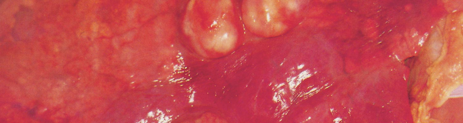
The most common clinical manifestation of disease caused by R. equi infections in foals is a chronic suppurative bronchopneumonia with extensive abscessation (Figure 1). The slow spread of the lung infection coupled with the ability of foals to compensate for the progressive loss of functional lung make early clinical diagnosis difficult.46 Early clinical findings of a slight increase in respiratory rate and a mild fever may be either missed or ignored, allowing the condition to progress. Therefore, even in the chronic form of the disease, respiratory signs are often of apparently acute onset. If pneumonia progresses, clinical signs may include decreased appetite, lethargy, fever, cough, tachypnea, and labored breathing characterized by nostril flaring and increased abdominal effort. Bilateral nasal discharge is an inconsistent finding. In a report of 161 foals affected with R. equi pneumonia, the most common clinical signs were cough (71 per cent), fever (68 per cent), lethargy (53 per cent), and increased respiratory effort (43 per cent).28 A smaller percentage of affected foals present with a more devastating subacute form. These foals may be found dead or, more commonly, present in acute respiratory distress with a high fever and no previous history of clinical respiratory disease, and often die within several days despite aggressive treatment. At necropsy, this subacute form of the disease is characterized by the presence of a diffuse miliary pyogranulomatous pneumonia and the nature of the lesions suggests extensive lung involvement before recognition of the clinical signs.102 On auscultation, the lung sounds of foals affected by both the chronic or subacute form of the disease vary considerably and the findings can be confused by referred sounds from the upper airway.
Because ultrasonographic screening for early detection of pneumonia caused by R. equi has become routine practice at some endemic farms, the most frequently recognized manifestation of R. equi infection at those farms is a subclinical form in which foals develop peripheral pulmonary consolidation or abscessation without manifesting clinical signs.166 At those farms, the cumulative frequency of pulmonary lesions considerably exceeds the historical frequency of clinical pneumonia attributed to R. equi, indicating that many foals with mild subclinical disease recover spontaneously without therapy. In adult horses, the disease caused by R. equi is rare but there are sporadic reports of infection involving primarily the lungs or abdominal lymph nodes (similar to infection observed in foals) and, less common, of wound infection.
Extrapulmonary manifestations of rhodococcal infections are common. In a retrospective study of 150 foals with R. equi infections, at least 1 of 39 different extrapulmonary disorders (EPDs) were recognized in 74 per cent of foals.124 Survival was significantly lower in foals with EPDs (43 per cent; 48/111) relative to foals without EPDs (82 per cent; 32/39).124 This must be interpreted with caution because some EPDs, particularly abdominal lesions, can be difficult to recognize antemortem and were recognized only during necropsy.124 Some EPDs (e.g. polysynovitis, uveitis) might be the first clinical manifestation of R. equi-associated disease detected prior to clinical signs of pneumonia.
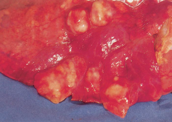
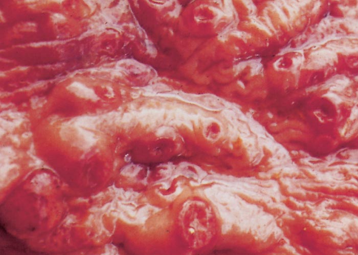
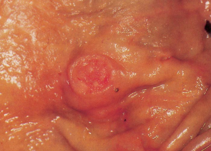
Intestinal lesions are present in approximately 50 per cent of foals with R. equi pneumonia presented for necropsy.179 Clinical signs associated with the abdominal form of the disease may include fever, depression, anorexia, weight loss, colic, and diarrhea.124 Diarrhea might occur in foals infected with R. equi either as an EPD caused by pyogranulomatous typhlocolitis or as a result of antimicrobial treatment. However, the majority of foals with R. equi pneumonia do not show clinical signs of intestinal disease. The intestinal form of R. equi infection is characterized by a multifocal ulcerative enterocolitis (Figures 2 and 3) and typhlitis over the area of the Peyer’s patches with granulomatous or suppurative inflammation of the mesenteric and/or colonic lymph nodes.179 In some cases, the only abdominal lesion noted may be a single large abscess (usually in a mesenteric lymph node) that often adheres to the large or small bowel.
Polysynovitis, characterized by effusion of multiple synovial structures and absence of lameness, occurs in approximately one fourth to one third of foals with naturally acquired R. equi infections.124, 143 Uveitis has also been described in foals naturally infected with R. equi.124 Foals with uveitis might display epiphora, photophobia, aqueous flare, hypopyon, iris discoloration, miosis, and other ophthalmic abnormalities. Experimental infection with virulent R. equi consistently results in polysynovitis and uveitis and development of these conditions is associated with the severity of pulmonary disease.51, 69 Culture of synovial fluid and aqueous humor within days of the onset of clinical signs in experimentally infected foals often yields R. equi and histologic examination of the synovial membrane reveals suppurative inflammation with bacteria.51, 69 These findings demonstrate that polysynovitis and uveitis are typically septic processes resulting from bacteraemia. However, the infection is eventually cleared from the synovial structures and the eye, resulting in nonseptic inflammation at these sites at the time of diagnosis or necropsy in chronic cases. Bacteraemic spread of the organism from the lungs or gastrointestinal tract may occasionally result in severe septic arthritis or vertebral osteomyelitis and/or discospondylitis.29, 53, 115 Other less common EPDs associated with metastatic spread of R. equi include endocarditis, pericarditis, pleuritis, sinusitis, cellulitis, dermatitis, subcutaneous abscesses, peripheral lymphadenopathy, guttural pouch empyema, ulcerative lymphangitis, myositis, stomatitis, omphalitis, and pyometra.76, 124
Diagnosis
It is important not to assume that every coughing foal with an elevated temperature is suffering from R. equi infection. Streptococcus zooepidemicus infection may cause bronchopneumonia in young foals and is far more readily treated than R. equi infection. However, the distinction between lower respiratory tract infections caused by R. equi and those caused by other pathogens is problematic especially at farms without previous history of R. equi infections. Thoracic radiography and ultrasonography are useful in evaluating the severity of pneumonia and in assessing response to therapy. Ultrasonography is also a useful tool for detection of some abdominal abscesses and for screening for R. equi-infected foals on farms where the disease is endemic (see Control). Ultrasonographic or radiographic detection of lung abscesses raises the degree of suspicion that pneumonia in a given foal is caused by R. equi. However, detection of pulmonary abscesses, despite being commonly used as a screening test, is not a definitive diagnostic test.
The definitive diagnosis of bronchopneumonia caused by R. equi should be based on bacteriologic culture or amplification of the vapA gene by polymerase chain reaction (PCR) from a tracheobronchial aspirate obtained from a foal meeting at least one of the following criteria:
- clinical signs of pneumonia,
- septic airway inflammation on cytologic examination, or
- radiographic or ultrasonographic evidence of bronchopneumonia.
If performed, amplification of vapA by PCR should be done in conjunction with bacterial culture because PCR does not permit identification of other bacterial pathogens and in vitro antimicrobial susceptibility testing of R. equi isolates.47 The definitive diagnosis of extrapulmonary infections (e.g. abdominal abscess, osteomyelitis) caused by R. equi must rely on bacteriologic culture or PCR amplification of vapA from samples obtained from the site of infection. The diagnosis of extrapulmonary disorders from sites at which R. equi cannot be detected (e.g. uveitis or polysynovitis) should be based on isolation of R. equi from a TBA or other primary sites of infection. The diagnosis of enterocolitis caused by R. equi is problematic because isolation of R. equi from faeces cannot be taken as evidence of enterocolitis caused by R. equi.47
Currently available serologic tests should not be used for the diagnosis of pneumonia caused by R. equi. Independent studies evaluating the performance of serologic tests available for diagnosis of infection caused by R. equi at endemic farms have demonstrated that these tests have either low sensitivity, low specificity, or both.49, 50, 100, 120
Control
Rhodococcus equi pneumonia is often not recognized until it is well advanced and, therefore, difficult to treat. Even severely affected foals might appear normal to a casual observer. Thus, in the absence of an effective commercially available vaccine or other effective methods for preventing this devastating disease, screening is a reasonable approach to control R. equi at endemic farms. Implementing screening testing at farms that are only sporadically affected by R. equi pneumonia may not be warranted. Over the past decade, control of R. equi infections at many farms where the disease is endemic has relied on early detection of pulmonary disease using thoracic ultrasonography and initiation of therapy before development of clinical disease.166 Thoracic ultrasonography has been reported to decrease mortality due to R. equi pneumonia at some farms relative to historical controls but controlled studies are lacking.166 Historically, antimicrobial treatment was recommended for all foals with pulmonary lesions ≥ 1 cm in diameter.166 The temporal association between widespread use of macrolides and rifampin resulting from ultrasonographic screening and a perceived increase in the frequency of detection of isolates of R. equi resistant to macrolides and rifampin suggest that this practice might not be innocuous. Emergence of widespread macrolide- and rifampin-resistance at a farm after widespread use of these drugs was implemented on the basis of an ultrasonographic screening program has been documented.21 Blinded and randomized, placebo-controlled studies indicate that 80 to 90 per cent of foals with lesion scores (defined as the sum of the largest diameter of all pulmonary lesions ≥ 1cm) between 1 and 10 cm recover without antimicrobial therapy. In addition, antimicrobial treatment does not significantly hasten lesion resolution in these foals compared to administration of a placebo.164, 167 By being more selective and treating only foals with larger lesion scores combined with weekly or twice weekly clinical and ultrasonographic monitoring of the foals, one farm was able to decrease antimicrobial drug use considerably without an increase in mortality.63, 130
Intravenous administration of HIP obtained from horses vaccinated against R. equi using various antigens has generally proved effective in significantly reducing the severity of R. equi pneumonia in foals following experimental challenge.67, 104, 122, 135 However, studies evaluating the efficacy of various HIP preparations under field conditions have given equivocal results. Although the data are conflicting and not all trials have shown a statistically significant reduction in the cumulative incidence of R. equi pneumonia, five of seven studies demonstrated reduction of relative risk, suggesting some benefit of HIP.27, 28, 48, 62, 71, 97 The optimal amount of plasma to be transfused and the optimal age at which transfusion should occur remain to be determined. Because of evidence that many foals become infected early in life,68 it is commonly recommended that foals receive transfusion of at least one litre of commercially produced and licensed HIP no later than the second day of life. Because early administration may result in the decline of passively transferred antibody to a non-protective level at a time when foals might still be susceptible to R. equi and when environmental challenge is high, it is common practice to administer a second litre of HIP at two to four weeks of age. Transfusion of HIP is not completely effective and therefore does not eliminate the need for careful monitoring of foals at risk. Thus, the cost-effectiveness of transfusion depends on the value of the foals and the prevalence of disease at a given farm.
Treatment
Treatment of even severely affected foals is worthwhile, since studies have established that foals that recover from R. equi pneumonia perform in races as well as expected, although foals that contract infection might be slightly less likely to race.2, 3 It is important to consider treatment of subclinically affected foals and foals with clinical disease separately because ultrasonographic lesions in many foals that are clinically healthy will resolve without treatment (see Screening).
Treatment of foals with severe clinical pneumonia
A wide variety of antimicrobial agents are active against R. equi in vitro. However, many of these drugs are reported to be ineffective in vivo, possibly because of poor cellular uptake and resulting low intracellular concentrations. The combination of a macrolide antimicrobial agent with rifampin has been the mainstay of therapy in foals with clinical pneumonia caused by R. equi although randomized clinical trials comparing efficacy of various therapies are lacking. The combination of a macrolide and rifampin is synergistic both in vitro and in vivo, and the use of the two classes of drugs in combination reduces the likelihood of development of resistance to either drug.55, 113, 121 In addition, studies in immunodeficient mice indicate that the combination of a macrolide with rifampin is superior to either drug used in monotherapy.20, 21, 113 The antimicrobial agents commonly used for the treatment of R. equi infections in foals are listed in Table 1. Macrolides and rifampin are highly active against R. equi in vitro but only exert bacteriostatic activity.55, 113 The three macrolides that have been used for the treatment of pneumonia caused by R. equi are erythromycin, clarithromycin and azithromycin. Clarithromycin is the most active against R. equi in vitro and against intracellular R. equi in cell culture.11, 55 The minimum inhibitory concentration at which 90 per cent of R. equi isolates are inhibited (MIC90) is 0.06, 0.5 and 1.0 µg/ml, for clarithromycin, erythromycin, and azithromycin, respectively.74, 125 The combination clarithromycin-rifampin was significantly more effective than erythromycin-rifampin or azithromycin-rifampin in a retrospective study, especially in foals with severe pulmonary lesions.52 Despite data analysis being adjusted for disease severity and age, these results must be interpreted cautiously because of potential biases inherent to retrospective data.
Table 1 Recommended dosages, oral bioavailability and serum half-lives of antimicrobial agents commonly used for the treatment of R. equi infections in foals.
| Class | Drug | Dose (mg/kg) | Route | Frequency | References |
| Macrolides | Erythromycin* | 25 | POa | 6-8 | 86, 87 |
|
| Clarithromycin | 7.5 | PO | 12 | 172 |
|
| Azithromycin | 10 | PO | 24-48b | 39, 73 |
| Rifamycins | Rifampin | 5 | PO | 12 | 19, 24, 83 |
* Erythromycin base, stearate, phosphate, estolate, or ethylsuccinate.
a Oral bioavailability is low unless the foal is fasted
b Administration q 24 h for 5 days and q 48 h thereafter
The most common adverse effect in foals treated with macrolides alone or in combination with rifampin is diarrhoea. Hyperthermia and tachypnoea have been described in foals treated with macrolides during periods of very hot or humid weather.140 Recent studies demonstrate that azithromycin, clarithromycin and erythromycin suppress sweating in foals, with erythromycin inducing more severe sweating dysfunction.137, 138 Another rare complication of macrolides is severe enterocolitis in mares whose foals are treated with erythromycin, presumably by disrupting the mare's normal colonic microflora following ingestion of small amounts of active drug during coprophagia or by contamination of feeders or water buckets with drug present on the foal's muzzle.7
Foals with mild to moderate subclinical lesions
Only studies enrolling foals with lesions scores ≥ 10 cm (see Screening) have documented a significant difference in the rate of recovery between foals treated with antimicrobials and a placebo group.63, 165 In two separate, blinded, placebo-controlled studies, azithromycin alone (10 mg/kg PO q 24 h) or gamithromycin alone (6 mg/kg IM q 7 days) were non-inferior to azithromycin with rifampin. 63, 165 In another study, therapy with tulathromycin was more effective than a placebo but not as effective as the combination of azithromycin with rifampin.130 It is important to note that up to 78 per cent of the foals receiving the placebo in the aforementioned studies recovered without the need for antimicrobial treatment.63 Therefore, the apparent efficacy of macrolide monotherapy in foals with mild lesions should not be interpreted as evidence of similar efficacy in foals with severe pneumonia. It is also unknown if macrolide monotherapy is more likely to select for resistance in vivo than combination therapy.
Emergence of resistance to macrolides and rifampin
Although resistance has increased in recent years, the vast majority of R. equi isolates from foals are still highly susceptible to macrolides and rifampin in vitro. Failure to respond to antimicrobial therapy does not provide evidence that a given foal is infected with an isolate resistant to antimicrobial agents. Therefore, the presence of an isolate resistant to macrolides or rifampin can only be confirmed by in vitro susceptibility testing done by a laboratory following the criteria established by the Clinical and Laboratory Standards Institute. In a recent study, the overall prevalence of macrolide and rifampin-resistant isolates in Texas and Florida over a 10-year-period was 4 per cent and the odds of death were seven times higher in foals infected with resistant isolates compared with foals infected with susceptible isolates.54 More recently, it was documented that mass antimicrobial treatment of subclinically affected foals selected for antimicrobial resistance over time with isolates of R. equi resistant to all macrolides and rifampin being cultured from as many as 40 per cent of the foals at some farms.21 Macrolide resistance in R. equi is conferred by acquisition of a rRNA methylase gene designated erm(46). This gene confers resistance to all macrolides, lincosamides, and streptogramins type B, but not to other classes of antimicrobial agents. The gene can be transferred by conjugation from resistant to susceptible isolates of R. equi.4 Most macrolide resistant isolates of R. equi identified to date are also resistant to rifampin. Resistance to rifampin in R. equi is the result of mutations in the rpoB gene.5, 43, 125, 157 Therapy of foals infected with isolates confirmed to be resistant to macrolides, rifampin, or both is problematic because of the limited range of effective alternatives. Macrolide- and rifampin-resistant isolates of R. equi are susceptible in vitro to fluoroquinolones, gentamicin, linezolid, and vancomycin.10, 22, 54 In one study, 18 of 24 isolates were also susceptible to chloramphenicol, minocycline, and trimethoprim-sulfamethoxazole.54 Currently, there are no data to indicate the preferred antimicrobial agent(s) for the treatment of foals infected with resistant isolates. Vancomycin, imipenem or linezolid in foals should be used only for the treatment of life threatening R. equi infections caused by isolates confirmed to be resistant to all other possible alternatives. The use of systemic and/or nebulized gentamicin is a reasonable consideration for foals infected with macrolide- and rifampin-resistant isolates.56
Duration of therapy and prognosis
Resolution of clinical signs and radiographic or ultrasonographic resolution of lung lesions are commonly used to guide the duration of therapy which generally ranges between two and eight weeks, depending on the severity of the initial lesions and response to therapy. Foals treated based on subclinical lesions identified during ultrasonographic screening typically require a shorter treatment duration. In fact, as addressed below, many foals with subclinical ultrasonographic lesions clear the infection without therapy.164, 167
The prognosis of R. equi-infected foals was poor prior to the introduction of the combination of erythromycin and rifampin as the treatment of choice in the early 1980s, with survival rates as low as 20 per cent.41 Using erythromycin and rifampin, Hillidge reported a successful outcome in 50 (88 per cent) of 57 foals with confirmed R. equi pneumonia.64 Studies at various referral centres have shown survival proportions ranging between 59 and 72 per cent.3, 52, 124 The prognosis for foals with abdominal abscesses is poor, although rare cases have responded to prolonged antimicrobial therapy.124, 161
Currently, there is inadequate evidence to recommend active immunization of mares or foals to control or prevent R. equi pneumonia but there are promising vaccine candidates under development.
References
- ADKINS, B., LECLERC, C. & MARSHALL-CLARKE, S., 2004. Neonatal adaptive immunity comes of age. Nature Reviews Immunology, 4, 553-64.
- AINSWORTH, D. M., BECK, K. A., BOATWRIGHT, C. E., SNEDDEN, K. A. & REBHUN, W. C., 1993. Lack of residual lung damage in horses in which Rhodococcus equi-induced pneumonia had been diagnosed. American Journal of Veterinary Research, 54, 2115-20.
- AINSWORTH, D. M., EICKER, S. W., YEAGAR, A. E., SWEENEY, C. R., VIEL, L., TESAROWSKI, D., LAVOIE, J. P., HOFFMAN, A., PARADIS, M. R., REED, S. M., ERB, H. N., DAVIDOW, E. & NALEVANKO, M., 1998. Associations between physical examination, laboratory, and radiographic findings and outcome and subsequent racing performance of foals with Rhodococcus equi infection: 115 cases (1984-1992). Journal of the American Veterinary Medical Association, 213, 510-5.
- ANASTASI, E., GIGUERE, S., BERGHAUS, L. J., HONDALUS, M. K., WILLINGHAM-LANE, J. M., MACARTHUR, I., COHEN, N. D., ROBERTS, M. C. & VASQUEZ-BOLAND, J. A., 2016. Novel transferable erm(46) determinant responsible for emerging macrolide resistance in Rhodococcus equi. Journal of Antimicrobial Chemotherapy, 71, 1746.
- ASOH, N., WATANABE, H., FINES-GUYON, M., WATANABE, K., OISHI, K., KOSITSAKULCHAI, W., SANCHAI, T., KUNSUIKMENGRAI, K., KAHINTAPONG, S., KHANTAWA, B., THARAVICHITKUL, P., SIRISANTHANA, T. & NAGATAKE, T., 2003. Emergence of rifampin-resistant Rhodococcus equi with several types of mutations in the rpoB gene among AIDS patients in northern Thailand. Journal of Clinical Microbiology, 41, 2337-2340.
- BARTON, M. D. & EMBURY, D. H., 1987. Studies of the pathogenesis of Rhodococcus equi infection in foals. Australian Veterinary Journal, 64, 332-9.
- BAVERUD, V., FRANKLIN, A., GUNNARSSON, A., GUSTAFSSON, A. & HELLANDER-EDMAN, A., 1998. Clostridium difficile associated with acute colitis in mares when their foals are treated with erythromycin and rifampicin for Rhodococcus equi pneumonia. Equine Veterinary Journal, 30, 482-8.
- BELL, K. S., PHILP, J. C., CHRISTOFI, N. & AW, D. W., 1996. Identification of Rhodococcus equi using the polymerase chain reaction. Lett Appl Microbiol, 23, 72-4.
- BERGHAUS, L. J., GIGUERE, S., BORDIN, A. I. & COHEN, N. D., 2018. Effects of priming with cytokines on intracellular survival and replication of Rhodococcus equi in equine macrophages. Cytokine, 102, 7-11.
- BERGHAUS, L. J., GIGUERE, S., GULDBECH, K., WARNER, E., UGORJI, U. & BERGHAUS, R. D., 2015. Comparison of Etest, disk diffusion, and broth macrodilution for in vitro susceptibility testing of Rhodococcus equi. Journal of Clinical Microbiology, 53, 314-8.
- BERGHAUS, L. J., GIGUERE, S., STURGILL, T. L., BADE, D., MALINSKI, T. J. & HUANG, R., 2012. Plasma pharmacokinetics, pulmonary distribution, and in vitro activity of gamithromycin in foals. The Journal of Veterinary Pharmacology and Therapeutics, 35, 59-66.
- BIDAUD, P., HEBERT, L., BARBEY, C., APPOURCHAUX, A. C., TORELLI, R., SANGUINETTI, M., LAUGIER, C. & PETRY, S., 2012. Rhodococcus equi's extreme resistance to hydrogen peroxide is mainly conferred by one of its four catalase genes. PLoS One, 7, e42396.
- BOLAND, C. A. & MEIJER, W. G., 2000. The iron dependent regulatory protein IdeR (DtxR) of Rhodococcus equi. FEMS Microbiology Letters, 191, 1-5.
- BOLTON, T., KUSKIE, K., HALBERT, N., CHAFFIN, K., HEALY, M., LAWHON, S., JACKSON, A. & COHEN, N., 2010. Detection of strain variation in isolates of Rhodococcus equi from an affected foal using repetitive sequence-based polymerase chain reaction. The Journal of Veterinary Diagnostic Investigation, 22, 611-5.
- BOYD, N. K., COHEN, N. D., LIM, W. S., MARTENS, R. J., CHAFFIN, M. K. & BALL, J. M., 2003. Temporal changes in cytokine expression of foals during the first month of life. Veterinary Immunology and Immunopathology, 92, 75-85.
- BREATHNACH, C. C., STURGILL-WRIGHT, T., STILTNER, J. L., ADAMS, A. A., LUNN, D. P. & HOROHOV, D. W., 2006. Foals are interferon gamma-deficient at birth. Veterinary Immunology and Immunopathology, 112, 199-209.
- BRYAN, L. K., ALEXANDER, E. R., LAWHON, S. D. & COHEN, N. D., 2018. Detection of vapN in Rhodococcus equi isolates cultured from humans. PLoS One, 13, e0190829.
- BRYAN, L. K., CLARK, S. D., DIAZ-DELGADO, J., LAWHON, S. D. & EDWARDS, J. F., 2017. Rhodococcus equi Infections in Dogs. Veterinary Pathology, 54, 159-163.
- BURROWS, G. E., MACALLISTER, C. G., EWING, P., STAIR, E. & TRIPP, P. W., 1992. Rifampin disposition in the horse: effects of age and method of oral administration. The Journal of Veterinary Pharmacology and Therapeutics, 15, 124-32.
- BURTON, A. J., GIGUERE, S., BERGHAUS, L. J. & HONDALUS, M. K., 2015. Activity of clarithromycin or rifampin alone or in combination against experimental Rhodococcus equi infection in mice. Antimicrob Agents Chemother, 59, 3633-6.
- BURTON, A. J., GIGUERE, S., STURGILL, T. L., BERGHAUS, L. J., SLOVIS, N. M., WHITMAN, J. L., LEVERING, C., KUSKIE, K. R. & COHEN, N. D., 2013. Macrolide- and rifampin-resistant Rhodococcus equi on a horse breeding farm, Kentucky, USA. Emerging Infectious Diseases, 19, 282-5.
- CARLSON, K. L., KUSKIE, K. R., CHAFFIN, K. M., LIBAL, M. C., GIGUERE, S., LAWHON, S. D. & COHEN, N. D., 2010. Antimicrobial activity of tulathromycin and 14 other antimicrobials against virulent Rhodococcus equi in vitro. The Veterinary Therapeutics, 11, E1-9.
- CASTON, S. S., MCCLURE, S. R., MARTENS, R. J., CHAFFIN, M. K., MILES, K. G., GRIFFITH, R. W. & COHEN, N. D., 2006. Effect of hyperimmune plasma on the severity of pneumonia caused by Rhodococcus equi in experimentally infected foals. The Veterinary Therapeutics, 7, 361-75.
- CASTRO, L. A., BROWN, M. P., GRONWALL, R., HOUSTON, A. E. & MILES, N., 1986. Pharmacokinetics of rifampin given as a single oral dose in foals. American Journal of Veterinary Research, 47, 2584-6.
- CHAFFIN, M. K., COHEN, N. D. & MARTENS, R. J., 2003. Evaluation of equine breeding farm characteristics as risk factors for development of Rhodococcus equi pneumonia in foals. Journal of the American Veterinary Medical Association, 222, 467-75.
- CHAFFIN, M. K., COHEN, N. D. & MARTENS, R. J., 2003. Evaluation of equine breeding farm management and preventative health practices as risk factors for development of Rhodococcus equi pneumonia in foals. Journal of the American Veterinary Medical Association, 222, 476-85.
- CHAFFIN, M. K., COHEN, N. D., MARTENS, R. J., EDWARDS, R. F. & NEVILL, M., 2003. Foal-related risk factors associated with development of Rhodococcus equi pneumonia on farms with endemic infection. Journal of the American Veterinary Medical Association, 223, 1791-9.
- CHAFFIN, M. K., COHEN, N. D., MARTENS, R. J., O'CONOR, M. & BERNSTEIN, L. R., 2011. Evaluation of the efficacy of gallium maltolate for chemoprophylaxis against pneumonia caused by Rhodococcus equi infection in foals. American Journal of Veterinary Research, 72, 945-57.
- CHAFFIN, M. K., HONNAS, C. M., CRABILL, M. R., SCHNEITER, H. L., BRUMBAUGH, G. W. & BRINER, R. P., 1995. Cauda equina syndrome, diskospondylitis, and a paravertebral abscess caused by Rhodococcus equi in a foal. Journal of the American Veterinary Medical Association, 206, 215-20.
- CHICKEN, C., MUSCATELLO, G., FREESTONE, J., ANDERSON, G. A., BROWNING, G. F. & GILKERSON, J. R., 2012. Air sampling in the breathing zone of neonatal foals for prediction of subclinical Rhodococcus equi infection. Equine Veterinary Journal, 44, 203-6.
- CHIRINO-TREJO, J. M., PRESCOTT, J. F. & YAGER, J. A., 1987. Protection of foals against experimental Rhodococcus equi pneumonia by oral immunization. Canadian Journal of Veterinary Research, 51, 444-7.
- COHEN, N. D., CARTER, C. N., SCOTT, H. M., CHAFFIN, M. K., SMITH, J. L., GRIMM, M. B., KUSKIE, K. R., TAKAI, S. & MARTENS, R. J., 2008. Association of soil concentrations of Rhodococcus equi and incidence of pneumonia attributable to Rhodococcus equi in foals on farms in central Kentucky. American Journal of Veterinary Research, 69, 385-95.
- COHEN, N. D., CHAFFIN, M. K., KUSKIE, K. R., SYNDERGAARD, M. K., BLODGETT, G. P. & TAKAI, S., 2013. Association of perinatal exposure to airborne Rhodococcus equi with risk of pneumonia caused by R equi in foals. American Journal of Veterinary Research, 74, 102-9.
- COHEN, N. D., KUSKIE, K. R., SMITH, J. L., SLOVIS, N. M., BROWN, S. E., 2ND, STEPUSIN, R. S., CHAFFIN, M. K., TAKAI, S. & CARTER, C. N., 2012. Association of airborne concentration of virulent Rhodococcus equi with location (stall versus paddock) and month (January through June) on 30 horse breeding farms in central Kentucky. American Journal of Veterinary Research, 73, 1603-9.
- COHEN, N. D., O'CONOR, M. S., CHAFFIN, M. K. & MARTENS, R. J., 2005. Farm characteristics and management practices associated with development of Rhodococcus equi pneumonia in foals. Journal of the American Veterinary Medical Association, 226, 404-13.
- COHEN, N. D., SMITH, K. E., FICHT, T. A., TAKAI, S., LIBAL, M. C., WEST, B. R., DELROSARIO, L. S., BECU, T., LEADON, D. P., BUCKLEY, T., CHAFFIN, M. K. & MARTENS, R. J., 2003. Epidemiologic study of results of pulsed-field gel electrophoresis of isolates of Rhodococcus equi obtained from horses and horse farms. American Journal of Veterinary Research, 64, 153-61.
- COULSON, G. B., AGARWAL, S. & HONDALUS, M. K., 2010. Characterization of the role of the pathogenicity island and vapG in the virulence of the intracellular actinomycete pathogen Rhodococcus equi. Infection and Immunity, 78, 3323-34.
- DARRAH, P. A., HONDALUS, M. K., CHEN, Q., ISCHIROPOULOS, H. & MOSSER, D. M., 2000. Cooperation between reactive oxygen and nitrogen intermediates in killing of Rhodococcus equi by activated macrophages. Infection and Immunity, 68, 3587-93.
- DAVIS, J. L., GARDNER, S. Y., JONES, S. L., SCHWABENTON, B. A. & PAPICH, M. G., 2002. Pharmacokinetics of azithromycin in foals after i.v. and oral dose and disposition into phagocytes. The Journal of Veterinary Pharmacology and Therapeutics, 25, 99-104.
- DAWSON, D. R., NYDAM, D. V., PRICE, C. T., GRAHAM, J. E., CYNAMON, M. H., DIVERS, T. J. & FELIPPE, M. J., 2011. Effects of opsonization of Rhodococcus equi on bacterial viability and phagocyte activation. American Journal of Veterinary Research, 72, 1465-75.
- ELISSALDE, G. S., RENSHAW, H. W. & WALBERG, J. A., 1980. Corynebacterium equi: an interhost review with emphasis on the foal. Comparative Immunology, Microbiology & Infectious Diseases, 3, 433-45.
- FERNANDEZ-MORA, E., POLIDORI, M., LUHRMANN, A., SCHAIBLE, U. E. & HAAS, A., 2005. Maturation of Rhodococcus equi-containing vacuoles is arrested after completion of the early endosome stage. Traffic, 6, 635-53.
- FINES, M., PRONOST, S., MAILLARD, K., TAOUJI, S. & LECLERCQ, R., 2001. Characterization of mutations in the rpoB gene associated with rifampin resistance in Rhodococcus equi isolated from foals. Journal of Clinical Microbiology, 39, 2784-7.
- FLAMINIO, M. J., NYDAM, D. V., MARQUIS, H., MATYCHAK, M. B. & GIGUERE, S., 2009. Foal monocyte-derived dendritic cells become activated upon Rhodococcus equi infection. Clinical and Vaccine Immunology, 16, 176-83.
- GARTON, N. J., GILLERON, M., BRANDO, T., DAN, H. H., GIGUERE, S., PUZO, G., PRESCOTT, J. F. & SUTCLIFFE, I. C., 2002. A novel lipoarabinomannan from the equine pathogen Rhodococcus equi. Structure and effect on macrophage cytokine production. Journal of Biological Chemistry, 277, 31722-33.
- GIGUERE, S., COHEN, N. D., CHAFFIN, M. K., HINES, S. A., HONDALUS, M. K., PRESCOTT, J. F. & SLOVIS, N. M., 2011. Rhodococcus equi: clinical manifestations, virulence, and immunity. Journal of Veterinary Internal Medicine, 25, 1221-30.
- GIGUERE, S., COHEN, N. D., CHAFFIN, M. K., SLOVIS, N. M., HONDALUS, M. K., HINES, S. A. & PRESCOTT, J. F., 2011. Diagnosis, treatment, control, and prevention of infections caused by Rhodococcus equi in foals. Journal of Veterinary Internal Medicine, 25, 1209-20.
- GIGUERE, S., GASKIN, J. M., MILLER, C. & BOWMAN, J. L., 2002. Evaluation of a commercially available hyperimmune plasma product for prevention of naturally acquired pneumonia caused by Rhodococcus equi in foals. Journal of the American Veterinary Medical Association, 220, 59-63.
- GIGUERE, S., HERNANDEZ, J., GASKIN, J., MILLER, C. & BOWMAN, J. L., 2003. Evaluation of white blood cell concentration, plasma fibrinogen concentration, and an agar gel immunodiffusion test for early identification of foals with Rhodococcus equi pneumonia. Journal of the American Veterinary Medical Association, 222, 775-81.
- GIGUERE, S., HERNANDEZ, J., GASKIN, J., PRESCOTT, J. F., TAKAI, S. & MILLER, C., 2003. Performance of five serological assays for diagnosis of Rhodococcus equi pneumonia in foals. Clinical and Diagnostic Laboratory Immunology, 10, 241-5.
- GIGUERE, S., HONDALUS, M. K., YAGER, J. A., DARRAH, P., MOSSER, D. M. & PRESCOTT, J. F., 1999. Role of the 85-kilobase plasmid and plasmid-encoded virulence-associated protein A in intracellular survival and virulence of Rhodococcus equi. Infection and Immunity, 67, 3548-57.
- GIGUERE, S., JACKS, S., ROBERTS, G. D., HERNANDEZ, J., LONG, M. T. & ELLIS, C., 2004. Retrospective comparison of azithromycin, clarithromycin, and erythromycin for the treatment of foals with Rhodococcus equi pneumonia. Journal of Veterinary Internal Medicine, 18, 568-73.
- GIGUERE, S. & LAVOIE, J. P., 1994. Rhodococcus equi vertebral osteomyelitis in 3 quarter horse colts. Equine Veterinary Journal, 26, 74-7.
- GIGUERE, S., LEE, E., WILLIAMS, E., COHEN, N. D., CHAFFIN, M. K., HALBERT, N., MARTENS, R. J., FRANKLIN, R. P., CLARK, C. C. & SLOVIS, N. M., 2010. Determination of the prevalence of antimicrobial resistance to macrolide antimicrobials or rifampin in Rhodococcus equi isolates and treatment outcome in foals infected with antimicrobial-resistant isolates of R equi. Journal of the American Veterinary Medical Association, 237, 74-81.
- GIGUERE, S., LEE, E. A., GULDBECH, K. M. & BERGHAUS, L. J., 2012. In vitro synergy, pharmacodynamics, and postantibiotic effect of 11 antimicrobial agents against Rhodococcus equi. Veterinary Microbiology, 160, 207-13.
- GIGUERE, S. C., ND., 2018. Controversies in therapy of infections caused by Rhodococcus equi in foals. Equine Veterinary Education, 30, 336-341.
- GOTOH, K., MITSUYAMA, M., IMAIZUMI, S., KAWAMURA, I. & YANO, I., 1991. Mycolic acid-containing glycolipid as a possible virulence factor of Rhodococcus equi for mice. Microbiol Immunol, 35, 175-85.
- HARRIS, S. P., FUJIWARA, N., MEALEY, R. H., ALPERIN, D. C., NAKA, T., GODA, R. & HINES, S. A., 2010. Identification of Rhodococcus equi lipids recognized by host cytotoxic T lymphocytes. Microbiology, 156, 1836-47.
- HARRIS, S. P., HINES, M. T., MEALEY, R. H., ALPERIN, D. C. & HINES, S. A., 2011. Early development of cytotoxic T lymphocytes in neonatal foals following oral inoculation with Rhodococcus equi. Veterinary Immunology and Immunopathology, 141, 312-6.
- HIETALA, S. K. & ARDANS, A. A., 1987. Interaction of Rhodococcus equi with phagocytic cells from R. equi-exposed and non-exposed foals. Veterinary Microbiology, 14, 307-20.
- HIETALA, S. K. & ARDANS, A. A., 1987. Neutrophil phagocytic and serum opsonic response of the foal to Corynebacterium equi. Veterinary Immunology and Immunopathology, 14, 279-94.
- HIGUCHI, T., ARAKAWA, T., HASHIKURA, S., INUI, T., SENBA, H. & TAKAI, S., 1999. Effect of prophylactic administration of hyperimmune plasma to prevent Rhodococcus equi infection on foals from endemically affected farms. Zentralbl Veterinarmed B, 46, 641-8.
- HILDEBRAND, F., VENNER, M. & GIGUERE, S., 2015. Efficacy of gamithromycin for the treatment of foals with mild to moderate bronchopneumonia. Journal of Veterinary Internal Medicine, 29, 333-8.
- HILLIDGE, C. J., 1987. Use of erythromycin-rifampin combination in treatment of Rhodococcus equi pneumonia. Veterinary Microbiology, 14, 337-42.
- HINES, M. T., PAASCH, K. M., ALPERIN, D. C., PALMER, G. H., WESTHOFF, N. C. & HINES, S. A., 2001. Immunity to Rhodococcus equi: antigen-specific recall responses in the lungs of adult horses. Veterinary Immunology and Immunopathology, 79, 101-14.
- HONDALUS, M. K., DIAMOND, M. S., ROSENTHAL, L. A., SPRINGER, T. A. & MOSSER, D. M., 1993. The intracellular bacterium Rhodococcus equi requires Mac-1 to bind to mammalian cells. Infection and Immunity, 61, 2919-29.
- HOOPER-MCGREVY, K. E., WILKIE, B. N. & PRESCOTT, J. F., 2005. Virulence-associated protein-specific serum immunoglobulin G-isotype expression in young foals protected against Rhodococcus equi pneumonia by oral immunization with virulent R-equi. Vaccine, 23, 5760-5767.
- HOROWITZ, M. L., COHEN, N. D., TAKAI, S., BECU, T., CHAFFIN, M. K., CHU, K. K., MAGDESIAN, K. G. & MARTENS, R. J., 2001. Application of Sartwell's model (lognormal distribution of incubation periods) to age at onset and age at death of foals with Rhodococcus equi pneumonia as evidence of perinatal infection. Journal of Veterinary Internal Medicine, 15, 171-5.
- HUBER, L., GIGUERE, S., BERGHAUS, L. J., HANAFI, A., VITOSH-SILLMAN, S. & CZERWINSKI, S. L., 2018. Development of septic polysynovitis and uveitis in foals experimentally infected with Rhodococcus equi. PLoS One, 13, e0192655.
- HUGHES, K. L. & SULAIMAN, I., 1987. The ecology of Rhodococcus equi and physicochemical influences on growth. Veterinary Microbiology, 14, 241-50.
- HURLEY, J. R. & BEGG, A. P., 1995. Failure of hyperimmune plasma to prevent pneumonia caused by Rhodococcus equi in foals. Australian Veterinary Journal, 72, 418-20.
- JACKS, S., GIGUERE, S., CRAWFORD, P. C. & CASTLEMAN, W. L., 2007. Experimental infection of neonatal foals with Rhodococcus equi triggers adult-like gamma interferon induction. Clinical and Vaccine Immunology, 14, 669-77.
- JACKS, S., GIGUERE, S., GRONWALL, P. R., BROWN, M. P. & MERRITT, K. A., 2001. Pharmacokinetics of azithromycin and concentration in body fluids and bronchoalveolar cells in foals. American Journal of Veterinary Research, 62, 1870-5.
- JACKS, S. S., GIGUERE, S. & NGUYEN, A., 2003. In vitro susceptibilities of Rhodococcus equi and other common equine pathogens to azithromycin, clarithromycin, and 20 other antimicrobials. Antimicrob Agents Chemother, 47, 1742-5.
- JAIN, S., BLOOM, B. R. & HONDALUS, M. K., 2003. Deletion of vapA encoding Virulence Associated Protein A attenuates the intracellular actinomycete Rhodococcus equi. Molecular Microbiology, 50, 115-28.
- JANICEK, J. C., KRAMER, J., COATES, J. R., LATTIMER, J. C., LACARRUBBA, A. M. & MESSER, N. T., 2006. Intracranial abscess caused by Rhodococcus equi infection in a foal. Journal of the American Veterinary Medical Association, 228, 251-3.
- JOHNSON, J. A., PRESCOTT, J. F. & MARKHAM, R. J., 1983. The pathology of experimental Corynebacterium equi infection in foals following intragastric challenge. Veterinary Pathology, 20, 450-9.
- KAKUDA, T., HIROTA, T., TAKEUCHI, T., HAGIUDA, H., MIYAZAKI, S. & TAKAI, S., 2014. VirS, an OmpR/PhoB subfamily response regulator, is required for activation of vapA gene expression in Rhodococcus equi. BMC Microbiology, 14, 243.
- KANALY, S. T., HINES, S. A. & PALMER, G. H., 1993. Failure of pulmonary clearance of Rhodococcus equi infection in CD4+ T-lymphocyte-deficient transgenic mice. Infection and Immunity, 61, 4929-32.
- KANALY, S. T., HINES, S. A. & PALMER, G. H., 1995. Cytokine modulation alters pulmonary clearance of Rhodococcus equi and development of granulomatous pneumonia. Infection and Immunity, 63, 3037-41.
- KANALY, S. T., HINES, S. A. & PALMER, G. H., 1996. Transfer of a CD4+ Th1 cell line to nude mice effects clearance of Rhodococcus equi from the lung. Infection and Immunity, 64, 1126-32.
- KASUGA-AOKI, H., TAKAI, S., SASAKI, Y., TSUBAKI, S., MADARAME, H. & NAKANE, A., 1999. Tumour necrosis factor and interferon-gamma are required in host resistance against virulent Rhodococcus equi infection in mice: cytokine production depends on the virulence levels of R. equi. Immunology, 96, 122-7.
- KOHN, C. W., SAMS, R., KOWALSKI, J. J., POWERS, J. & WALLACE, S., 1993. Pharmacokinetics of single intravenous and single and multiple dose oral administration of rifampin in mares. The Journal of Veterinary Pharmacology and Therapeutics, 16, 119-31.
- KUSKIE, K. R., SMITH, J. L., WANG, N., CARTER, C. N., CHAFFIN, M. K., SLOVIS, N. M., STEPUSIN, R. S., CATTOI, A. E., TAKAI, S. & COHEN, N. D., 2011. Effects of location for collection of air samples on a farm and time of day of sample collection on airborne concentrations of virulent Rhodococcus equi at two horse breeding farms. American Journal of Veterinary Research, 72, 73-9.
- LADRON, N., FERNANDEZ, M., AGUERO, J., GONZALEZ ZORN, B., VAZQUEZ-BOLAND, J. A. & NAVAS, J., 2003. Rapid identification of Rhodococcus equi by a PCR assay targeting the choE gene. Journal of Clinical Microbiology, 41, 3241-5.
- LAKRITZ, J., WILSON, W. D., MARSH, A. E. & MIHALYI, J. E., 2000. Effects of prior feeding on pharmacokinetics and estimated bioavailability after oral administration of a single dose of microencapsulated erythromycin base in healthy foals. American Journal of Veterinary Research, 61, 1011-5.
- LAKRITZ, J., WILSON, W. D., MARSH, A. E. & MIHALYI, J. E., 2000. Pharmacokinetics of erythromycin estolate and erythromycin phosphate after intragastric administration to healthy foals. American Journal of Veterinary Research, 61, 914-9.
- LETEK, M., et al., 2010. The genome of a pathogenic rhodococcus: cooptive virulence underpinned by key gene acquisitions. PLoS Genet, 6, e1001145.
- LETEK, M., OCAMPO-SOSA, A. A., SANDERS, M., FOGARTY, U., BUCKLEY, T., LEADON, D. P., GONZALEZ, P., SCORTTI, M., MEIJER, W. G., PARKHILL, J., BENTLEY, S. & VAZQUEZ-BOLAND, J. A., 2008. Evolution of the Rhodococcus equi vap pathogenicity island seen through comparison of host-associated vapA and vapB virulence plasmids. Journal of Bacteriology, 190, 5797-805.
- LINDER, R. & BERNHEIMER, A. W., 1997. Oxidation of macrophage membrane cholesterol by intracellular Rhodococcus equi. Veterinary Microbiology, 56, 269-76.
- LIU, M., BORDIN, A., LIU, T., RUSSELL, K. & COHEN, N., 2011. Gene expression of innate Th1-, Th2-, and Th17-type cytokines during early life of neonatal foals in response to Rhodococcus equi. Cytokine, 56, 356-64.
- LIU, T., NERREN, J., LIU, M., MARTENS, R. & COHEN, N., 2009. Basal and stimulus-induced cytokine expression is selectively impaired in peripheral blood mononuclear cells of newborn foals. Vaccine, 27, 674-83.
- LOPEZ, A. M., HINES, M. T., PALMER, G. H., ALPERIN, D. C. & HINES, S. A., 2002. Identification of pulmonary T-lymphocyte and serum antibody isotype responses associated with protection against Rhodococcus equi. Clinical and Diagnostic Laboratory Immunology, 9, 1270-6.
- LUHRMANN, A., MAUDER, N., SYDOR, T., FERNANDEZ-MORA, E., SCHULZE-LUEHRMANN, J., TAKAI, S. & HAAS, A., 2004. Necrotic death of Rhodococcus equi-infected macrophages is regulated by virulence-associated plasmids. Infection and Immunity, 72, 853-62.
- MACARTHUR, I., PARREIRA, V. R., LEPP, D., MUTHARIA, L. M., VAZQUEZ-BOLAND, J. A. & PRESCOTT, J. F., 2011. The sensor kinase MprB is required for Rhodococcus equi virulence. Veterinary Microbiology, 147, 133-41.
- MADARAME, H., TAKAI, S., MATSUMOTO, C., MINAMIYAMA, K., SASAKI, Y., TSUBAKI, S., HASEGAWA, Y. & NAKANE, A., 1997. Virulent and avirulent Rhodococcus equi infection in T-cell deficient athymic nude mice: pathologic, bacteriologic and immunologic responses. FEMS Immunology and Medical Microbiology, 17, 251-62.
- MADIGAN, J. E., HIETALA, S. & MULLER, N., 1991. Protection against naturally acquired Rhodococcus equi pneumonia in foals by administration of hyperimmune plasma. Journal of reproduction and fertility, 44, 571-8.
- MAKRAI, L., FODOR, L., VENDEG, I., SZIGETI, G., DENES, B., REICZIGEL, J. & VARGA, J., 2005. Comparison of selective media for the isolation of Rhodococcus equi and description of a new selective plating medium. Acta Veterinaria Hungarica, 53, 275-85.
- MARTENS, J. G., MARTENS, R. J. & RENSHAW, H. W., 1988. Rhodococcus (Corynebacterium) equi: bactericidal capacity of neutrophils from neonatal and adult horses. American Journal of Veterinary Research, 49, 295-9.
- MARTENS, R. J., COHEN, N. D., CHAFFIN, M. K., TAKAI, S., DOHERTY, C. L., ANGULO, A. B. & EDWARDS, R. E., 2002. Evaluation of 5 serologic assays to detect Rhodococcus equi pneumonia in foals. Journal of the American Veterinary Medical Association, 221, 825-33.
- MARTENS, R. J., COHEN, N. D., JONES, S. L., MOORE, T. A. & EDWARDS, J. F., 2005. Protective role of neutrophils in mice experimentally infected with Rhodococcus equi. Infection and Immunity, 73, 7040-2.
- MARTENS, R. J., FISKE, R. A. & RENSHAW, H. W., 1982. Experimental subacute foal pneumonia induced by aerosol administration of Corynebacterium equi. Equine Veterinary Journal, 14, 111-6.
- MARTENS, R. J., MARTENS, J. G. & FISKE, R. A., 1990. Rhodococcus-Equi Foal Pneumonia - Pathogenesis and Immunoprophylaxis. Proceedings of the Thirty-Fifth Annual Convention of the American Association of Equine Practitioners, 199-213.
- MARTENS, R. J., MARTENS, J. G., FISKE, R. A. & HIETALA, S. K., 1989. Rhodococcus equi foal pneumonia: protective effects of immune plasma in experimentally infected foals. Equine Veterinary Journal, 21, 249-55.
- MARTENS, R. J., TAKAI, S., COHEN, N. D., CHAFFIN, M. K., LIU, H., SAKURAI, K., SUGIMOTO, H. & LINGSWEILER, S. W., 2000. Association of disease with isolation and virulence of Rhodococcus equi from farm soil and foals with pneumonia. Journal of the American Veterinary Medical Association, 217, 220-5.
- MCQUEEN, C. M., DINDOT, S. V., FOSTER, M. J. & COHEN, N. D., 2015. Genetic Susceptibility to Rhodococcus equi. Journal of Veterinary Internal Medicine, 29, 1648-59.
- MIRANDA-CASOLUENGO, R., COULSON, G. B., MIRANDA-CASOLUENGO, A., VAZQUEZ-BOLAND, J. A., HONDALUS, M. K. & MEIJER, W. G., 2012. The Hydroxamate Siderophore Rhequichelin Is Required for Virulence of the Pathogenic Actinomycete Rhodococcus equi. Infection and Immunity, 80, 4106-4114.
- MORTON, A. C., BEGG, A. P., ANDERSON, G. A., TAKAI, S., LAMMLER, C. & BROWNING, G. F., 2001. Epidemiology of Rhodococcus equi strains on Thoroughbred horse farms. Applied and Environmental Microbiology, 67, 2167-75.
- MUSCATELLO, G., ANDERSON, G. A., GILKERSON, J. R. & BROWNING, G. F., 2006. Associations between the ecology of virulent Rhodococcus equi and the epidemiology of R-equi pneumonia on Australian thoroughbred farms. Applied and Environmental Microbiology, 72, 6152-6160.
- MUSCATELLO, G., GILKERSON, J. R. & BROWNING, G. F., 2009. Detection of virulent Rhodococcus equi in exhaled air samples from naturally infected foals. Journal of Clinical Microbiology, 47, 734-7.
- NAVAS, J., GONZALEZ-ZORN, B., LADRON, N., GARRIDO, P. & VAZQUEZ-BOLAND, J. A., 2001. Identification and mutagenesis by allelic exchange of choE, encoding a cholesterol oxidase from the intracellular pathogen Rhodococcus equi. Journal of Bacteriology, 183, 4796-805.
- NERREN, J. R., MARTENS, R. J., PAYNE, S., MURRELL, J., BUTLER, J. L. & COHEN, N. D., 2009. Age-related changes in cytokine expression by neutrophils of foals stimulated with virulent Rhodococcus equi in vitro. Veterinary Immunology and Immunopathology, 127, 212-9.
- NORDMANN, P. & RONCO, E., 1992. In-vitro antimicrobial susceptibility of Rhodococcus equi. Journal of Antimicrobial Chemotherapy, 29, 383-93.
- NORDMANN, P., RONCO, E. & NAUCIEL, C., 1992. Role of T-lymphocyte subsets in Rhodococcus equi infection. Infection and Immunity, 60, 2748-52.
- OLCHOWY, T. W., 1994. Vertebral body osteomyelitis due to Rhodococcus equi in two Arabian foals. Equine Veterinary Journal, 26, 79-82.
- PARGASS, I. S., WILLS, T. B., DAVIS, W. C., WARDROP, K. J., ALPERIN, D. C. & HINES, S. A., 2009. The influence of age and Rhodococcus equi infection on CD1 expression by equine antigen presenting cells. Veterinary Immunology and Immunopathology, 130, 197-209.
- PATTON, K. M., MCGUIRE, T. C., HINES, M. T., MEALEY, R. H. & HINES, S. A., 2005. Rhodococcus equi-specific cytotoxic T lymphocytes in immune horses and development in asymptomatic foals. Infection and Immunity, 73, 2083-93.
- PEI, Y., PARREIRA, V., NICHOLSON, V. M. & PRESCOTT, J. F., 2007. Mutation and virulence assessment of chromosomal genes of Rhodococcus equi 103. Canadian Journal of Veterinary Research, 71, 1-7.
- PERKINS, G. A., YEAGER, A., ERB, H. N., NYDAM, D. V., DIVERS, T. J. & BOWMAN, J. L., 2002. Survival of foals with experimentally induced Rhodococcus equi infection given either hyperimmune plasma containing R. equi antibody or normal equine plasma. The Veterinary Therapeutics, 3, 334-46.
- PHUMOONNA, T., MUSCATELLO, G., CHICKEN, C., GILKERSON, J. R., BROWNING, G. F., BARTON, M. D. & HEUZENROEDER, M. W., 2006. Clinical evaluation of a peptide-ELISA based upon N-terminal B-cell epitope of the VapA protein for diagnosis of Rhodococcus equi pneumonia in foals. The Journal of Veterinary Medicine B Infect Dis Vet Public Health, 53, 126-32.
- PRESCOTT, J. F. & NICHOLSON, V. M., 1984. The Effects of Combinations of Selected Antibiotics on the Growth of Corynebacterium-Equi. Journal of Veterinary Pharmacology and Therapeutics, 7, 61-64.
- PRESCOTT, J. F., NICHOLSON, V. M., PATTERSON, M. C., MELEIRO, M. C. Z., DEARAUJO, A. C., YAGER, J. A. & HOLMES, M. A., 1997. Use of Rhodococcus equi virulence-associated protein for immunization of foals against R-equi pneumonia. American Journal of Veterinary Research, 58, 356-359.
- REN, J. & PRESCOTT, J. F., 2004. The effect of mutation on Rhodococcus equi virulence plasmid gene expression and mouse virulence. Veterinary Microbiology, 103, 219-30.
- REUSS, S. M., CHAFFIN, M. K. & COHEN, N. D., 2009. Extrapulmonary disorders associated with Rhodococcus equi infection in foals: 150 cases (1987-2007). Journal of the American Veterinary Medical Association, 235, 855-63.
- RIESENBERG, A., FESSLER, A. T., EROL, E., PRENGER-BERNINGHOFF, E., STAMM, I., BOSE, R., HEUSINGER, A., KLARMANN, D., WERCKENTHIN, C. & SCHWARZ, S., 2014. MICs of 32 antimicrobial agents for Rhodococcus equi isolates of animal origin. Journal of Antimicrobial Chemotherapy, 69, 1045-9.
- ROCHA, J. N., CYWES-BENTLEY, C., BORDIN, A. I., BRAKE, C. N., PIER, G. B. & COHEN, N. D., 2016. Maternal vaccination with dPNAG protects foals against the intracellular pathogen Rhodococcus equi. Journal of Immunology, 196,
- ROFE, A. P., DAVIS, L. J., WHITTINGHAM, J. L., LATIMER-BOWMAN, E. C., WILKINSON, A. J. & PRYOR, P. R., 2017. The Rhodococcus equi virulence protein VapA disrupts endolysosome function and stimulates lysosome biogenesis. Microbiologyopen, 6,
- ROSS, T. L., BALSON, G. A., MINERS, J. S., SMITH, G. D., SHEWEN, P. E., PRESCOTT, J. F. & YAGER, J. A., 1996. Role of CD4+, CD8+ and double negative T-cells in the protection of SCID/beige mice against respiratory challenge with Rhodococcus equi. Canadian Journal of Veterinary Research, 60, 186-92.
- RUSSELL, D. A., BYRNE, G. A., O'CONNELL, E. P., BOLAND, C. A. & MEIJER, W. G., 2004. The LysR-type transcriptional regulator VirR is required for expression of the virulence gene vapA of Rhodococcus equi ATCC 33701. Journal of Bacteriology, 186, 5576-84.
- RUTENBERG, D., VENNER, M. & GIGUERE, S., 2017. Efficacy of Tulathromycin for the Treatment of Foals with Mild to Moderate Bronchopneumonia. Journal of Veterinary Internal Medicine, 31, 901-906.
- RYAN, C. & GIGUERE, S., 2010. Equine neonates have attenuated humoral and cell-mediated immune responses to a killed adjuvanted vaccine compared to adult horses. Clinical and Vaccine Immunology, 17, 1896-902.
- RYAN, C., GIGUERE, S., HAGEN, J., HARTNETT, C. & KALYUZHNY, A. E., 2010. Effect of age and mitogen on the frequency of interleukin-4 and interferon gamma secreting cells in foals and adult horses as assessed by an equine-specific ELISPOT assay. Veterinary Immunology and Immunopathology, 133, 66-71.
- SANGKANJANAVANICH, N., KAWAI, M., KAKUDA, T. & TAKAI, S., 2017. Rescue of an intracellular avirulent Rhodococcus equi replication defect by the extracellular addition of virulence-associated protein A. The Journal of Veterinary Medical Science, 79, 1323-1326.
- SANZ, M., LOYNACHAN, A., SUN, L., OLIVEIRA, A., BREHENY, P. & HOROHOV, D. W., 2013. The effect of bacterial dose and foal age at challenge on Rhodococcus equi infection. Veterinary Microbiology, 167, 623-31.
- SANZ, M. G., LOYNACHAN, A. & HOROHOV, D. W., 2016. Rhodococcus equi hyperimmune plasma decreases pneumonia severity after a randomised experimental challenge of neonatal foals. Veterinary Record, 178, 261.
- SANZ, M. G., VILLARINO, N., FERREIRA-OLIVEIRA, A. & HOROHOV, D. W., 2015. VapA-specific IgG and IgG subclasses responses after natural infection and experimental challenge of foals with Rhodococcus equi. Veterinary Immunology and Immunopathology, 164, 10-5.
- STIELER, A. L., SANCHEZ, L. C., MALLICOTE, M. F., MARTABANO, B. B., BURROW, J. A. & MACKAY, R. J., 2016. Macrolide-induced hyperthermia in foals: Role of impaired sweat responses. Equine Veterinary Journal, 48, 590-4.
- STIELER STEWART, A. L., SANCHEZ, L. C., MALLICOTE, M. F., MUNIZ, A. L., WESTERTERP, M. S., BURROW, J. A. & MAC, K. R., 2017. Effects of clarithromycin, azithromycin and rifampicin on terbutaline-induced sweating in foals. Equine Veterinary Journal, 49, 624-628.
- STRANAHAN, L. W., PLUMLEE, Q. D., LAWHON, S. D., COHEN, N. D. & BRYAN, L. K., 2018. Rhodococcus equi Infections in Goats: Characterization of Virulence Plasmids. Veterinary Pathology, 55, 273-276.
- STRATTON-PHELPS, M., WILSON, W. D. & GARDNER, I. A., 2000. Risk of adverse effects in pneumonic foals treated with erythromycin versus other antibiotics: 143 cases (1986-1996). Journal of the American Veterinary Medical Association, 217, 68-73.
- SUTCLIFFE, I. C., 1997. Macroamphiphilic cell envelope components of Rhodococcus equi and closely related bacteria. Veterinary Microbiology, 56, 287-99.
- SUTCLIFFE, I. C., 1998. Cell envelope composition and organisation in the genus Rhodococcus. Antonie Van Leeuwenhoek, 74, 49-58.
- SWEENEY, C. R., SWEENEY, R. W. & DIVERS, T. J., 1987. Rhodococcus equi pneumonia in 48 foals: response to antimicrobial therapy. Veterinary Microbiology, 14, 329-36.
- SYDOR, T., VON BARGEN, K., HSU, F. F., HUTH, G., HOLST, O., WOHLMANN, J., BECKEN, U., DYKSTRA, T., SOHL, K., LINDNER, B., PRESCOTT, J. F., SCHAIBLE, U. E., UTERMOHLEN, O. & HAAS, A., 2013. Diversion of phagosome trafficking by pathogenic Rhodococcus equi depends on mycolic acid chain length. Cellular Microbiology, 15, 458-73.
- TAKAI S, A. T., YAMAGUCHI K, ET AL., 1994. Prevalence of virulence plasmids in environmental isolates of Rhodococcus equi from horse-breeding farms in hokkaido. Journal of Equine Veterinary Science, 5,
- TAKAI, S., FUJIMORI, T., KATSUZAKI, K. & TSUBAKI, S., 1987. Ecology of Rhodococcus equi in horses and their environment on horse-breeding farms. Veterinary Microbiology, 14, 233-9.
- TAKAI, S., FUKUNAGA, N., OCHIAI, S., IMAI, Y., SASAKI, Y., TSUBAKI, S. & SEKIZAKI, T., 1996. Identification of intermediately virulent Rhodococcus equi isolates from pigs. Journal of Clinical Microbiology, 34, 1034-7.
- TAKAI, S., FUKUNAGA, N., OCHIAI, S., SAKAI, T., SASAKI, Y. & TSUBAKI, S., 1996. Isolation of virulent and intermediately virulent Rhodococcus equi from soil and sand on parks and yards in Japan. The Journal of Veterinary Medical Science, 58, 669-72.
- TAKAI, S., HINES, S. A., SEKIZAKI, T., NICHOLSON, V. M., ALPERIN, D. A., OSAKI, M., TAKAMATSU, D., NAKAMURA, M., SUZUKI, K., OGINO, N., KAKUDA, T., DAN, H. & PRESCOTT, J. F., 2000. DNA sequence and comparison of virulence plasmids from Rhodococcus equi ATCC 33701 and 103. Infection and Immunity, 68, 6840-7.
- TAKAI, S., IIE, M., WATANABE, Y., TSUBAKI, S. & SEKIZAKI, T., 1992. Virulence-associated 15- to 17-kilodalton antigens in Rhodococcus equi: temperature-dependent expression and location of the antigens. Infection and Immunity, 60, 2995-7.
- TAKAI, S., IMAI, Y., FUKUNAGA, N., UCHIDA, Y., KAMISAWA, K., SASAKI, Y., TSUBAKI, S. & SEKIZAKI, T., 1995. Identification of virulence-associated antigens and plasmids in Rhodococcus equi from patients with AIDS. The Journal of Infectious Diseases, 172, 1306-11.
- TAKAI, S., MARTENS, R. J., JULIAN, A., GARCIA RIBEIRO, M., RODRIGUES DE FARIAS, M., SASAKI, Y., INUZUKA, K., KAKUDA, T., TSUBAKI, S. & PRESCOTT, J. F., 2003. Virulence of Rhodococcus equi isolated from cats and dogs. Journal of Clinical Microbiology, 41, 4468-70.
- TAKAI, S., MOROZUMI, Y., HIGASHIYAMA, S. & TSUBAKI, S., 1986. Nitroblue tetrazolium reduction by neutrophils of newborn foals, adult horses, and a foal infected with Rhodococcus (Corynebacterium) equi. Nihon Juigaku Zasshi, 48, 405-8.
- TAKAI, S., OHBUSHI, S., KOIKE, K., TSUBAKI, S., OISHI, H. & KAMADA, M., 1991. Prevalence of virulent Rhodococcus equi in isolates from soil and feces of horses from horse-breeding farms with and without endemic infections. Journal of Clinical Microbiology, 29, 2887-9.
- TAKAI, S., SASAKI, Y., IKEDA, T., UCHIDA, Y., TSUBAKI, S. & SEKIZAKI, T., 1994. Virulence of Rhodococcus equi isolates from patients with and without AIDS. Journal of Clinical Microbiology, 32, 457-60.
- TAKAI, S., SEKIZAKI, T., OZAWA, T., SUGAWARA, T., WATANABE, Y. & TSUBAKI, S., 1991. Association between a large plasmid and 15- to 17-kilodalton antigens in virulent Rhodococcus equi. Infection and Immunity, 59, 4056-60.
- TAKAI, S., TAKEDA, K., NAKANO, Y., KARASAWA, T., FURUGOORI, J., SASAKI, Y., TSUBAKI, S., HIGUCHI, T., ANZAI, T., WADA, R. & KAMADA, M., 1997. Emergence of rifampin-resistant Rhodococcus equi in an infected foal. Journal of Clinical Microbiology, 35, 1904-8.
- TAN, C., PRESCOTT, J. F., PATTERSON, M. C. & NICHOLSON, V. M., 1995. Molecular characterization of a lipid-modified virulence-associated protein of Rhodococcus equi and its potential in protective immunity. Canadian Journal of Veterinary Research, 59, 51-9.
- TKACHUK-SAAD, O. & PRESCOTT, J., 1991. Rhodococcus equi plasmids: isolation and partial characterization. Journal of Clinical Microbiology, 29, 2696-700.
- TOYOOKA, K., TAKAI, S. & KIRIKAE, T., 2005. Rhodococcus equi can survive a phagolysosomal environment in macrophages by suppressing acidification of the phagolysosome. Journal of Medical Microbiology, 54, 1007-15.
- VALDES, A. & JOHNSON, J. R., 2005. Septic pleuritis and abdominal abscess formation caused by Rhodococcus equi in a foal. Journal of the American Veterinary Medical Association, 227, 960-3, 919.
- VALERO-RELLO, A., HAPESHI, A., ANASTASI, E., ALVAREZ, S., SCORTTI, M., MEIJER, W. G., MACARTHUR, I. & VAZQUEZ-BOLAND, J. A., 2015. An Invertron-Like Linear Plasmid Mediates Intracellular Survival and Virulence in Bovine Isolates of Rhodococcus equi. Infection and Immunity, 83, 2725-37.
- VAN DER GEIZE, R., GROMMEN, A. W., HESSELS, G. I., JACOBS, A. A. & DIJKHUIZEN, L., 2011. The steroid catabolic pathway of the intracellular pathogen Rhodococcus equi is important for pathogenesis and a target for vaccine development. PLoS Pathog, 7, e1002181.
- VENNER, M., ASTHEIMER, K., LAMMER, M. & GIGUERE, S., 2013. Efficacy of mass antimicrobial treatment of foals with subclinical pulmonary abscesses associated with Rhodococcus equi. Journal of Veterinary Internal Medicine, 27, 171-6.
- VENNER, M., CREDNER, N., LAMMER, M. & GIGUERE, S., 2013. Comparison of tulathromycin, azithromycin and azithromycin-rifampin for the treatment of mild pneumonia associated with Rhodococcus equi. Veterinary Record, 173, 397.
- VENNER, M., KERTH, R. & KLUG, E., 2007. Evaluation of tulathromycin in the treatment of pulmonary abscesses in foals. Journal of Veterinary Science, 174, 418-21.
- VENNER, M., RODIGER, A., LAEMMER, M. & GIGUERE, S., 2012. Failure of antimicrobial therapy to accelerate spontaneous healing of subclinical pulmonary abscesses on a farm with endemic infections caused by Rhodococcus equi. Journal of Veterinary Science, 192, 293-8.
- VON BARGEN, K., POLIDORI, M., BECKEN, U., HUTH, G., PRESCOTT, J. F. & HAAS, A., 2009. Rhodococcus equi virulence-associated protein A is required for diversion of phagosome biogenesis but not for cytotoxicity. Infection and Immunity, 77, 5676-81.
- WAGNER, B., BURTON, A. & AINSWORTH, D., 2010. Interferon-gamma, interleukin-4 and interleukin-10 production by T helper cells reveals intact Th1 and regulatory TR1 cell activation and a delay of the Th2 cell response in equine neonates and foals. Vet Res, 41, 47.
- WALL, D. M., DUFFY, P. S., DUPONT, C., PRESCOTT, J. F. & MEIJER, W. G., 2005. Isocitrate lyase activity is required for virulence of the intracellular pathogen Rhodococcus equi. Infection and Immunity, 73, 6736-41.
- WILLINGHAM-LANE, J. M., BERGHAUS, L. J., GIGUERE, S. & HONDALUS, M. K., 2016. Influence of Plasmid Type on the Replication of Rhodococcus equi in Host Macrophages. mSphere, 1,
- WOMBLE, A. Y., GIGUERE, S., LEE, E. A. & VICKROY, T. W., 2006. Pharmacokinetics of clarithromycin and concentrations in body fluids and bronchoalveolar cells of foals. American Journal of Veterinary Research, 67, 1681-1686.
- WOOLCOCK, J. B., FARMER, A. M. & MUTIMER, M. D., 1979. Selective medium for Corynebacterium equi isolation. Journal of Clinical Microbiology, 9, 640-2.
- WRIGHT, L. M., CARPINONE, E. M., BENNETT, T. L., HONDALUS, M. K. & STARAI, V. J., 2018. VapA of Rhodococcus equi binds phosphatidic acid. Molecular Microbiology, 107, 428-444.
- YAGER, J. A., DUDER, C. K., PRESCOTT, J. F. & ZINK, M. C., 1987. The interaction of Rhodococcus equi and foal neutrophils in vitro. Veterinary Microbiology, 14, 287-94.
- YAGER, J. A., FOSTER, S. F., ZINK, M. C., PRESCOTT, J. F. & LUMSDEN, J. H., 1986. In vitro bactericidal efficacy of equine polymorphonuclear leukocytes against Corynebacterium equi. American Journal of Veterinary Research, 47, 438-40.
- YAGER, J. A., PRESCOTT, C. A., KRAMAR, D. P., HANNAH, H., BALSON, G. A. & CROY, B. A., 1991. The effect of experimental infection with Rhodococcus equi on immunodeficient mice. Veterinary Microbiology, 28, 363-76.
- ZINK, M. C., YAGER, J. A., PRESCOTT, J. F. & FERNANDO, M. A., 1987. Electron microscopic investigation of intracellular events after ingestion of Rhodococcus equi by foal alveolar macrophages. Veterinary Microbiology, 14, 295-305.
- ZINK, M. C., YAGER, J. A. & SMART, N. L., 1986. Corynebacterium equi Infections in Horses, 1958-1984: A Review of 131 Cases. The Canadian Veterinary Journal, 27, 213-7.


