- Infectious Diseases of Livestock
- Part 3
- Tetanus
- GENERAL INTRODUCTION: SPIROCHAETES
- Swine dysentery
- Borrelia theileri infection
- Borrelia suilla infection
- Lyme disease in livestock
- Leptospirosis
- GENERAL INTRODUCTION: AEROBIC ⁄ MICRO-AEROPHILIC, MOTILE, HELICAL ⁄ VIBROID GRAM-NEGATIVE BACTERIA
- Genital campylobacteriosis in cattle
- Proliferative enteropathies of pigs
- Campylobacter jejuni infection
- GENERAL INTRODUCTION: GRAM-NEGATIVE AEROBIC OR CAPNOPHILIC RODS AND COCCI
- Moraxella spp. infections
- Bordetella bronchiseptica infections
- Pseudomonas spp. infections
- Glanders
- Melioidosis
- Brucella spp. infections
- Bovine brucellosis
- Brucella ovis infection
- Brucella melitensis infection
- Brucella suis infection
- Brucella infections in terrestrial wildlife
- GENERAL INTRODUCTION: FACULTATIVELY ANAEROBIC GRAM NEGATIVE RODS
- Klebsiella spp. infections
- Escherichia coli infections
- Salmonella spp. infections
- Bovine salmonellosis
- Ovine and caprine salmonellosis
- Porcine salmonellosis
- Equine salmonellosis
- Yersinia spp. infections
- Haemophilus and Histophilus spp. infections
- Haemophilus parasuis infection
- Histophilus somni disease complex in cattle
- Actinobacillus spp. infections
- Actinobacillus equuli infections
- Gram-negative pleomorphic infections: Actinobacillus seminis, Histophilus ovis and Histophilus somni
- Porcine pleuropneumonia
- Actinobacillus suis infections
- Pasteurella and Mannheimia spp. infections
- Pneumonic mannheimiosis and pasteurellosis of cattle
- Haemorrhagic septicaemia
- Pasteurellosis in sheep and goats
- Porcine pasteurellosis
- Progressive atrophic rhinitis
- GENERAL INTRODUCTION: ANAEROBIC GRAM-NEGATIVE, IRREGULAR RODS
- Fusobacterium necrophorum, Dichelobacter (Bacteroides) nodosus and Bacteroides spp. infections
- GENERAL INTRODUCTION: GRAM-POSITIVE COCCI
- Staphylococcus spp. infections
- Staphylococcus aureus infections
- Exudative epidermitis
- Other Staphylococcus spp. infections
- Streptococcus spp. infections
- Strangles
- Streptococcus suis infections
- Streptococcus porcinus infections
- Other Streptococcus spp. infections
- GENERAL INTRODUCTION: ENDOSPORE-FORMING GRAM-POSITIVE RODS AND COCCI
- Anthrax
- Clostridium perfringens group infections
- Clostridium perfringens type A infections
- Clostridium perfringens type B infections
- Clostridium perfringens type C infections
- Clostridium perfringens type D infections
- Malignant oedema⁄gas gangrene group of Clostridium spp.
- Clostridium chauvoei infections
- Clostridium novyi infections
- Clostridium septicum infections
- Other clostridial infections
- Tetanus
- Botulism
- GENERAL INTRODUCTION: REGULAR, NON-SPORING, GRAM-POSITIVE RODS
- Listeriosis
- Erysipelothrix rhusiopathiae infections
- GENERAL INTRODUCTION: IRREGULAR, NON-SPORING, GRAM-POSITIVE RODS
- Corynebacterium pseudotuberculosis infections
- Corynebacterium renale group infections
- Bolo disease
- Actinomyces bovis infections
- Trueperella pyogenes infections
- Actinobaculum suis infections
- Actinomyces hyovaginalis infections
- GENERAL INTRODUCTION: MYCOBACTERIA
- Tuberculosis
- Paratuberculosis
- GENERAL INTRODUCTION: ACTINOMYCETES
- Nocardiosis
- Rhodococcus equi infections
- Dermatophilosis
- GENERAL INTRODUCTION: MOLLICUTES
- Contagious bovine pleuropneumonia
- Contagious caprine pleuropneumonia
- Mycoplasmal pneumonia of pigs
- Mycoplasmal polyserositis and arthritis of pigs
- Mycoplasmal arthritis of pigs
- Bovine genital mycoplasmosis
- Neurotoxin-producing group of Clostridium spp.
- Contagious equine metritis
- Tyzzer's disease
- MYCOTIC AND ALGAL DISEASES: Mycoses
- MYCOTIC AND ALGAL DISEASES: Pneumocystosis
- MYCOTIC AND ALGAL DISEASES: Protothecosis and other algal diseases
- DISEASE COMPLEXES / UNKNOWN AETIOLOGY: Epivag
- DISEASE COMPLEXES / UNKNOWN AETIOLOGY: Ulcerative balanoposthitis and vulvovaginitis of sheep
- DISEASE COMPLEXES / UNKNOWN AETIOLOGY: Ill thrift
- Eperythrozoonosis
- Bovine haemobartonellosis
Tetanus
This content is distributed under the following licence: Attribution-NonCommercial CC BY-NC  View Creative Commons Licence details here
Tetanus
M W ODENDAAL AND N P J KRIEK
Introduction
Tetanus is a non-contagious, almost invariably fatal neurointoxication, particularly of horses and sheep, occasionally of cattle, goats, pigs and humans, and rarely of dogs and cats. The disease is caused by Clostridium tetani and usually develops after deep, penetrating wounds have been contaminated by the organism. Necrotic tissue in such tissue injuries creates the necessary anaerobic environment which allows the organism to grow vegetatively and produce its potent neurotoxin, tetanospasmin, which causes the rigidity and muscle spasms characteristic of the disease.
Owing to its distinctive clinical features, tetanus is usually readily recognizable, particularly in the later stages of the disease. It was described by Hippocrates in the early records of medical history and, even in modern-day human and veterinary medicine, it remains an important clinical entity that requires prophylactic vaccination. In the developing countries it is a serious public health problem, with 50 000 human fatalities being reported each year.27 Sporadic cases or outbreaks of tetanus occur in humans and animals throughout southern Africa.15, 39
Aetiology
Clostridium tetani is a Gram-positive bacillus in young cultures, becoming Gram-negative in cultures that have been incubated for longer than 24 hours. The bacteria occur singly or in pairs, and measure 0,5–1,7 × 2,1–18,1 μm.7
The bacterium is moderately fastidious in its anaerobic requirements. After two days’ incubation on blood agar, the colonies are 2 to 5mm in diameter, slightly raised, feathery, semitranslucent and grey. They have an irregular or rhizoid margin and are surrounded by a zone of haemolysis.7, 33 Most C. tetani cells are motile due to the presence of peritrichous flagella which allow them to swarm on media with a moist surface. Non-motile strains also occur. The most characteristic microscopic morphological feature of C. tetani is the development of round to spherical terminal spores which cause the bacterium to bulge, giving it a typical ‘drumstick’ appearance. The spores are unable to retain Gram’s stain, and are resistant to many standard disinfection procedures, including steam heat at 100 °C for 30 minutes, but can be destroyed by autoclaving at 115 °C for 20 to 30 minutes. Spores germinate under anaerobic and aerobic conditions in the presence of certain substrates.33
Clostridium tetani grows well on most laboratory media, its nutritional requirements being satisfied by various growth factors in the peptones and tissue extracts present in the media. The optimum temperature for growth is 37 °C, though there is still moderate growth at 30 °C, but no growth at 25 °C. Its growth in cultures is also inhibited by the presence of 20 per cent bile or 6,5 per cent sodium chloride.
The organism is metabolically relatively inactive and does not produce acid from carbohydrates, lecithinase or lipase, and neither does it hydrolyse esculin or reduce nitrate. 7 It is a typical example of a non-saccharolytic and only slightly proteolytic Clostridium. Certain strains may, however, ferment glucose and inositol, and most strains hydrolyse gelatine34 and produce indole.25
Clostridium tetani is susceptible to metronidazole, penicillin, tetracycline, erythromycin, chloramphenicol, clindamycin and augmentin, but it is resistant to the aminoglycosides and most of the cephalosporins.7, 25
The vegetative forms of C. tetani synthesize two very potent toxins: tetanospasmin and tetanolysin. A structural gene on a plasmid codes for the production of the toxins, the absence of the gene being the reason for the occurrence of non-toxigenic strains of C. tetani.11 Tetanolysin (a haemolysin) is an oxygen-labile, membrane-damaging toxin that plays no role in the pathogenesis of tetanus.35 The main toxin, tetanospasmin, is a potent neurotoxin and is produced intracellularly in the bacterium, being discharged into the surrounding medium as the organisms die, undergo autolysis and disintegrate. It is very susceptible to heat, being destroyed by 20 minutes’ exposure at 60 °C,9 and can be produced in vitro and purified within six days.24 Tetanospasmin has a molecular weight of 150 000 Daltons, and is composed of a heavy chain (100 000 Daltons) and a light chain (50 000 Daltons) linked by a single, disulphide bond. It appears that the light chain is the toxic portion of the molecule and that the interchain disulphide bond plays an important role in membrane translocation of the toxin into nerve cells.28 In mice, tetanospasmin is lethal at a dosage of 1 ng/kg body weight, whereas the lethal dose is 0,3 ng/kg in guinea pigs and varies from 0,5 to 5 ng/kg in rabbits.12 More details of the structure and actions of tetanus toxin can be obtained from a number of reviews.21, 22
Epidemiology
Tetanus occurs wherever animals are farmed. Susceptibility varies considerably among the different mammalian and avian species, with horses and humans being the most sensitive to the effects of the toxin, followed, in order of sensitivity, by sheep and goats, mice, rats, rabbits, monkeys, dogs, cats, pigs and cattle, while birds are the most resistant.1, 16, 34
The spores of C. tetani are present in soil, dust, and the faeces of most herbivores6, 39 but their distribution is unequal, which probably explains why tetanus is more common in some districts than in others. The general perception that horse manure is the most common source of the organism is erroneous.33 The fact that C. tetani is part of the normal intestinal flora of many animal species plays an important role in the dissemination of the organism. It appears that the vegetative organisms in the intestinal tract of animals may replicate and produce toxin, leading to the formation of tetanus antitoxin following its absorption.37 The possibility of autointoxication from the gut has been suggested,26 but the occurrence of tetanus as a consequence of absorption of the toxin in this way has not been confirmed in animals.
Though wounds of any nature are prone to become contaminated with C. tetani, deep, penetrating wounds such as those caused by nails penetrating the hooves of horses are most often associated with the development of tetanus. In addition, wounds associated with castration and tail-docking operations, bites, permanent-tooth eruptions, compound fractures, shearing, the umbilical stump of neonates and frost-bite may also support the growth of C. tetani. In lambs, tissue necrosis following the application of elastic rings for tail-docking and castration creates ideal conditions for proliferation of the organism and toxin production, resulting in the development of tetanus, sometimes in outbreak form (Figure 185.1). In cattle, it rarely occurs after penetration of the wall of the reticulum by foreign bodies, and as a result of uterine retention of the placenta after birth. In some cases it may be difficult or impossible to locate the portal of entry of the organism, as the wounds in which it was located may have healed, leaving the spores to lie dormant in the tissue for varying lengths of time—from months to years — before germinating if and when conditions become favourable.
Pathogenesis
The spores of C. tetani are introduced into a wound in soil, faeces or other contaminated material. Tissue necrosis and infection of wounds with other micro-organisms predispose to the germination and growth of the organism by creating an area within the tissues with a low redox potential.4, 32 Clostridium tetani is non-invasive and remains localized at the site of entry, where it produces neurotoxin (tetanus toxin, tetanospasmin) for as long as conditions are favourable. It is assumed that tetanolysin is also produced in the wound, but that it does not play a major role in the pathogenesis of tetanus.35
Several factors influence the length of the incubation period of tetanus: the amount of tetanospasmin formed in the lesion, the toxinogenicity of the infecting strain, the amount of the toxin entering the blood circulation or neural pathways, and the susceptibility of receptors in the neural pathways. The incubation period may be very long, as spores of C. tetani may remain dormant in tissues for months or years until sporulation is triggered by the development of a necrotic area in their vicinity.40 Weak toxin-producing strains do not produce sufficient neurotoxin to induce clinical tetanus. Unlike the botulinum toxin, tetanus toxin cannot be absorbed from the intestinal tract, although it has been suggested that it can.26 This is because it lacks the complexing proteins that are required for the toxin to cross the intestinal epithelial barrier.30
Tetanospasmin produced in wounds reaches the central nervous system by one of two routes, either humorally through the lymph or blood streams, or by way of the peripheral nerves. In the latter instance, also referred to as ascending tetanus, it enters the nervous system by endocytosis at the presynaptic terminals of motor axons, and then ascends by retrograde intra-axonal transport to reach the ventral horns of the spinal cord, from where it diffuses cranially.40 Ascending tetanus is demonstrated under experimental conditions by injecting a sub-lethal dose of tetanus neurotoxin intramuscularly into the hind leg of a rabbit. The leg into which the toxin was injected goes into spasm, followed by the muscles of the opposite leg, and then the muscles of the back and abdomen.34
Descending or generalized tetanus is the form that usually occurs in livestock and humans. In this form, neurotoxin is produced in amounts too great to be fully absorbed locally by the motor nerve endings. These excess amounts of toxin are taken up into the lymphatic and blood vascular systems, but are prevented by the blood–brain and blood– peripheral nerve barriers from directly entering the nervous system. Circulating toxin gains access by endocytosis at nerve endings throughout the body, from where it is transported up the axons to the central nervous system. Certain motor nerve centres such as those of the head and neck have short nerves supplying them, resulting in clinical signs appearing there first, followed by those in the upper part of the body.34, 40
Tetanus toxin acts on the central nervous system mainly at synapses,21 primarily by irreversibly blocking transmission of impulses by inhibitory neurons which prevent the contraction of a muscle or groups of muscles when the antagonistic muscles contract. Tetanus neurotoxin, a zinc endopeptidase that is structurally similar to the botulinum toxins, is composed of two disulphide-linked polypeptide chains. The larger of the subunits is responsible for binding to neurons. The smaller subunit binds to the VAMP/synaptobrevin, causing inhibition of the transmission of impulses by preventing the release of neurotransmitters.21 No structural lesions are induced by the toxin in nervous tissue; the lesion being purely biochemical and the result of a presynaptic blockade of gamma-aminobutyric acid or glycine release at inhibitory synapses on motor neurons.
These effects result in a state of constant muscular spasticity, and give rise to exaggerated responses to normally innocuous stimuli. Sustained spasm of the respiratory muscles eventually causes death by asphyxiation, cardiac arrest and general exhaustion. 3, 14, 29 Excitatory transmission is also inhibited at a late stage of the intoxication.4
Clinical signs
The incubation period of tetanus may be as short as three days, but it usually varies between one to three weeks after infection has occurred; in exceptional cases it may be as long as several months or even years.
Initially, a general increase in stiffness of voluntary muscles becomes evident and is followed by tetanic spasms of all muscles, particularly when affected animals are disturbed in some way by external stimuli, such as handling, noise (e.g. that emitted by the clapping of hands), or even sight. Trismus with restriction of jaw movements, spasms of the nostrils and tail, prolapse of the third eyelids, hyperaesthesia and stiffness of the hind legs arecommonsigns. Death usually follows within 12 to 72 hours, but the course of the disease may occasionally be prolonged in the more subacute to chronic disease, from which some animals may even recover.26
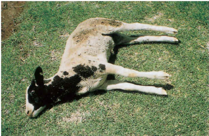
Lambs, older sheep, and goats usually develop the acute disease. Affected lambs do not suckle, have grinning facial expressions due to contractions of the facial muscles, and hold their heads high and their legs wide apart. Owing to the spasms of the muscles of the limbs, flexion of the joints becomes virtually impossible and affected animals move with great difficulty. If the animals are startled, they fall down in lateral recumbency and go into spasms, showing opisthotonus with the legs stiffened and outstretched due to more forcible contraction of the extensor muscles (Figure 185.2 and Figure 185.3). Death occurs after three to ten days. The case fatality rate is approximately 100 per cent in animals manifesting clinical signs.18
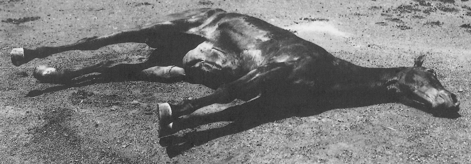
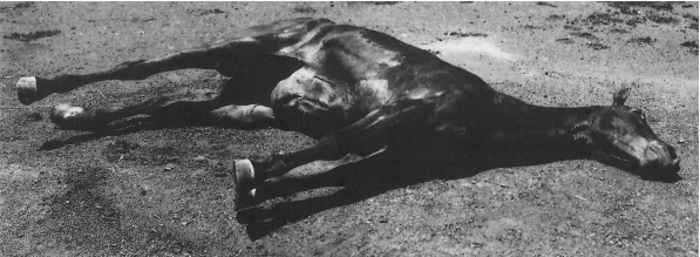
In horses the clinical signs are characterized by the presence of flared nostrils, a sawhorse stance, severe muscle spasms, trismus and prolapse of the third eyelids. When trismus occurs, the mouth is held tightly closed and manual attempts to separate the jaws are generally unsuccessful. Other signs may include constipation, colic, retention of urine, inability to eat feed off the ground, opisthotonus, sweating, dyspnoea, hyperaesthesia with an anxious facial expression as a result of spasms of the facial muscles, and staring eyes. The ears are erect and the tail is rigid and is usually held sideways. The pharyngeal muscles are also similarly affected and saliva dribbles profusely from the mouth.15 Affected animals eventually go down, assume a position of lateral recumbency and, as in the case of lambs and other animals, go into spasms if stimulated (Figure 185.4). The muscular spasms occur with greater frequency and have longer durations as death approaches.
The clinical signs of tetanus in cattle include an extended head, a raised tail, holding the legs in a slightly abducted position (sawhorse stance), prolapse of the third eyelids, ruminal stasis, constipation, marked stiffness and, in some animals, bloating. There is pronounced dullness and depression, with the reflex nervous irritability being less marked than in horses.15, 26
Pathology
There is an initial respiratory alkalosis followed by acidosis in animals suffering from tetanus, and this increases progressively as a consequence of lactic acid formation resulting from muscular hyperactivity. During the period of alkalosis, hyperkalaemia is present, but this changes to hypokalaemia when acidosis develops. The hypoxia that follows the decreased pulmonary ventilation due to respiratory muscle spasms causes microcirculatory disturbances in pulmonary tissue and is a major cause of mortality. Glucose metabolism is impaired but is not stimulated by insulin, lipolysis is triggered, energy expenditure increases, and glucocorticoids and catecholamine blood levels are reduced. Aldolase and creatine phosphokinase levels are elevated, the extent being directly related to the severity of the disease.4
Specific macroscopic and microscopic lesions are absent, except for the probable presence of an infected wound such as those seen when elastic rings are used to castrate or dock the tails of lambs or kids.

Diagnosis
A diagnosis of tetanus is usually based on the history and clinical findings.36 The isolation of C. tetani from necrotic wound tissue in conjunction with the demonstration of the toxin in tissue specimens may be used to confirm a diagnosis of tetanus, but this is rarely done.36 Specimens are obtained from wounds or any site at which necrosis of tissue has occurred and which manifest signs of infection. For bacteriological investigation part of this material is inoculated into cooked meat or liver broth before the medium is heated to 80 °C for 20 minutes to destroy all possible contaminants.2 It is then incubated for two to three days. Subcultures are made onto solid agar plates and specific fluid medium. The culture supernatant may be used for animal inoculation tests to assess toxigenicity. Laboratory animal inoculation of saline suspensions of wound material in order to determine its toxigenicity is possibly of greater diagnostic meaning than the bacterial isolation of C. tetani from such material. As a control, one or more of the animals should be given a prophylactic dose of antitoxin before inoculation with the wound suspension. 36 The presence in smears prepared from wounds of Gram-positive rods or Gram-negative filamentous rods with round, terminal spores giving them a ‘drumstick’ appearance can only be regarded as indicative of a possible C. tetani infection, as other Clostridium spp., such as C. tetanomorphum and C. tetanoides, have a similar appearance. The isolation of C. tetani from specimens of intestinal content taken at necropsy is meaningless for diagnostic purposes as the organism normally occurs in the intestinal tract of animals.
Tetanus toxin may be demonstrated in the serum of some affected animals, especially during the early stages of the disease. Such specimens are divided for test purposes into three portions, one being treated with antitoxin, the second is subjected to heat treatment at 100 °C for 30 minutes, and the third left untreated. A portion of each is then injected intramuscularly in 0,5 to 1,0 ml quantities intramuscularly into mice, which are observed for seven days. If the serum does contain tetanospasmin in detectable amounts by this method, signs of tetanus will develop only in the mouse or mice which received the untreated serum.2
Differential diagnosis
In South Africa, the clinical signs caused by a number of neurotoxic plants may be confused with those of tetanus. These include Sarcostemma viminale spp. in cattle, sheep and goats, Cynanchum spp. in sheep, goats and horses, and Euphorbia mauritanica in sheep and goats. In horses, tetanus in its early stages may also be confused with eclampsia (hypocalcaemic tetany), acute laminitis, hypomagnesaemia and meningitis.1 Eclampsia is confined to lactating mares and responds to treatment with calcium salts. Rigidity of the neck is also caused by cerebrospinal meningitis, and although the animal shows hyperaesthesia, it is generally depressed and hardly excitable.
Lactation tetany of cattle is associated with tetanic muscle spasms and convulsions while white muscle disease in young ruminants may cause a marked stiffness but no muscle spasms. The clinical signs in recumbent cattle, sheep and goats suffering from cerebrocortical necrosis may resemble those of tetanus, but the absence of prolapse of the third eyelids and of rigidity of leg muscles excludes tetanus. The nervous signs caused by heartwater in cattle may also be confused with tetanus, although they are much less pronounced and a fever commonly occurs.15 Although strychnine poisoning is uncommon in farm animals, its clinical signs may be confused with the early signs of tetanus.15
Control
The prognosis of an animal with tetanus is always guarded. It is most unfavourable when there is a short time period between infection and the onset of clinical signs, and when the development of generalized tetanic spasms is rapid. Smith describes in detail the general medical principles for the treatment of tetanus in livestock.31 Animals should be sedated, placed in a quiet, dark stable, and treated with a muscle relaxant. The stable should contain a floor which provides good footing as affected animals have difficulty in rising, and it should also be bedded deeply with straw or wood shavings to minimize the development of decubitus. The C. tetani infection should be eliminated by surgical debridement of the wound, the local infiltration of penicillin G around the wound, and the institution of a course of penicillin administered parenterally.
In order to neutralize unbound tetanus toxin, antitoxin can be infiltrated into the tissues around the affected area as well as administered intravenously (if the value of the animal warrants the expense). Active immunity to tetanus should be established by the administration of a tetanus toxoid vaccine which should be repeated after about a month. The application of general nursing principles is important. This consists of such procedures as the maintenance of hydration, electrolyte balance and nutrition of the affected animal and relieving ruminal tympany should it arise. Feed should be placed in a position which allows easy access.
In one survey the injection of tetanus antitoxin intravenously, intramuscularly or into the epidural space in horses suffering from tetanus resulted in a 5 per cent recovery rate, while that in animals receiving injections into the subarachnoid space increased to 77 per cent.23 In addition, intravenous alimentation,13 and acupuncture,38 may be effective.
Passive immunization with hyperimmune serum obtained from actively immunized sheep8 or horses20 confers effective protection in unimmunized animals and humans. It is effective within hours of administration but its protection does not persist for longer than three weeks.19 It is used either for the protection of animals in which the possible development of tetanus can be anticipated because they are suffering from wounds in which the local conditions in the damaged tissues are conducive to the multiplication of C. tetani, or for the treatment of animals suffering from tetanus. Administered antitoxin will only neutralize the tetanus toxin which is in circulation, and, as antibodies do not cross the blood–brain barrier, this treatment is without effect once the clinical manifestations of tetanus become evident. Following a possible initial beneficial response to the administration of the antitoxin, animals may suffer a relapse, in which case the treatment should be repeated. Cognizance must be taken of the danger of repeating this form of treatment. If it is repeated after 24 hours of the first dose, the development of antiphylactic shock should be anticipated.
The antitoxin, which in South Africa is available from the Onderstepoort Veterinary Institute, is costly and its use should only be considered when the value of the animal justifies the expense.
Tetanus is best controlled by vaccination. The tetanus toxin is highly immunogenic when it is administered as a toxoid in association with an adjuvant. Adjuvants such as waterin- oil emulsion17, 20 and aluminium phosphate5 are used.
As horses are highly susceptible to tetanus, they should be vaccinated on a regular basis, whereas other species, particularly sheep, should be routinely vaccinated only in those instances where the disease has become a consistent problem on a property.
When first vaccinated, horses should be inoculated with the vaccine twice with an interval of about one month. Active immunization is effective 12 to 14 days after the initial injection, but a second vaccination three to four weeks later is required to sustain the immunity at a protective level. Thereafter they should be given an annual booster vaccination. Mares with high levels of circulating tetanus antitoxin titres will provide protective passive colostral immunity to their foals up to the age of 10 weeks.17 The combined use of active-passive immunization in horses has definite advantages and provides an immediate short-term protection and long-term immunity.20 This method is most often employed in horses in which the development of the disease can be anticipated because of an injury, and it should be combined with the systemic administration of a course of antibiotic therapy.
Ewes that have not been vaccinated previously should be vaccinated twice during pregnancy to afford high levels of maternal antibodies to their new-born lambs. The first injection should be given approximately eight weeks before lambing and the second injection three to four weeks later. The tetanus vaccine manufactured and distributed by Onderstepoort Biological Products consists of an aluminium phosphate- adsorbed toxoid.5, 10 High antibody titres are obtained when the first administration of vaccine consists of an oil-adjuvanted toxoid, if this is available, and the second or regular boosters of aluminium phosphate-absorbed toxoid.5 A single booster is required annually 7 to 14 days before lambing.
Vaccination against tetanus does not preclude the application of standard hygiene principles during castration, taildocking and other surgical procedures.
References
- BEROZA, G.A., 1980. Tetanus in a horse. Journal of the American Veterinary Medical Association, 177, 1152–1154.
- BISPING, W. & AMTSBERG, G., 1988. Colour Atlas for the Diagnosis of Bacterial Pathogens in Animals. Berlin & Hamburg: Paul Parey Scientific Publishers.
- BIZZINI, B., 1979. Tetanus toxin. Microbiological Reviews, 43, 224–240.
- BIZZINI, B., 1986. Clostridium tetani. In: gyles, c.l. & thoen, c.o., (eds). Pathogenesis of Bacterial Infections in Animals. Ames: Iowa State University Press.
- CAMERON, C.M., VAN BILJON, B.J., BOTHA, W.J.S. & KNOETZE, P.C., 1983. Comparison of oil adjuvant and aluminium phosphate-adsorbed toxoid for the passive immunization of lambs against tetanus. Onderstepoort Journal of Veterinary Research, 50, 229–231.
- CARMAN, R.J., 1985. Horse diarrhoea: Clostridium tetani as a cause of misdiagnosis of enterotoxaemia. The Veterinary Record, 117, 445.
- CATO, E.P., GEORGE, W.L. & FINEGOLD, S.M., 1986. Genus Clostridium Prazmowski 1880. In: sneath, p.h.a., mair, n.s., sharpe, m.e. & holt, j.g., (eds). Bergey’s Manual of Systematic Bacteriology. Vol. II. Baltimore & London: Williams & Wilkens.
- COX, J.C., LIEFMAN, C.E., PREMIER, R.R., CHANDLER, H.M., HERRINGTON, R.W., MIDDLETON, H.D. & HURRELL, J.G.R., 1984. Immune response and reactions to various dose regimens for raising hyperimmune antisera in sheep. Veterinary Immunology and Immunopathology, 7, 65–72.
- DUNCAN, C.L., 1975. Role of clostridial toxins in pathogenesis. In: Microbiology. Washington, D.C.: American Society for Microbiology.
- ERASMUS, B.J., CAMERON, C.M., HUNTER, P., CILLIERS, J.A., OBEREM, P.T., STOLTSZ, W.H. & DE WAAL, D.T., 1990. Onderstepoort Vaccines. Booklet issued by the Department of Agriculture and Development and obtained from the Directorate Agricultural Information, Private Bag X144, Pretoria 0001.
- FINN, C.W., SILVER, R.P., HABIG, W.H., HARDEGREE, M.C., ZON, G. & GARON, C.F., 1984. The structural gene for tetanus neurotoxin is on a plasmid. Science, 224, 882–884.
- GILL, M., 1987. Bacterial toxins. In: laskin, a.i. & lechevalier, h.a., (eds).CRC Handbook of Microbiology. 2nd edn. Vol. VIII: Toxins and Enzymes. Florida: CRC Press, Inc.
- GREATOREX, J.C., 1975. Intravenous nutrition in the treatment of tetanus in horses. The Veterinary Record, 97, 498.
- HABERMANN, E. & DREYER, F., 1986. Clostridial neurotoxins: Handling and action at the cellular and molecular level. Current Topics in Microbiology and Immunology, 129, 93–179.
- HENNING, M.W., 1956. Animal Diseases in South Africa., 3rd edn. South Africa: Central News Agency.
- HUNGERFORD, T.G., 1990. Diseases of Livestock. Sydney, New York: McGraw-Hill Book Company.
- JANSEN, B.C. & KNOETZE, P.C., 1979. The immune response of horses to tetanus toxoid. Onderstepoort Journal of Veterinary Research, 46, 211–216.
- JENSEN, R. & SWIFT, B.L., 1982. Diseases of Sheep. 2nd edn. Philadelphia: Lea & Febiger.
- LIEFMAN, C.E., 1976. Prophylaxis of tetanus. Australian Veterinary Journal, 52, 51–52.
- LIEFMAN, C.E., 1980. Combined active-passive immunization of horses against tetanus. Australian Veterinary Journal, 56, 119–122.
- MONTECUCCO, C. & SCHIAVO, G., 1995. Structure and function of tetanus and botulinum neurotoxins. Quarterly Review of Biophysics, 28, 423– 427.
- MONTECUCCO, C., SCHIAVO, G. & ROSSETTO, O., 1996. The mechanism of action of tetanus and botulinum neurotoxins. Archives of Toxicology, (supplement), 18, 342–354.
- MUYLLE, E., OYAERT, W., OOMS, L. & DECRAEMERE, H., 1975. Treatment of tetanus in the horse by injections of tetanus antitoxin into the subarachnoid space. Journal of the American Veterinary Medical Association, 167, 47–48.
- OZUTSUMI, K., SUGIMOTO, N. & MATSUDA, M., 1985. Rapid simplified method for production and purification of tetanus toxin. Applied and Environmental Microbiology, 49, 939–943.
- PHILLIPS, K.D., BRAZIER, J.S., LEVETT, P.N. & WILLIS, A.T., 1985. Clostridia. In: collins, c.h. & grange, j.m., (eds). Isolation and Identification of Micro-organisms of Medical and Veterinary Importance. London: Academic Press.
- RAMSAY, W.R., 1973. An outbreak of tetanus-like disease in cattle. Australian Veterinary Journal, 49, 188–189.
- RUBIN, E., 1989. The clostridia. In: schaechter, m., medoff, g. & schlessinger, d., (eds). Mechanisms of Microbial Disease. Baltimore & London: Williams & Wilkens.
- SCHIAVO, G., PAPINI, E., GENNA, G. & MONTECUCCO, C., 1990. An interchain disulfide bond is required for the neurotoxicity of tetanus toxin. Infection and Immunity, 58, 4136–4141.
- SIMPSON, L.L., 1986. Molecular pharmacology of botulinum toxin and tetanus toxin. Annual Review of Pharmacology and Toxicology, 26, 427–453.
- SINGH, B.R., LI, B. & READ, D., 1995. Botulinum versus tetanus neurotoxins: Why is botulinum neurotoxin but not tetanus neurotoxin a food poison? Toxicon, 33, 1541–1547.
- SMITH, B.B., 1990. Large Animal Internal Medicine. St Louis: The C.V. Mosby Co.
- SMITH, J.W.G., 1984. Tetanus. In: wilson, g., miles, a. & parker, m.t., (eds). Topley and Wilson’s Principles of Bacteriology, Virology and Immunology. 7th edn. Vol. III. London: Arnold.
- SMITH, L.D.S, 1975. The Pathogenic Anaerobic Bacteria. 2nd edn. Springfield, Illinois: Charles C. Thomas.
- SMITH, L.D.S. & WILLIAMS, B.L., 1984. The Pathogenic Anaerobic Bacteria. 3rd edn. Springfield, Illinois: Charles C. Thomas.
- STEPHEN, J. & PIETROWSKI, R.A., 1986. Bacterial Toxins. 2nd edn. Washington: American Society for Microbiology.
- STERNE, M. & BATTY, I., 1975. Pathogenic Clostridia. Boston & London: Butterworths.
- WELLS, C.L. & BALISH, E., 1983. Clostridium tetani growth and toxin production in the intestines of germ-free rats. Infection and Immunity, 41, 826–828.
- WHITE, S.S. & CHRISTIE, M.P., 1985. Acupuncture used as an adjunct in the treatment of a horse with tetanus. Australian Veterinary Journal, 62, 25–26.
- WILKENS, C.A., RICHTER, M.B., HOBBS, W.B., WHITCOMB, M., BERGH, N. & CARSTENS, J., 1988. Occurrence of Clostridium tetani in soil and horses. South African Medical Journal, 73, 718–720.
- WOOLCOCK, J.B., 1979. Bacterial Infection and Immunity in Domestic Animals. Oxford & New York: Elsevier Scientific Publishing Company.


