- Infectious Diseases of Livestock
- Part 3
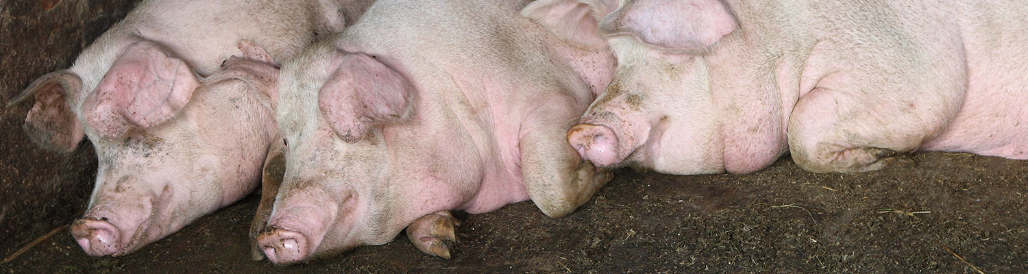
- Mycoplasmal pneumonia of pigs
- GENERAL INTRODUCTION: SPIROCHAETES
- Swine dysentery
- Borrelia theileri infection
- Borrelia suilla infection
- Lyme disease in livestock
- Leptospirosis
- GENERAL INTRODUCTION: AEROBIC ⁄ MICRO-AEROPHILIC, MOTILE, HELICAL ⁄ VIBROID GRAM-NEGATIVE BACTERIA
- Genital campylobacteriosis in cattle
- Proliferative enteropathies of pigs
- Campylobacter jejuni infection
- GENERAL INTRODUCTION: GRAM-NEGATIVE AEROBIC OR CAPNOPHILIC RODS AND COCCI
- Moraxella spp. infections
- Bordetella bronchiseptica infections
- Pseudomonas spp. infections
- Glanders
- Melioidosis
- Brucella spp. infections
- Bovine brucellosis
- Brucella ovis infection
- Brucella melitensis infection
- Brucella suis infection
- Brucella infections in terrestrial wildlife
- GENERAL INTRODUCTION: FACULTATIVELY ANAEROBIC GRAM NEGATIVE RODS
- Klebsiella spp. infections
- Escherichia coli infections
- Salmonella spp. infections
- Bovine salmonellosis
- Ovine and caprine salmonellosis
- Porcine salmonellosis
- Equine salmonellosis
- Yersinia spp. infections
- Haemophilus and Histophilus spp. infections
- Haemophilus parasuis infection
- Histophilus somni disease complex in cattle
- Actinobacillus spp. infections
- Actinobacillus equuli infections
- Gram-negative pleomorphic infections: Actinobacillus seminis, Histophilus ovis and Histophilus somni
- Porcine pleuropneumonia
- Actinobacillus suis infections
- Pasteurella and Mannheimia spp. infections
- Pneumonic mannheimiosis and pasteurellosis of cattle
- Haemorrhagic septicaemia
- Pasteurellosis in sheep and goats
- Porcine pasteurellosis
- Progressive atrophic rhinitis
- GENERAL INTRODUCTION: ANAEROBIC GRAM-NEGATIVE, IRREGULAR RODS
- Fusobacterium necrophorum, Dichelobacter (Bacteroides) nodosus and Bacteroides spp. infections
- GENERAL INTRODUCTION: GRAM-POSITIVE COCCI
- Staphylococcus spp. infections
- Staphylococcus aureus infections
- Exudative epidermitis
- Other Staphylococcus spp. infections
- Streptococcus spp. infections
- Strangles
- Streptococcus suis infections
- Streptococcus porcinus infections
- Other Streptococcus spp. infections
- GENERAL INTRODUCTION: ENDOSPORE-FORMING GRAM-POSITIVE RODS AND COCCI
- Anthrax
- Clostridium perfringens group infections
- Clostridium perfringens type A infections
- Clostridium perfringens type B infections
- Clostridium perfringens type C infections
- Clostridium perfringens type D infections
- Malignant oedema⁄gas gangrene group of Clostridium spp.
- Clostridium chauvoei infections
- Clostridium novyi infections
- Clostridium septicum infections
- Other clostridial infections
- Tetanus
- Botulism
- GENERAL INTRODUCTION: REGULAR, NON-SPORING, GRAM-POSITIVE RODS
- Listeriosis
- Erysipelothrix rhusiopathiae infections
- GENERAL INTRODUCTION: IRREGULAR, NON-SPORING, GRAM-POSITIVE RODS
- Corynebacterium pseudotuberculosis infections
- Corynebacterium renale group infections
- Bolo disease
- Actinomyces bovis infections
- Trueperella pyogenes infections
- Actinobaculum suis infections
- Actinomyces hyovaginalis infections
- GENERAL INTRODUCTION: MYCOBACTERIA
- Tuberculosis
- Paratuberculosis
- GENERAL INTRODUCTION: ACTINOMYCETES
- Nocardiosis
- Rhodococcus equi infections
- Dermatophilosis
- GENERAL INTRODUCTION: MOLLICUTES
- Contagious bovine pleuropneumonia
- Contagious caprine pleuropneumonia
- Mycoplasmal pneumonia of pigs
- Mycoplasmal polyserositis and arthritis of pigs
- Mycoplasmal arthritis of pigs
- Bovine genital mycoplasmosis
- Neurotoxin-producing group of Clostridium spp.
- Contagious equine metritis
- Tyzzer's disease
- MYCOTIC AND ALGAL DISEASES: Mycoses
- MYCOTIC AND ALGAL DISEASES: Pneumocystosis
- MYCOTIC AND ALGAL DISEASES: Protothecosis and other algal diseases
- DISEASE COMPLEXES / UNKNOWN AETIOLOGY: Epivag
- DISEASE COMPLEXES / UNKNOWN AETIOLOGY: Ulcerative balanoposthitis and vulvovaginitis of sheep
- DISEASE COMPLEXES / UNKNOWN AETIOLOGY: Ill thrift
- Eperythrozoonosis
- Bovine haemobartonellosis
Mycoplasmal pneumonia of pigs
This content is distributed under the following licence: Attribution-NonCommercial CC BY-NC  View Creative Commons Licence details here
NJ Maclachlan and M-L Penrith (Editors). P Wallgren, Mycoplasmal pneumonia of pigs, 2019.
Mycoplasmal pneumonia of pigs
Previous authors: P WALLGREN
P WALLGREN - Professor, State Veterinarian, Dipl ECPHM, Department of Animal Health and Antimicrobial Strategies, National Veterinary Institute, Uppsala, 75195, Sweden
Introduction
Mycoplasmal pneumonia of swine (MPS), caused by Mycoplasma hyopneumoniae,39 is an insidious, subacute to chronic pneumonia characterized clinically by a non-productive cough, loss of condition, growth retardation and low mortality. The causative agent was first isolated in 1965 in the USA30 and Europe.18 Mycoplasmal pneumonia of swine occurs worldwide causing great losses to pig producers due to decreased growth of fatteners, either as a result of the disease itself or in combination with secondary infections.36, 51
Aetiology
Mycoplasma hyopneumoniae is a small, fastidious, slow-growing organism that can only be propagated on special media (see General Introduction: Mollicutes).12, 35 Overgrowth by other faster growing mycoplasmas may jeopardize cultivation. Mycoplasma hyopneumonia should be differentiated from other closely related mycoplasmas (see Diagnosis), such as M. flocculare.14, 39, 40
It may survive in the environment for up to one month under humid conditions.17 but as it is sensitive to heat, sunlight and dehydration it is, in general, rapidly inactivated in the environment.15
Epidemiology
The disease is typically introduced into uninfected herds by subclinically infected pigs which are usually adult breeding animals. Herds a short distance from an infected herd(s) may become infected by airborne transmission of M. hyopneumoniae, particularly during conditions of cold outdoor temperature and high humidity.22 Infections with M. hyopneumoniae have not been reported in minimal- disease herds located two miles from the closest infected herd.17
Within herds, M. hyopneumoniae is transmitted either in aerosols45 or by direct contact between pigs, and is often spread from older to younger pigs. Mycoplasma hyopneumoniae infections are most common among fatteners, i.e. those in units housing high numbers of young pigs. The disease is exacerbated by poor hygienic conditions, which also pave the way for secondary infections such as Pasteurella multocida is frequently isolated from fattening pigs with pneumonia.8, 12, 56
Piglets are susceptible to M. hyopneumoniae infection because of their poor antibody response.54 Nevertheless, piglets of conventional dams (especially older sows16) generally possess some immunity to M. hyopneumoniae due to protective, colostrum-derived antibodies.23, 54 In contrast, poorly immunized dams or those with no immunity (especially gilts) may infect their offspring.8
Multiple strains of M. hyopneumoniae are found in pig populations, but certain strains appear to be responsible for outbreaks.32 Limited genetic variation was found among M. hyopneumoniae isolates in a in a herd with commingled pigs from different sources.37 Nevertheless, infections with multiple strains may occur within herds associated with increased severity of pneumonic lesions at slaughter, suggesting that reducing the number of different strains may lead to fewer lung lesions at slaughter and better respiratory health of pigs.35
Pathogenesis
After inhalation, M. hyopneumoniae colonizes the lungs extracellularly in the vicinity of ciliated epithelial cells and provokes an inflammatory reaction that may develop as early as one week post-infection.23, 27 The extent of the reaction correlates with the severity of infection. Circulating antibodies to M. hyopneumoniae have been demonstrated 14 days post-infection.13, 28, 47 It has been suggested that cell-mediated immune responses may play a role in the pneumonic process.33, 48 This contention is supported by the fact that an increased ability of mononuclear cells to produce antibodies in vitro after exposure to M. hyopneumoniae has been demonstrated.53 Antibodies provide protection to animals following challenge, even when pigs are exposed to stressors that affect the general immune response negatively.59
Clinical signs
Mycoplasmal pneumonia of pigs is a chronic, often insidious, disease with high morbidity and low mortality, at least when uncomplicated. The incubation period is one to three weeks43 and the clinical signs comprise a dry, unproductive cough that increases in intensity on physical exertion, unthriftiness and a decrease in appetite. If established early during the fattening period in a group of pigs, MPS will affect a great majority of them before they reach market weight.42, 44, 51, 61 However, the disease may go unnoticed since pigs with active infections decrease their physical activities and show no overt evidence that they are infected. Their inappetence is generally hidden from the farmer since pen mates of the affected pigs will consume the pooled feed ration. As individual pigs in a herd become diseased at different times, this ‘feed stealing’ can, at least to some extent, explain the phenomenon often referred to as ‘compensatory growth’.58
In uncomplicated cases of MPS, the clinical signs gradually decrease and disappear within 30 to 40 days after onset of the disease,43 and the lesions in the lungs heal completely within approximately 10 to 12 weeks.52 However, if the lung lesions are secondarily infected by bacteria, the course of the disease can be considerably prolonged. The most important secondary invader within the respiratory disease complex of pigs appears to be Pasteurella multocida.8, 12, 56
Pathology
In uncomplicated cases of MPS the pneumonic lesions occur predominantly in the ventral aspects of the cranial, accessory and middle lobes but may also be present in the cranio-ventral parts of the caudal lobes. They comprise clearly demarcated areas of consolidation varying in colour from dark reddish-blue to pinkish-grey, and are often associated with some degree of atelectasis. Older lesions are greyer, and often become secondarily invaded by bacteria, the identification of which determines the nature of the subsequent inflammatory reaction which is often purulent or serofibrinous, and may extend to involve the pleura with the development of pleuritis.27, 29, 39 Mycoplasma hyopneumoniae may, however, in itself cause fibrinous to serofibrinous pleuritis. Oedema and lymphoid hyperplasia of the mediastinal lymph nodes may be present.
Microscopically the nature of the lung reaction varies because the lesions in most field cases are secondarily invaded by bacteria, and the inflammatory reaction will reflect that provoked by both the secondary and primary pathogens. Two to three weeks after infection in uncomplicated experimentally induced disease, the lesions comprise alveolar interstitial thickening due mainly to lymphocyte and neutrophil infiltrations and to proliferation of cells in the septa, giant cell formation and perivascular lymphoid hyperplasia.27, 29, 39 In older cases, six to eight weeks after infection, the predominant lesion is pronounced peribronchial and peribronchiolar lymphoid hyperplasia.
Diagnosis
A presumptive diagnosis can be made on the basis of the clinical signs and macro-and microscopic pathology, but as other pathogens can elicit similar signs and lesions, it is necessary to confirm the diagnosis.29
Mycoplasma hyopneumoniae grows slowly in cultures and is easily overgrown by other mycoplasmal species, therefore bacteriological culture of diseased lung tissue is not a reliable method for diagnosing MPS. Other more efficient methods for detecting the organism in lung tissue include immunofluorescence,19 DNA probes,1, 46 oligonucleotide probes complementary to ribosomal RNA,15, 21 and the use of the polymerase chain reaction (PCR).31, 43, 45 Also, gene sequencing can be used to type M. hyopneumoniae directly from clinical material without prior isolation and culture.11, 32, 35
Mycoplasma hyopneumonia may, however, be absent from the lungs of immune pigs, which remain seropositive for a long period. Therefore, the detection of circulating antibodies has been found to be superior to the diagnostic methods described above when performing epidemiological studies in pig herds. Circulating antibodies to M. hyopneumoniae can be detected using several methods, but the indirect or blocking enzyme-linked immunosorbent assay (ELISA) is preferred. Cross-reactions with other Mycoplasma spp. are minimized either by treating the antigen with Tween6 or by performing blocking ELISAs based on monoclonal antibodies to the microbe.13
Differential diagnosis
The differential diagnosis should include pneumonia caused by Bordetella bronchiseptica and Mycoplasma hyorhinis, and swine influenza virus.
Control
Because MPS is a chronic, insidious disease, chemotherapy has generally not been successful in controlling it. Nevertheless, M. hyopneumoniae is sensitive to lincomycin, quinolones, tetracyclines and tiamulin.2, 62 These drugs can be used for individual treatment during outbreaks of MPS in naive herds or strategically in control programmes. The long-term use of antibiotics aimed is not recommended due to increasing minimal inhibitory concentration (MIC) values toward the antimicrobials, which has been demonstrated for drugs such as tetracyclines.62
As M. hyopneumoniae is generally transmitted in aerosols within herds, appropriate management practices should be employed to prevent its spread, including an appropriate stocking density and acceptable air quality, ventilation, and temperature. Since the disease is often spread from older to younger pigs, the introduction of age segregated rearing systems is important.9, 20 Despite the presence of M. hyopneumoniae within the breeding stock, some farrow-to-finish herds have been reared to market size without any pigs developing either macroscopic pneumonic lesions or becoming seropositive to M. hyopneumoniae by housing only one age category of growers per unit.25, 56
Inactivated vaccines9, 22, 32 may be used to control the disease but do not totally prevent the development of lung lesions following experimental challenge with M. hyopneumoniae.10, 11, 24, 38, 60 Similar results, together with increased daily weight gains, have been reported in vaccination trials in conventional pig herds.23, 27 The vaccines have been shown to stimulate the cellular immune response.5, 7 The vaccination of piglets during the first week of life has been recommended by some researchers, but others obtained better results when older piglets were vaccinated,49 possibly due to an increased age-related ability to mount an immune response towards M. hyopneumoniae.54 Under field conditions a higher level of protection was obtained when piglets nine weeks of age were vaccinated compared to littermates vaccinated for the first time at three weeks of age.59 If piglets are vaccinated once at the age of nine to ten weeks but before they have been exposed to M. hyopneumoniae, subsequent natural infection may act as a form of a ‘booster vaccination’, after which they will possess a high level of protection against MPS. However, if vaccinated twice, i.e. at nine to ten weeks and again at 13 weeks of age, they will be even better protected.55
The protective role of colostrum from older sows has been investigated.16 By transferring older sows to new facilities prior to farrowing and not restocking the previously used buildings for a year, it was possible to eradicate MPS.50 The administration of antibiotics has considerably increased the efficiency of such programmes, and successful eradication attempts have been carried out in a number of countries.4, 26, 34, 41, 57, 64 Generally, such programmes include the isolation of pigs older than 10 months and the strategic treatment of sows before farrowing and their transfer to cleaned and disinfected units.
Another method of protecting the piglets from infection by M. hyopneumoniae (and certain other infections) in infected herds is the introduction of medicated early weaning (MEW).3 Medicated piglets are weaned from their medicated dams at the age of 14 days, and the piglets are transferred to new facilities that have been disinfected. If the sows are also transferred to disinfected facilities, this method may be used to establish disease-free herds.
The ultimate way to eradicate M. hyopneumoniae is to establish specific pathogen-free (SPF) or ‘minimal disease’ herds.63 The first generation of pigs within these systems is obtained by caesarean section of the dams. The subsequent generations of pigs may be mated by artificial insemination, which also ensures the maintenance of genetic improvement of the breeding stock. However, careful consideration should be given to the introduction of new pure-bred stock. Specific pathogen-free herds should be monitored continuously with respect to freedom from infection.
References
- AHRENS, P. & FRIIS, N. F., 1991. Identification of Mycoplasma hyopneumoniae with a DNA probe. Letter of Applied Microbiology, 12, 249–253.
- AITKEN, I. A., MORGAN, J. H., DALZIEL, R., BURCH, D. G. S. & RIPLEY, P. H., 1999. Comparative in vitro activity of valnemulin against porcine bacterial pathogens. The Veterinary Record, 144, 128.
- ALEXANDER, T. J. L., THORNTON, K., BOON, G., LYSONS, R. J. & GUSH, A. F., 1980. Medicated early weaning to obtain pigs free from pathogens endemic in the herd of origin. The Veterinary Record, 106, 114–119.
- BAEKBO, P., MADSEN, K. S., AAGÅRD, M. & SZANCER, J., 1994. Eradication of Mycoplasma hyopneumoniae from infected herds without restocking. Proceedings of the International Pig Veterinary Society Congress, Bangkok, Thailand. 13, 135.
- BANDRICK, M., THEIS, K. & MOLITOR, T. W., 2014. Maternal immunity enhances Mycoplasma hyopneumoniae vaccination induced cell-mediated immune response in piglets. BMC Veterinary Research, 10:124.
- BEREITER, M., YOUNG, T. F., JOO, H. S. & ROSS, R. F., 1990. Evaluation of the ELISA, and the comparison to the complement fixation test and radial immunodiffusion enzyme assay for detection of antibodies against Mycoplasma hyopneumoniae in swine serum. Veterinary Microbiology, 25, 177–192.
- BHOGAL, B. S., DAYALU, K. I., KEICH, R. L., ROGERS, A. M. & GERBER, J. D., 1992. Preferential stimulation of cell mediated immune (CMI) responses in bronchial lymph node (BLN) of piglets vaccinated with a Mycoplasma hyopneumoniae vaccine. In: Proceedings of the International Pig Veterinary Society Congress, The Hague, The Netherlands. 11, 298.
- BÖLSKE, G., MARTINSSON, K. & PERSSON, N., 1980. The incidence of mycoplasma and bacteria from lungs of swine with enzootic pneumonia in Sweden. Proceedings of the International Pig Veterinary Society Congress, Copenhagen, Denmark. 6, 213.
- CLARK, L. K., SCHEIDT, A. B., ARMSTRONG, C. H., KNOX, K. & MAYROSE, V. B., 1986. The effect of all in-all out management on pigs from a herd affected by enzootic pneumonia. Veterinary Medicine, 86, 948–951.
- DAYALU, K. I. & ROSS, R. F., 1990. Evaluation of experimental vaccines for control of porcine pneumonia induced by Mycoplasma hyopneumonia. Proceedings of the International Pig Veterinary Society Congress, Lausanne, Switzerland. 11, 83.
- DEL POZO SACRISTÁN, R., SIERENS, A., MARCHIORO, S. B., VANGROENWEGHE, F., JOURQUIN, J., LABARQUE, G., HAESEBROUCK, F. & MAES, D., 2014. Efficacy of early Mycoplasma hyopneumoniae vaccination against mixed respiratory infections in older fattening pigs. The Veterinary Record, 174, 197.
- FALK, K. & LIUM, B., 1990. Enzootic pneumonia of pigs—studies on field material on the relationship between the extent of lung lesions and the demonstration of Pasteurella multocida and Mycoplasma hyorhinis. Proceedings of the International Pig Veterinary Society Congress, Lausanne, Switzerland. 11, 93.
- FELD, N. C., QVIST, P., AHRENS, P., FRIIS, N. F. & MEYLING, A., 1992. A monoclonal blocking ELISA detecting serum antibodies to Mycoplasma hyopneumoniae. Veterinary Microbiology, 30, 35–46.
- FRIIS, N. F., 1975. Some recommendations concerning primary isolation of Mycoplasma suipneumoniae and Mycoplasma flocculare. Nordisk Veterinaermedicin, 27, 337–339.
- FUTO, S., SETO, Y., MITSUSE, S. & MORI, Y., 1992. Detection of Mycoplasma hyopneumoniae by using rRNA-oligonucleotide hybridization. Journal of Clinical Microbiology, 30, 1509–1513.
- GOODWIN, R. F. W., 1965. The phenomenon of suppressed respiratory disease in litters of older sows. The Veterinary Record, 77, 383–387.
- GOODWIN, R. F. W., 1985. Apparent reinfection of enzooticpneumonia- free pig herds: Search for possible causes. The Veterinary Record, 116, 690–694.
- GOODWIN, R. F. W., POMEROY, A. P. & WHITTLESTONE, P., 1965. Production of enzootic pneumonia in pigs with a mycoplasma. The Veterinary Record, 77, 1247–1249.
- HOLMGREN, N., 1974. Swine enzootic pneumonia: Immunological studies in infected herds. Research in Veterinary Science, 17, 145–153.
- HOLMGREN, N., GERTH-LÖFSTEDT, M., J., B. & WALLGREN, P., 1994. Prevalence of some respiratory pathogens in different piglet breeding systems. Proceedings of the International Pig Veterinary Society Congress, Bangkok, Thailand. 13, 130.
- JOHANSSON, K. E., MATTSSON, J. G., JAKOBSSON, K., FERNANDEZ, C., BERGSTRÖM, K., WALLGREN, P. & GÖBEL, U. B., 1992. Specificity of oligonucleotide probes complementary to evolutionarily variable regions of 16S rRNA from Mycoplasma hyopneumoniae and Mycoplasma hyorhinis. Research in Veterinary Science, 52, 195–204.
- JORSAL, S. E. & THOMSEN, B. L., 1988. A cox regression analysis related to Mycoplasma suipneumoniae reinfection in Danish SPF herds. Acta Veterinaria Scandinavica Supplement, 84, 436–438.
- KOBISH, M., BLANCHARD, B. & LE POTIER, M. F., 1993. Mycoplasma hyopneumoniae infection in pigs: Duration of the disease and resistance to reinfection. Veterinary research, 24, 67–77.
- KRISTENSEN, C. S., VINTHER, J., SVENSMARK, K. & BAEKBO, P., 2014. A field evaluation of two vaccines against Mycoplasma hyopneumoniae infection in pigs. Acta veterinaria Scandinavica, 58, 24.
- LINDAHL, E. & WALLGREN, P., 1997. Respiratory infections in pigs: Effects of age segregation at a building level. Svensk Veterinärtidning, 49, 219–223.
- LIUM, B., SKOMSØY, A., JØRGENSEN, A. L. O. E. B. & SCANZER, J., 1992. An attempt to eradicate Mycoplasma hyopneumoniae from selected Norwegian farrowing to finishing herds. Proceedings of the International Pig Veterinary Society Congress, The Hague, The Netherlands. 12, 300.
- LIVINGSTONE, C. W., STAIR, E. L., UNDERDAHL, N. R. & MEBUS, C. A., 1972. Pathogenesis of mycoplasmal infection in swine. American Journal of Veterinary Research, 33, 2249–2258.
- LOYD, L. C., COTTEW, G. S. & ANDERSON, D. A., 1987. Early serological responses to Mycoplasma hyopneumoniae infection. Israel Journal of Medical Sciences, 23, 647–649.
- MARÉ, C. J. & LOVEDAY, R. K., 1994. Mycoplasmal pneumonia of pigs. In: COETZER, J.A.W., THOMSON, G.R. & TUSTIN, R.C., (eds). Infectious Diseases of Livestock with Special Reference to Southern Africa. Cape Town: Oxford University Press, Southern Africa.
- MARÉ, C. J. & SWITZER, W. P., 1965. Mycoplasma hyopneumoniae: A causative agent of virus pig pneumonia. Veterinary Medicine, 60, 841–846.
- MATTSON, J. G., BERGSTRÖM, K., P., W. & JOHANSSON, K. E., 1995. Detection of Mycoplasma hyopneumoniae in nose swabs from pigs by in vitro amplification of the 16S rRNA gene. Journal of Clinical Microbiology, 33, 893–897.
- MAYOR, D., ZEEH, F., FREY, J. & KUNNERT, P., 2007. Diversity of Mycoplasma hyopneumoniae in pig farms revealed by direct molecular typing of clinical material. Veterinary research, 38, 391-398.
- MESSIER, S., ROSS, R. F. & PAUL, P. S., 1990. Humoral and cellular immune responses of pigs inoculated with Mycoplasma hyopneumoniae. American Journal of Veterinary Research, 51, 52–58.
- MÉSZAROS, J., STIPKOVITS, L., ANTAL, T., SZABO, I. & VESZELY, P., 1985. Eradication of some infectious pig diseases by perinatal tiamulin treatment and early weaning. The Veterinary Record, 116, 8–12.
- MICHIELS, A., VRANCKX, K., PIEPERS, S. D. E. L., POSO SACRISTÁN, R., ARSENAKIS, I., BOYEN, F., HAESEBROUCK, F. & MAES, D., 2017. Impact of diversity of Mycoplasma hyopneumoniae strains on lung lesions in slaughter pigs. Veterinary research, 48, (1),2.
- NOYES, E. P., FEENEY, D. A. & PIJOAN, C., 1990. Comparison of the effect of pneumonia detected during lifetime with pneumonia detected at slaughter on growth in swine. Journal of American Veterinary Medical Association, 197, 1025–1029.
- PANTOJA, L. G., PETTIT, K., DOS SANTOS, L. F., TUBBS, R. & PIETERS, M., 2016. Mycoplasma hyopneumoniae genetic variability within a swine operation. Journal of Veterinary Diagnostic Investigations, 28, 175-179.
- PETERSEN, G., WEISS, D., EGAN, J., KORSHUS, J., PETERS, R. & NIRON, M., 1990. Response to Mycoplasma hyopneumoniae vaccination in nursing piglets. Proceedings of the International Pig Veterinary Society Congress, Lausanne, Switzerland. 11, 84.
- ROSS, R. F., 1999. Mycoplasmal diseases. In: STRAW, B.E., D’ALLAIRE, S., MENGELING, W.L. & TAYLOR, D.J., (eds). Diseases of Swine. 8th edn. Ames, Iowa: Iowa State University Press.
- ROSS, R. F. & WHITTLESTONE, P., 1983. Recovery of, identification of and serological responses to porcine mycoplasmas. In: TULLY, J.G. & RAZIN, S., (eds). Methods in Mycoplasmology. 2nd vol. New York: Academic Press.
- SCHULLER, W., NEUMEISTER, E. & VOGEL, D., 1977. Zur Sanierung von mit enzootischer Pneumonie Verseuchten Schweinbestanden. Wiener Tierärzliche Monatschrift, 64, 156–160.
- SHELDRAKE, R., GARDNER, M., SAUNDERS, M. & ROMALIS, L., 1990. Serum antibody response to Mycoplasma hyopneumoniae measured by enzyme-linked immunosorbent assay after experimental and natural infection of pigs. Australian Veterinary Journal, 67, 39–42.
- SØRENSEN, V., AHRENS, P., BARFOD, K., FEENSTRA, A. A., FELD, N. C., FRIIS, N. F., BILLE-HANSEN, V., JENSEN, N. E. & PEDERSEN, M. W., 1997. Mycoplasma hyopneumoniae infection in pigs: Duration of disease and evaluation of four diagnostic assays. Veterinary Microbiology, 54, 23–34.
- SØRENSEN, V., BARFOD, K., FELD, N. C. & VRAA-ANDERSEN, L., 1993. Application of enzyme-linked immunosorbent assay for the surveillence of Mycoplasma hyopneumoniae infection in pigs. Revue Scientifique et Technique de l’Office International des Épizooties, 12, 593–604.
- STÄRK, K. D. C., NICOLET, J. & FREY, J., 1998. Detection of Mycoplasma hyopneumoniae by air sampling with a nested PCR assay. Applied and Environmental Microbiology, 64, 543–548.
- STEMKE, G. W., 1989. A gene probe to detect Mycoplasma hyopneumoniae, the etiological agent to enzootic porcine pneumonia. Molecular Cell Probes, 3, 225–232.
- STRASSER, M., ABIVEN, P., KOBISCH, M. & NICOLET, J., 1992. Immunological and pathological reactions in piglets experimentally infected with Mycoplasma hyopneumoniae and/or Mycoplasma flocculare. Veterinary Immunology and Immunopathology, 31, 141–153.
- TAJIMA, M., YAGIHASHI, T., NUMAYA, T., TAKEUCHI, A. & OKASHI, F., 1984. Mycoplasma hyopneumoniae infection in pigs immunosuppressed by thymectomy and treatment with antithymocyte serum. American Journal of Veterinary Research, 45, 1928–1932.
- VRAA-ANDERSEN, L. & CHRISTENSEN, G., 1993. The prevention of enzootic pneumonia. A clinical trial of Suvaxyn M. hyo-vaccine. I. The effect of production parameters in 5 herds. Dansk Veterinaertidskrift, 76, 129–130.
- WALDMANN, O. & RADTKE, G., 1937. Erster Bericht über Erfolge der Bekämpfung der Ferkegrippe durch die riemser Einzelhüttenanlage. Berliner Tierärzliche Wochenschrift, 53, 241–248.
- WALLGREN, P., ARTURSSON, K., FOSSUM, C. & ALM, G. V., 1993. Incidence of infections in pigs bred for slaughter revealed by elevated serum levels of interferon and development of antibodies to Mycoplasma hyopneumoniae and Actinobacillus pleuropneumoniae. Journal of Veterinary Medicine B, 40, 1–12.
- WALLGREN, P., BESKOW, P., FELLSTRÖM, C. & RENSTRÖM, H. M. L., 1994. Porcine lung lesions at slaughter and their correlation to the incidence of infections with Mycoplasma hyopneumoniae and Actinobacillus pleuropneumoniae during the rearing period. Journal of Veterinary Medicine B, 41, 441–452.
- WALLGREN, P., BÖLSKE, G. & FOSSUM, C., 1992. In vitro stimulation of antibody production to Mycoplasma hyopneumoniae by porcine peripheral blood mononuclear cells. Veterinary Microbiology, 32, 363–374.
- WALLGREN, P., BÖLSKE, G., GUSTAFSSON, S., MATTSSON, S. & FOSSUM, C., 1998. Humoral response to Mycoplasma hyopneumoniae in sows and offspring following an outbreak of mycoplasmosis. Veterinary Microbiology, 60, 193–205.
- WALLGREN, P., LÖFSTEDT, M. & HELDMER, E., 1998. Strategic vaccination against Mycoplasma hyopneumoniae to avoid merchandise of contagious animals from multiplying herds. Proceedings of the International Pig Veterinary Society Congress. Birmingham, UK. 15 (2): 149.
- WALLGREN, P., NÖRREGÅRD, E., MOLANDER, B., PERSSON, M. & EHLORSSON, C. J., 2016. Serological patterns of Actinobacillus pleuropneumoniae, Mycoplasma hyopneumoniae, Pasteurella multocida and Streptococcus suis in pig herds affected by pleuritis. Acta veterinaria Scandinavica, 4;58(1):71.
- WALLGREN, P., SAHLANDER, P., HASSLEBÄCK, G. & HELDMER, E., 1993. Control of infections with Mycoplasma hyopneumoniae in swine herds by disrupting the chain of infection, disinfection of buildings and strategic medical treatment. Journal of Veterinary Medicine B, 40, 157–169.
- WALLGREN, P., SEGALL, T., PEDERSEN, M., A. & GUNNARSSON, A., 1999. Experimental infections with Actinobacillus pleuropneumoniae in pigs. I. Comparison of five different parenteral antibiotic treatments. Journal of Veterinary Medicine B, 46, 249–260.
- WALLGREN, P., WILÉN, I. L. & FOSSUM, C., 1994. Influence of experimentally induced endogenous production of cortisol on the immune capacity of pigs. Veterinary Immunology and Immunopathology, 42, 301–316.
- WENG, C. N., TZAN, Y. L., LIU, S. D., LIN, S. Y. & LEE, C. J., 1992. Protective effects of an oral microencapsulated Mycoplasma hyopneumoniae vaccine against experimental infections in pigs. Research in Veterinary Science, 53, 42–46.
- YAGIHASHI, T., KAZAMA, S. & TAJIMA, M., 1993. Seroepidemiology of mycoplasmal pneumonia of swine in Japan as surveyed by an enzyme-linked immunosorbent assay. Veterinary Microbiology, 34, 155–166.
- YAMAMOTO, K., KOSHIMIZU, K. & OGATA, M., 1986. In vitro susceptibility of Mycoplasma hyopneumoniae to antibiotics. Japanese Journal of Veterinary Sciences, 48, 1–5.
- YOUNG, G. A., UNDERDAHL, N. R., SUMPTION, L. J., PEO, E. R., OLSEN, L. S., KELLY, G. W., HUDMAN, D. B., CALDWELL, J. D. & ADAMS, C. H., 1959. Swine repopulation. I. Performance within a ‘disease-free’ experimental station herd. Journal of American Veterinary Medical Association, 134, 491–496.
- ZIMMERMANN, W., ODERMATT, W. & TSCHUDI, P., 1989. Enzootishe Pneumonie (EP): Dei Teilsanerung EP-Reinfizierter Schweinezuchtbetreibe als alternative zur Totalsanierung. Schweizer Archiv für Tierheilkunde, 131, 179–186, 191.


