- Infectious Diseases of Livestock
- Part 3
- Campylobacter jejuni infection
- GENERAL INTRODUCTION: SPIROCHAETES
- Swine dysentery
- Borrelia theileri infection
- Borrelia suilla infection
- Lyme disease in livestock
- Leptospirosis
- GENERAL INTRODUCTION: AEROBIC ⁄ MICRO-AEROPHILIC, MOTILE, HELICAL ⁄ VIBROID GRAM-NEGATIVE BACTERIA
- Genital campylobacteriosis in cattle
- Proliferative enteropathies of pigs
- Campylobacter jejuni infection
- GENERAL INTRODUCTION: GRAM-NEGATIVE AEROBIC OR CAPNOPHILIC RODS AND COCCI
- Moraxella spp. infections
- Bordetella bronchiseptica infections
- Pseudomonas spp. infections
- Glanders
- Melioidosis
- Brucella spp. infections
- Bovine brucellosis
- Brucella ovis infection
- Brucella melitensis infection
- Brucella suis infection
- Brucella infections in terrestrial wildlife
- GENERAL INTRODUCTION: FACULTATIVELY ANAEROBIC GRAM NEGATIVE RODS
- Klebsiella spp. infections
- Escherichia coli infections
- Salmonella spp. infections
- Bovine salmonellosis
- Ovine and caprine salmonellosis
- Porcine salmonellosis
- Equine salmonellosis
- Yersinia spp. infections
- Haemophilus and Histophilus spp. infections
- Haemophilus parasuis infection
- Histophilus somni disease complex in cattle
- Actinobacillus spp. infections
- Actinobacillus equuli infections
- Gram-negative pleomorphic infections: Actinobacillus seminis, Histophilus ovis and Histophilus somni
- Porcine pleuropneumonia
- Actinobacillus suis infections
- Pasteurella and Mannheimia spp. infections
- Pneumonic mannheimiosis and pasteurellosis of cattle
- Haemorrhagic septicaemia
- Pasteurellosis in sheep and goats
- Porcine pasteurellosis
- Progressive atrophic rhinitis
- GENERAL INTRODUCTION: ANAEROBIC GRAM-NEGATIVE, IRREGULAR RODS
- Fusobacterium necrophorum, Dichelobacter (Bacteroides) nodosus and Bacteroides spp. infections
- GENERAL INTRODUCTION: GRAM-POSITIVE COCCI
- Staphylococcus spp. infections
- Staphylococcus aureus infections
- Exudative epidermitis
- Other Staphylococcus spp. infections
- Streptococcus spp. infections
- Strangles
- Streptococcus suis infections
- Streptococcus porcinus infections
- Other Streptococcus spp. infections
- GENERAL INTRODUCTION: ENDOSPORE-FORMING GRAM-POSITIVE RODS AND COCCI
- Anthrax
- Clostridium perfringens group infections
- Clostridium perfringens type A infections
- Clostridium perfringens type B infections
- Clostridium perfringens type C infections
- Clostridium perfringens type D infections
- Malignant oedema⁄gas gangrene group of Clostridium spp.
- Clostridium chauvoei infections
- Clostridium novyi infections
- Clostridium septicum infections
- Other clostridial infections
- Tetanus
- Botulism
- GENERAL INTRODUCTION: REGULAR, NON-SPORING, GRAM-POSITIVE RODS
- Listeriosis
- Erysipelothrix rhusiopathiae infections
- GENERAL INTRODUCTION: IRREGULAR, NON-SPORING, GRAM-POSITIVE RODS
- Corynebacterium pseudotuberculosis infections
- Corynebacterium renale group infections
- Bolo disease
- Actinomyces bovis infections
- Trueperella pyogenes infections
- Actinobaculum suis infections
- Actinomyces hyovaginalis infections
- GENERAL INTRODUCTION: MYCOBACTERIA
- Tuberculosis
- Paratuberculosis
- GENERAL INTRODUCTION: ACTINOMYCETES
- Nocardiosis
- Rhodococcus equi infections
- Dermatophilosis
- GENERAL INTRODUCTION: MOLLICUTES
- Contagious bovine pleuropneumonia
- Contagious caprine pleuropneumonia
- Mycoplasmal pneumonia of pigs
- Mycoplasmal polyserositis and arthritis of pigs
- Mycoplasmal arthritis of pigs
- Bovine genital mycoplasmosis
- Neurotoxin-producing group of Clostridium spp.
- Contagious equine metritis
- Tyzzer's disease
- MYCOTIC AND ALGAL DISEASES: Mycoses
- MYCOTIC AND ALGAL DISEASES: Pneumocystosis
- MYCOTIC AND ALGAL DISEASES: Protothecosis and other algal diseases
- DISEASE COMPLEXES / UNKNOWN AETIOLOGY: Epivag
- DISEASE COMPLEXES / UNKNOWN AETIOLOGY: Ulcerative balanoposthitis and vulvovaginitis of sheep
- DISEASE COMPLEXES / UNKNOWN AETIOLOGY: Ill thrift
- Eperythrozoonosis
- Bovine haemobartonellosis
Campylobacter jejuni infection
This content is distributed under the following licence: Attribution-NonCommercial CC BY-NC  View Creative Commons Licence details here
Campylobacter jejuni infection
Previous authors: M L VAN DER WALT
Current authors:
E DI GIANNATALE - Head of the Bacteriology Department and National Reference Laboratory of Campylobacter, Microbiologist, via Campo Boario, Teramo, 64100, Italy
Introduction
Campylobacter jejuni is a common human pathogen and an important cause of diarrhoea worldwide.28, 77, 89 It is isolated frequently from the intestine of healthy animals and considered to be part of the normal intestinal flora. Birds and domestic ruminants play an integral role in the ecology of C. jejuni and may serve as a source of infection resulting in outbreaks of disease or sporadic cases in humans.34, 62 Poultry and cattle are the most important animal reservoirs. Infection in humans can be acquired via multiple routes, including direct contact with these animals, consumption of contaminated animal products, and environmental contamination such as water.17 Campylobacter jejuni alone, or in combination with other pathogens, may cause enteritis and diarrhoea in calves87 and sheep,83, 91 but its significance in such cases should be interpreted with care.64, 97 For many years it was considered to be the aetiological agent of ‘winter dysentery’ (caused by bovine coronavirus), characterized by profuse watery diarrhoea of sudden onset and short duration in adult cattle.57, 76 Campylobacter jejuni is also an occasional cause of mastitis in cattle41, 42, 52, 61 and abortion90 in cattle, sheep,20, 40, 92 goats5 and blesbok (Damaliscus dorcas).6
Aetiology
Campylobacter jejuni is micro-aerophilic and grows optimally in an atmosphere containing 5 per cent O2, 10 per cent CO2 and 85 per cent N2 and at 37-42°C. It is unable to ferment or oxidize carbohydrates. The organisms are slender, spirally curved, Gram-negative rods 0,2 - 0,8 μm wide and 0,5-6,0 μm long. They vary in morphology and may have an increased number of spirals or may be gull-wing-shaped, while coccoid forms occur in older cultures in response to deleterious conditions. The bacteria are motile, the movement being characteristically darting or corkscrew-like. Motility is caused by polar flagella at one or both ends of the organism51, 81 that propel it through viscous solutions, such as the mucus layer of the gastrointestinal tract.53
Campylobacter jejuni can survive in a moist environment for several weeks at 4 °C and maintain its metabolic activities39 but it tends to die more rapidly at ambient temperatures. It survives in bovine faeces for at least 20 days at 25 °C and for eight weeks at 4 °C.19 In cows’ milk or water C. jejuni survives for two to five weeks when kept at 4 °C.72, 94 The bacterium is sensitive to dehydration at room temperature,28 while freezing drastically reduces the number of organisms on meat.84 It does not proliferate below 30°C,68 perhaps due to the absence of cold shock proteins.39
The organism is inactivated by iodine udder washes within eight minutes and is rapidly killed by hydrochloric acid at pH 2,3.11 Campylobacters do not survive the temperatures of pasteurization: inactivation in skimmed milk and beef cubes requires 50 seconds at 92 °C and 40 seconds at 95,7 °C, respectively.19
Epidemiology
The organism is widely distributed particularly in avian species, farm animals, pets such as dogs and cats, and humans. Food animals, especially poultry, are reservoirs of C. jejuni. Fifty to 80 per cent of cases of campylobacteriosis in humans are attributed to chickens as the source of infection.7, 22, 28, 65, 80
Campylobacter jejuni is shed in the faeces of both diarrhoeic and healthy animals.63, 70, 86 Faecal shedding by healthy animals is intermittent and only few organisms are shed. There appears to be no difference in the carrier rates between healthy cattle, horses, pigs and dogs. Carrier rates do vary within a species and between animals from different sources.69
Organisms are commonly present in the rumen of calves and less so in adult cattle. In healthy sheep C. jejuni is frequently carried in the intestinal tract.1, 79
Isolation levels are five times higher from fresh than from frozen tissue. Refrigeration or frozen storage alone do not add a significant safety margin and therefore poultry contaminated with C. jejuni can cause infection if not properly handled and sufficiently cooked.10 In humans it is estimated that handling and consumption of broiler meat may account for 20 to 30 per cent of human cases of campylobacteriosis.22 Apart from chicken products, infection may follow consumption of untreated surface water, unpasteurized and incompletely cooked milk,37 and other contaminated food products such as meat from farm animals.24, 78 Carcasses of feedlot-fed cattle are more likely to be contaminated than those of pasture-fed cattle.27 Apart from milk no other dairy products act as a source of C. jejuni. 37
Flies may be important in the transmission of the bacterium, particularly to humans. On poultry and pig farms, 50 and 43 per cent respectively of Musca domestica may contain C. jejuni.73
Pathogenesis
Although C. jejuni mainly colonizes the gastrointestinal tract in animals, it may cross the intestinal epithelial barrier leading to bacteraemia.85 In pregnant animals C. jejuni may reach the gravid uterus, resulting in subsequent placentitis, foetal infection and abortion.14, 79
During a brief period of bacteraemia, which lasts no longer than 72 hours, organisms are disseminated to the mesenteric lymph nodes, gastrointestinal tract, gallbladder and spleen. Disappearance of the bacteraemia coincides with the appearance of circulating antibodies.88
The initial colonization of the entire intestinal tract by C. jejuni changes over time, and after 96 hours bacteria persist only in the ileum, caecum and colon. Lesions in calves develop mostly in the large intestine while in lambs they occur in the ileum.88 This intestinal localization is associated with scouring four to five days after infection,93 which also coincides with the development of severe oedema and inflammation in the colon. The inflammatory reaction is characterized by severe neutrophil infiltration, the development of microscopic ulcers, and focal goblet cell hyperplasia. The initial acute inflammatory reaction is followed by the infiltration of lymphocytes and macrophages.15, 93
In sheep the onset of the gastroenteritis can be precipitated by the stress of transport and a change in the diet.83
Different animal models have been developed to study the virulence and pathogenicity of C. jejuni.32 Studies on the mechanisms of virulence of C. jejuni showed that it is a unique pathogen, being able to execute N-linked glycosylation of more than 30 proteins related to colonization, adherence, and invasion. The cytolethal distending toxins (CdtA,B,C) contribute to the pathogenicity of C. jejuni.18 Host factors seem to play a major role in the pathogenesis of campylobacteriosis in humans.18
The organisms colonize the intestinal mucosal surface, adhere to and invade the epithelial cells48, 49 and the mucosa. After colonization, the bacteria multiply and produce toxins,58, 87 including an enterotoxin that is immunologically related to cholera toxin,43, 94 and other cytotoxic and cytolethal toxins.30, 44
Clinical signs
Cattle
Calves and older animals may suffer from an enteric syndrome.4 After an incubation period of one to three days, there is a rise in temperature to 41 °C and passage of dark, mucoid or haemorrhagic faeces70, 86 The diarrhoea may last up to 18 days. Experimental infections are self-limiting96 and the course of the disease is usually not longer than 20 days.87
Other syndromes in cattle associated with C. jejuni infections include abortion storms in late gestation, the birth of live but weak calves,90 and a varying degree of purulent mastitis.52
Sheep and goats
The morbidity in outbreaks of diarrhoea in fattening lambs may reach 30 per cent.83 In field outbreaks many ewes have mild diarrhoea before the first abortions occur. In an outbreak up to 10 per cent or more of the ewes may abort, and at the same time others may give birth to weak lambs.20 An abortion rate of 23.2 per cent in small ruminants has been reported in the USA,74, 99 where C. jejuni infection caused primarily by a highly virulent tetracycline-resistant C. jejuni clone has now replaced Campylobacter fetus subsp. fetus as an important cause of abortion in sheep.75
In goats, abortion may occur during late gestation. As in sheep, diarrhoea is seen in the does before or during the abortions.5
Pathology
Lesions in cattle suffering from the enteric syndrome occur primarily in the large intestine. Macroscopically the small intestine is flaccid and may contain fluid ingesta, whereas the large intestine has very fluid content containing strings of mucus and blood.87 The mesenteric lymph nodes are consistently enlarged.69, 86 In lambs the ileum is more commonly affected.
Bacteria occur close to the surface of the cells, sometimes in the lumen of crypts, and in the lamina propria surrounding the small blood vessels.3 In the ileum and the jejunum there is loss of superficial villar epithelium, stunting and fusion of villi, dilatation of crypts, thickening of the lamina propria, oedema and congestion and inflammatory cell infiltration of the mucosa,70 and prominent lymphoid hyperplasia.86 Scattered crypt abscesses occur, as well as excessive mucus production.88
In cattle that have aborted there is severe necrotizing placentitis and occasionally bronchopneumonia is present in the foetus.90
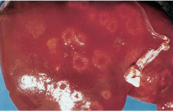
Ewes that abort suffer from severe purulent endometritis accompanied by vasculitis, and necropurulent placentitis. Bacterial colonies are present in the placenta.40 Aborted foetuses have severe purulent bronchopneumonia and multifocal hepatic necrosis (Figure 1).40
Diagnosis
Campylobacter jejuni is a labile organism and dies rapidly on exposure to air, heat and desiccation, and is also rapidly eliminated by putrefactive organisms. Specimens for diagnostic purposes should consist of fresh tissues, which must be placed in a transport medium such as Stuart’s or Amies’, kept cool, and should reach the laboratory as soon as possible.
For successful culture, prior filtering (pore size of 0,65 μm) and the use of commercially available selective media such as Campylobacter modified Charcoal Cefoperazone Deoxycholate Agar (mCCDA), Skirrow, Preston, Butzler, Blaser, Campy-BAP and others is recommended, and the organisms should be cultured in microaerophilic conditions. Preferential growth is also obtained if the cultures are incubated at the upper range of the optimal incubation temperature of 37 to 42 °C.81 After 48 hours of culture, the colonies are typically small (2 to 4 mm in diameter) and low-convex with entire edges. Swarming colonies may extend along the line of incubation when moist media are used. Colonies are further identified by biotyping.81
The identity of organisms may also be confirmed in tissue sections by immunofluorescent or immunoperoxidase techniques40 and by commercial latex agglutination tests.36 ELISA tests have also been applied successfully.29
When attempting the isolation of C. jejuni in live animals, it must be kept in mind that bacteraemia and faecal shedding of organisms may occur sporadically96 and that many animals are symptomless carriers. In fatal cases in cattle and sheep, organisms can be isolated from both normal and grossly inflamed intestinal mucosa.3, 83, 86 Organisms may be isolated from the gallbladder in chronically affected animals.87
In abortion storms, organisms may be isolated in pure culture and in large numbers from the placenta, as well as from the organs and stomach content of aborted foetuses.5, 6, 40, 90 Faecal cultures are of no help in the diagnosis of abortion, as C. jejuni can be isolated with the same frequency in infected herds from normal cows and from those that have aborted, as well as from normal calves and those with diarrhoea.90
Milk from cows with subclinical or mild mastitis due to infection with C. jejuni has an increased somatic cell count, and several strains of the bacterium, with different drug sensitivities, may be isolated from the affected milk of different animals on the same farm.61 Isolation of C. jejuni from milk is aided by the destruction of the lactoperoxidase system.9 Different strains of the organism may at the same time be isolated from the milk and the faeces of the same animal.41 Variable numbers of organisms are excreted from affected quarters for as long as 73 days after infection.50
Suspicious colonies of Campylobacter must be confirmed using different assays, including biochemical and molecular tests and matrix-assisted laser desorption/ionization (MALDI-TOF).8 Identification of C. jejuni can be achieved by means of multiplex PCR74, 95 or real- time PCR methods.45, 46 In sheep a standard PCR has been used to detect C. jejuni directly in aborted foetal tissues and placentas.33
Strains are differentiated by serotyping using heat-labile and heat-stable antigens,89 phage-typing, outer membrane protein-typing,12 pulsed field gel electrophoresis (PFGE) (Pulsenet USA), and multi locus sequence typing (MLST)21 that characterizes strains by their unique allelic profiles.59, 66 Strains of different pathogenicity may be identified in outbreaks,50 while serotypes also differ in pathogenicity and tropism.31, 40, 98 The first complete genome sequence of a C. jejuni strain was reported in 2001 for C. jejuni strain 11168.67 Currently whole-genome sequencing is used for C. jejuni typing.38, 54, 55
Differential diagnosis
Most cases of diarrhoea in cattle from which C. jejuni has been isolated in large numbers also yield other agents such as cryptosporidia, coccidia, rotavirus, coronavirus, enteropathogenic Escherichia coli, Salmonella serovars or nematodes. Clinical cases should in particular be differentiated from coccidiosis and paratyphoid.87
Control
No vaccines are commercially available to control C. jejuni infections in animals or birds, but an ovine C. fetus bacterin has been used in goats and sheep with good effect.5, 35
In general, most strains are susceptible to nitrofurans, gentamicin, and chloramphenicol.36 Multiple resistance to antibiotics may occur.16, 47 High resistance rates for quinolones, fluoroquinolones and tetracyclines have been reported in many countries but resistance to erythromycin and gentamicin in C. jejuni remains low.2, 25, 56, 100 An increased level of macrolide resistance is becoming a major public health concern in some countries in Europe and the USA.26
In sheep, abortions can be controlled by daily injections of erythromycin at a dosage of 4 mg/kg followed by an intramuscular injection of long-acting oxytetracycline.83 Orally administered chlortetracycline at a rate of 75 mg/kg per day for 14 days is also effective in controlling an outbreak of abortion.5
Permanent cure of intestinal infection appears to be difficult. In one report concerning the treatment of dogs with intestinal C. jejuni infection, both chloramphenicol and erythromycin failed to eliminate the infection, and the organism could again be isolated within nine days after discontinuation of treatment.60
References
- ACIK, M.N. & CETINKAYA, B., 2006. Heterogeneity of C.jejuni and C.coli strains from healthy sheeps. Veterinary Microbiology, 115, 370-375.
- ALFREDSON, D.A. & KOROLIK, V., 2007. Antibiotic resistance and resistance mechanisms in Campylobacter jejuni and Campylobacter coli. FEMS Microbiology Letters, 277(2), 123–132. [PubMed: 18031331].
- AL-MASHAT, R.R. & TAYLOR, D.J., 1980. Campylobacter spp. in enteric lesions in cattle. The Veterinary Record, 107, 31.
- AL-MASHAT, R.R. & TAYLOR, D.J., 1980. Production of diarrhoea and dysentery in experimental calves by feeding pure cultures of Campylobacter fetus subsp. jejuni. The Veterinary Record, 107, 459.
- ANDERSON, K.L., HAMOUD, M.M., URBANCE, J.W., RHOADES, H.E. & BRYNER, J.H., 1983. Isolation of Campylobacter jejuni from an aborted caprine fetus. Journal of the American Veterinary Association, 183, 90–92.
- ANON., 1984/1985. Diagnostics of infertility and abortion. Annual Report, Veterinary Research Institute, Onderstepoort.
- BANFFER, J.R.J., 1985. Biotypes and serotypes of Campylobacter jejuni and Campylobacter coli strains isolated from patients, pigs and chickens in the region of Rotterdam. Journal of Infection, 10, 277–281.
- BESSÉDE, E., SOLECKI O., SIFRE, E., LABADI, L. & MÉGRAUD, F., 2011. Identification of Campylobacter species and related organisms by matrix assisted laser desorption ionization–time of flight (MALDI-TOF) mass spectrometry. Clinical Microbiology and Infection, 17 (11), 1735–1739.
- BEUMER, R.R., CRUYSEN, J.J.M. & BIRTANTIE, I.R.K., 1988. The occurrence of Campylobacter jejuni in raw cows’ milk. Journal of Applied Bacteriology, 65, 93–96.
- BHADURI, S. & COTTRELL, B., 2004. Survival of cold-stressed Campylobacter jejuni on ground chicken and chicken skin during frozen storage. Applied and Environmental Microbiology, 70, 7103–7109.
- BLASER, M.J., HARDESTY, B., POWERS, B. & WANG, W.L., 1980. Survival of Campylobacter fetus subsp. jejuni in biological milieus. Journal of Clinical Microbiology, 11, 309–313.
- BLASER, M.J., HOPKINS, J.A., BERKA, R.M., VASIL, M.L. & WANG, W.L., 1983. Identification and characterization of Campylobacter jejuni outer membrane proteins. Infection and Immunity, 42, 276–284.
- BOLINGER, H. & KATHARIOU, S., 2017. The current state of macrolide resistance in Campylobacter spp.: Trends and impacts of resistance mechanisms. Applied and Environmental Microbiology, 83(12). pii: e00416-17. doi: 10.1128/AEM.00416-17. Print 2017 Jun 15.
- BOLTON, D.J., 2015. Campylobacter virulence and survival factors. Food Microbiology, 48, 99-108.
- BOOSINGER, T.R. & POWE, T.A., 1988. Campylobacter jejuni infections in gnotobiotic pigs. American Journal of Veterinary Research, 49, 456–458.
- BURRIDGE, R., WARREN, C. & PHILLIPS, I., 1986. Macrolide, lincosamide and streptogramin resistance in Campylobacter jejuni/coli. Journal of Antimicrobial Chemotherapy, 17, 315–321
- CENTERS FOR DISEASE CONTROL & PREVENTION., 2009. Campylobacter jejuni infection associated with unpasteurized milk and cheese—Kansas, 2007. MMWR, Morbidity and mortality weekly report, 57, 1377–1379.
- DASTI J.I., TAREEN M.A., LUGERT R., ZAUTNER A. E., UWE GROß U., 2010. Campylobacter jejuni: A brief overview on pathogenicity-associated factors and disease-mediating mechanisms. International Journal of Medical Microbiology, 300, 205-211.
- DEIBEL, K.E., 1985. Study of Campylobacter jejuni. Dissertation Abstracts International, 46, 1812–1813.
- DIKER, K.S. & ISTANBULLUOGLU, E., 1986. Ovine abortion associated with Campylobacter jejuni. The Veterinary Record, 118, 307.
- DINGLE, K.E., COLLES, F.M., WAREING, D.R., URE, R., FOX, A.J., BOLTON, F.E., BOOTSMA, H.J., WILLEMS, R.J., URWIN, R. & MAIDEN, M.C., 2001. Multilocus sequence typing system for Campylobacter jejuni. Journal of Clinical Microbiology, 39(1), 14-23.
- EFSA., 2010a Scientific quantification of the risk posed by broiler meat to human campylobacteriosis in the EU. EFSA Journal, 8(1), 1437.
- EPPS, S.V., HARVEY, R.B., HUME, M.E., PHILLIPS, T.D., ANDERSON, R.C. & NISBET, D.J., 2013. Foodborne Campylobacter: infections, metabolism, pathogenesis and reservoirs. International Journal of Environmental Research and Public Health, 26, 10, 6292-304.
- GARCIA, M.M., EAGLESOME, M.D. & RIGBY, C.,1983. Campylobacters important in veterinary medicine. Veterinary Bulletin, 53, 793–818.
- GE, B., WANG, F., JÖLUND-KARLSSON, M. & MC DERMOTT, P.F., 2013. Antimicrobial resistance in Campylobacter: susceptibility testing methods and resistance trends, Journal of Microbiological Methods, 95(1), 57-67.
- GIBREEL, A. & TAYLOR, D.E., 2006. Macrolide resistance in Campylobacter jejuni and Campylobacter coli. Journal of Antimicrobial Chemotherapy, 58(2), 243-255.
- GRAU, F.H.,1988. Campylobacter jejuni and Campylobacter hyointestinalis in the intestinal tract and on the carcasses of calves and cattle. Journal of Food Protection, 51, 857–861.
- GRIFFITHS, P.L. & PARK, R.W.A., 1990. Campylobacters associated with human diarrhoeal disease. Journal of Applied Bacteriology, 69, 281–301.
- GROHN, K. & GENIGEORGIS, C., 1985. Adaption of ELISA for the detection of Campylobacter antibodies and its application in seroepidemiological studies in sheep and cattle herds. Acta Veterinaria Scandinavica, 26, 30–48.
- GUERRANT, R.L., WANKE, C.A., PENNIE, R.A., BARRETT, L.J., LIMA, A.A.M. & O’BRIEN, A.D., 1987. Production of a unique cytotoxin by Campylobacter jejuni. Infection and Immunity, 55, 2526–2530.
- HAAJANEN, H., PUMMI, T., WERMUNDSEN, K., KATILA, M.L., SARKKINEN, H., MIETTINEN, I. & RAUTELIN, H., 2003. Detection and typing of Campylobacter jejuni and Campylobacter coli and analysis of indicator organisms in three waterborne outbreaks in Finland. Applied and Environmental Microbiology, 69, 1391–1396.
- HADDAC, N., MARCE, C., MAGRAS, C. & CAPPELIER, J.M., 2010. An overview of method used to clarify pathogenesis mechanisms of C. jejuni. Journal of Food Protection, 73(4), 786-802.
- HAMALI, H., FALLAH, S., JOOZANI, R.J., ZARE, P. & NOORSAADAT, G., 2014.Detection of Campylobcater spp., in sheep aborted fetus by PCR. Trends Life Science, 3, 49-56.
- HANNON, S.J., TABOADA, E.N., RUSSELL, M.L., ALLAN, B., WALDNER, C., WILSON, H.L., POTTER, A., BABIUK, L. & TOWNSEND, H.G., 2009. Genomics-based molecular epidemiology of Campylobacter jejuni isolates from feedlot cattle and from people in Alberta, Canada. Journal of Food Protection, 47,410–420.
- HANSEN, D.E., HEDSTROM, O.R., SONN, R.J. & SNYDER, S.P., 1990. Efficacy of a vaccine to prevent Chlamydia- or Campylobacter-induced abortions in ewes. Journal of the American Veterinary Medical Association, 196, 731–734.
- HARIHARAN, H., WRIGHT, T. & LONG, J.R., 1990. Isolation and antimicrobial susceptibility of Campylobacter coli and Campylobacter jejuni from slaughter hogs. Microbiologica (Italy), 13, 1–6.
- HARRIS, N.V., KIMBALL, T., WEISS, N.S. & NOLAN, C., 1986. Dairy products, produce and other non-meat foods as possible sources of Campylobacter jejuni and Campylobacter coli enteritis. Journal of Food Protection, 49, 347–351.
- HARRISON, E.M., PATERSON, G.K., HOLDEN, M.T., LARSEN, J., STEGGER, M., LARSEN, A.R., PETERSEN, A., SKOV, R.L., CHRISTENSEN, J.M., BAK ZEUTHEN, A., HELTBERG, O., HARRIS, S.R., ZADOKS, R.N., PARKHILL, J., PEACOCK, S.J. & HOLMES, M.A., 2013. Whole genome sequencing identifies zoonotic transmission of MRSA isolates with the novel mecA homologue mecC. EMBO Molecular Medicine, 5, 509–515.
- HAZELEGER, W.C., WOUTERS, J.A., ROMBOUTS, F.M. & ABEE, T., 1998. Physiological activity of Campylobacter jejuni far below the minimal growth temperature. Applied and Environmental Microbiology, 64, 3917–3922.
- HEDSTROM, O.R., SONN, R.J., LASSEN, E.D., HULTGREN, B.D., CRISMAN, R.O., SMITH, B.B. & SNYDER, S.P., 1987. Pathology of Campylobacter jejuni abortion in sheep. Veterinary Pathology, 24, 419–426.
- HUDSON, P.J., VOGT, R.L., BRONDUM, J. & PATTON, C.M., 1984. Isolation of Campylobacter jejuni from milk during an outbreak of campylobacteriosis. Journal of Infectious Diseases, 150, 789.
- HUTCHINSON, N.D., BOLTON, F.J., HINCHLIFFE, P.M., DAWKINS, H.C., HORSLEY, S.D., JESSOP, E.G., ROBERTSHAW, P.A. & COUNTER, D.E., 1985. Evidence of udder excretion of Campylobacter jejuni as the cause of milk-borne Campylobacter outbreak. Journal of Hygiene, 94, 205–215.
- JOHNSON, W.M. & LIOR, H., 1986. Cytotoxic and cytotonic factors produced by Campylobacter jejuni, C. coli and C. laridis. Journal of Clinical Microbiology, 24, 275–281.
- JOHNSON, W.M. & LIOR, H., 1988. A new heat-labile cytolethal toxin (CLDT) of Campylobacter spp. Microbial Pathogenesis, 4, 115–126.
- JOSEFSEN, M.H., JACOBSEN, N.R. & HOORFAR, J., 2004. Enrichment followed by quantitative PCR both for rapid detection and as a tool for quantitative risk assessment of food-borne thermotolerant campylobacters. Applied and Environmental Microbiology, 70, 3588-3592.
- JOSEFSEN, M.H., LÖFSTRÖM, C., HANSEN, T.B., CHRISTENSEN, L.S., OLSEN, J.E., HOORFAR, J., 2010. Rapid quantification of viable Campylobacter bacteria on chicken carcasses, using real-time PCR and propidium monoazide treatment, as a tool for quantitative risk assessment. Applied and Environmental Microbiology, 76, 5097-5104.
- KANEUCHI, C., ASHIHARA, M., SUGIYAMA, Y. & IMAIZUMI, T., 1988. Antimicrobial susceptibility of Campylobacter jejuni, Campylobacter coli, and Campylobacter laridis from cats, dogs, pigs and seagulls. Japanese Journal of Veterinary Science, 50, 685–691.
- KONKEL, M.E. & JOENS, L.A., 1989. Adhesion to and invasion of Hep-2 cells by Campylobacter spp. Infection and Immunity, 57, 2984–2990.
- KONKEL, M.E., BABAKHANI, F. & JOENS, L.A., 1990. Invasion-related antigens of Campylobacter jejuni. Journal of Infectious Diseases, 162, 888–895.
- LANDER, K.P. & BASKERVILLE, A., 1983. Campylobacter jejuni mastitis in cows: Bacteriology and pathology. Campylobacter. II. Proceedings of the Second International Workshop on Campylobacter Infections, Brussels, 6–9 September, 1983.
- LANDER, K.P. & GILL, K.P.W., 1985. Campylobacters. In: COLLINS, C.H. & GRANGE, J.M., (eds). Isolation and Identification of Microorganisms of Medical and Veterinary Importance. London: Academic Press.
- LANDER, K.P., 1984. Studies on Campylobacter jejuni infection of the bovine udder. PhD. Thesis, London School of Hygiene and Tropical Medicine, University of London.
- LERTSETHTAKARN, P., OTTEMANN, K.M. & HENDRIXSON, D.R., 2011. Motility and chemotaxis in Campylobacter and Helicobacter. Annual Review of Microbiology, 65, 389–410.
- LEWIS, T., LOMAN, N.J., BINGLE, L., JUMAA, P., WEINSTOCK, G.M., MORTIBOY, D. & PALLEN, M.J., 2010. High-throughput whole-genome sequencing to dissect the epidemiology of Acinetobacter baumannii isolates from a hospital outbreak. Journal of Hospital Infection, 75, 37–41.
- LLARENA, A.K., TABOADA, E. & ROSSI, M., 2017. Whole-genome sequencing in epidemiology of Campylobacter jejuni infections, Journal of Clinical Microbiology, 55, 1269-1275.
- LUANGTONGKUM, T., JEON, B., HAN, J., PLUMMER, P., LOGU, E.C.M. & ZHANG, Q., 2009. Antibiotic resistance in Campylobacter: emergence, transmission and persistence. Future Microbiology, 4(2), 189–200.
- MACPHERSON, L.W., 1957. Bovine virus enteritis (winter dysentery). Canadian Journal of Comparative Medicine, 21, 184–192.
- MANNINEN, K.I., PRESCOTT, J.F. & DOHOO, I.R., 1982. Pathogenicity of Campylobacter jejuni isolates from animals and humans. Infection and Immunity, 38, 46–52.
- MEANGER, J.D. & MARSHALL, R.B., 1989. Seasonal prevalence of thermophilic Campylobacter infections in dairy cattle and a study of infection of sheep. New Zealand Veterinary Journal, 37, 18–20.
- MONFORT, J.D., DONAHOE, J.P., STILLS, H.F. & BECH-NIELSEN, S., 1990. Efficacies of erythromycin and chloramphenicol in extinguishing fecal shedding of Campylobacter jejuni in dogs. Journal of the American Veterinary Medical Association, 196, 1069–1072.
- MORGAN, G., CHADWICK, P., LANDER, K.P. & GILL, K.P.W., 1985. Campylobacter jejuni mastitis in a cow: A zoonosis-related incident. The Veterinary Record, 116, 111.
- MULLNER, P., SHADBOLT, T., COLLINS-EMERSON, J.M., MIDWINTER, A.C., SPENCER, S.E., MARSHALL, J., CARTER, P.E., CAMPBELL, D.M., WILSON, D.J., HATHAWAY, S., PIRIE, R. & FRENCH, N.P., 2010. Molecular and spatial epidemiology of human campylobacteriosis: source association and enotype-related risk factors. Epidemiology & Infection, 138, 1372–1383.
- MYERS, L.L., FIREHAMMER, B.D., BORDER, M.M. & SHOOP, D.S., 1984. Prevalence of enteric pathogens in the faeces of healthy beef calves. American Journal of Veterinary Research, 45, 1544–1548.
- NIELSEN, E.M., 2002. Occurrence and strain diversity of thermophilic campylobacters in cattle of different age groups in dairy herds. Letters in Applied Microbiology, 35, 85–89.
- OOSTEROM, J., BANFFER, J.R.J., LAUWERS, S. & BUSSCHBACH, A.E., 1985. Serotyping of, and hippurate hydrolysis by, Campylobacter jejuni isolates from human patients, poultry and pigs in the Netherlands. Antonie van Leeuwenhoek, 51, 65–70
- OWEN, R.J., COSTAS, M. & DAWSON, C., 1989. Application of different chromosomal DNA restriction digest fingerprints to specific and subspecific identification of Campylobacter isolates. Journal of Clinical Microbiology, 27, 2338–2343.
- PARKHILL, J., WREN, B.W., MUNGALL, K., KETLEY, J.M. & CHURCHER, C., 2000. The genome sequence of the food-borne pathogen Campylobacter jejuni reveals hypervariable sequences. Nature, 403, 665–668.
- PENNER, J.L., 1988. The genus Campylobacter: a decade of progress. Clinical Microbiology, 1, 157–172.
- PRESCOTT, J.F. & BRUIN-MOSCH, C.W., 1981. Carriage of Campylobacter jejuni in healthy and diarrhoeic animals. American Journal of Veterinary Research, 42, 164–165.
- PRESCOTT, J.F. & MUNROE, D.L., 1982. Campylobacter jejuni enteritis in man and domestic animals. Journal of the American Veterinary Association, 181, 1524–1530.
- PULSENET USA-STANDARD OPERATING PROCEDURE FOR PULSENET PFGE OF CAMPYLOBACTER JEJUNI. CodePNL03,2013.http://www.pulsenetinternational.org/assets/PulseNet/uploads/pfge/PNL03_CampyPFGEprotocol.pdf
- REDWOOD, D.W., GILL, K.P.W. & LANDER, K.P.,1983. The survival of Campylobacter jejuni in milk. Campylobacter. II. Proceedings of the Second International Workshop on Campylobacter Infections, Brussels, 6–9 September 1983.
- ROSEF, O. & KAPPERUD, G., 1983. House flies (Musca domestica) as possible vectors of Campylobacter fetus subsp. jejuni. Applied and Environmental Microbiology, 5, 381–383.
- SAHIN, O., PLUMMER, P.J., JORDAN, D.M., SULAJ, K., PEREIRA, S., ROBBE-AUSTERMAN, S., WANG, L., YAEGER, M.J., HOFFMAN, L.J. & ZHANG, Q., 2008. Emergence of a tetracycline-resistant Campylobacter jejuni clone associated with outbreaks of ovine abortion in the United States. Journal of Clinical Microbiology, 46(5), 1663–1671.
- SAHIN, O., YAEGER, M., WU, Z. & ZHANG, Q., 2017. Campylobacter associated diseases in animals associated diseases in animals. Annual Review of Animal Biosciences, 5, 21-42
- SAIF, L.J., REDMAN, D.R., BROCK, K.V., KOHLER, E.M. & HECKERT, R.A., 1988. Winter dysentery in adult dairy cattle: Detection of coronavirus in the faeces. The Veterinary Record, 123, 300–301.
- SALAMA, S.M., BOLTON, F.J. & HUTCHINSON, D.N., 1990. Application of a new phagetyping scheme to campylobacters isolated during outbreaks. Epidemiology and Infection, 104, 405–411.
- SHANE, S.M. & MONTROSE, M.S., 1985. The occurrence and significance of Campylobacter jejuni in man and animals. Veterinary Research Communications, 9, 167–198.
- SKIRROW., M.B., 1994. Disesase due Campylobacter, Helicobacter and related bacteria. Journal of Comparative Pathology, 111, 113-149.
- SKIRROW, M.B.,1982. Campylobacter enteritis—the first five years. Journal of Hygiene, 89, 175–184.
- SMIBERT, R.M., 1984. Campylobacter. In: KRIEG, N.R. & HOLT, J.G., (eds). Bergey’s Manual of Systematic Bacteriology. Vol. I. Baltimore and London: Williams & Wilkins.
- STANLEY, K. & JONES, K., 2003. Cattle and sheep farms as reservoirs of Campylobacter. Journal of Applied Microbiology, 94(Suppl), 104S–113S.
- STANSFIELD, D.G., HUNT, B. & KEMBLE, P.R., 1986. Campylobacter gastroenteritis in fattening lambs. The Veterinary Record, 118, 210–211.
- STERN, N.J., GREEN, S.S., THAKER, N., KRONT, D.J. & CHIU, J., 1984. Recovery of Campylobacter jejuni from fresh and frozen meat and poultry collected at slaughter. Journal of Food Protection, 47, 372–374.
- TANG, Y., MEINERSMANN, R.J. & SAHIN, O., 2017. Wide but variable distribution of a hypervirulent Campylobacter jejuni Clone in beef and dairy cattle in the United States. In: DUDLEY, E.G., (ed.). Applied and Environmental Microbiology, 83(24), e01425-17. doi:10.1128/AEM.01425-17.
- TAYLOR, D.J. & AL-MASHAT, R.R., 1984. Enteric infections with catalase-positive campylobacters in cattle, sheep and pigs. In: BUTZLER, J.P.,(ed.). Campylobacter Infection in Man and Animals. Florida: CRC Press.
- TAYLOR, D.J., 1983. Campylobacter jejuni infections in calves. Veterinary Annual, 23, 60–64.
- TERZOLO, H.R., LAWSON, G.H.K., ANGUS, K.W. & SNODGRASS, D.R., 1987. Enteric Campylobacter infection in gnotobiotic calves and lambs. Research in Veterinary Science, 43, 72–77.
- TRESCHNAK, E., 1986. Thermophilic Campylobacter species in veterinary medicine—a review. Berliner und Münchener Tierärztliche Wochenschrift, 99, 52–57.
- VAN DONKERSGOED, J., JANZEN, E., SCHUMANN, F., CLARK, E.G., ORR, J., COPELAND, S., CHIRINO-TREJO, M., BERRY, C. & SPINATO, M., 1989. Campylobacter jejuni abortion in beef cattle. Canadian Veterinary Journal, 30, 680.
- VAN DEN BERGHE, J. & HOORENS, J., 1980. Campylobacter species and regional enteritis in lambs. Research in Veterinary Science, 29, 390–391.
- VARGA, J., MEZES, B., FODOR, L. & HAJTOS, I., 1990. Sero-groups of Campylobacter fetus and Campylobacter jejuni isolated in cases of ovine abortion. Journal of Veterinary Medicine, Series B, 37, 148–152.
- VITOVEC, J., KOUDELA, B., STERBA, J., TOMANCOVA, I., MATYAS, Z. & VLADIK, P., 1989. The gnotobiotic piglet as a model for the pathogenesis of Campylobacter jejuni infection. Zentralblatt für Bakteriologie, Mikrobiologie und Hygiene, A, 271, 91–103.
- WALKER, R.I., CALDWELL, M.B., LEE, E.C., GUERRY, P., TRUST, T.J. & RUIZ-PALACIOS, G.M., 1986. Pathophysiology of Campylobacter enteritis. Microbiological Reviews, 50, 81–94.
- WANG G, CLARK CG, TAYLOR MT, PUCKNELL C, BARTON C, PRICE L, 2002. Colony multiplex PCR Assay for identification and differentiation of Campylobacter jejuni, Campylobacter lari, Campylobacter upsaliensis, Campylobacter fetus subsp. fetus. Journal of Clinical Microbiology, 40, 4744-4747.
- WARNER, D.P. & BRYNER, J.H., 1984. Campylobacter jejuni and Campylobacter coli inoculation of neonatal calves. American Journal of Veterinary Research, 45, 1822–1824.
- WEIS, A.M., MILLER, W.A., BYRNE, B.A., CHOUICHA, N., BOYCE, W.M. & TOWNSEND, A.K., 2014. Prevalence and pathogenic potential of Campylobacter isolates from free-living, human-commensal American crows. Applied and Environmental Microbiology, 80, 1639–1644.
- WIECZOREK, K. & OSEK, J., 2017. Antimicrobial resistance and genotypes of Campylobacter jejuni from pig and cattle carcasses isolated in Poland during 2009-2016. Microbial Drug Resistance, Oct 10.
- WU, Z., SAHIN, O., SHEN, Z., LIU, P., MILLER, W.G. & ZHANG, Q., 2013. Multi-omics approaches to deciphering a hypervirulent strain of Campylobacter jejuni. Genome Biology and Evolution, 5, 2217–2230.
- ZHANG, Q., LIN, J. & PEREIRA, S., 2003. Fluoroquinolone-resistant Campylobacter in animal reservoirs: dynamics of development, resistance mechanisms and ecological fitness. Animal Health Research Reviews, 4(2), 63-71.


