- Infectious Diseases of Livestock
- Part 3
- Gram-negative pleomorphic infections: Actinobacillus seminis, Histophilus ovis and Histophilus somni
- GENERAL INTRODUCTION: SPIROCHAETES
- Swine dysentery
- Borrelia theileri infection
- Borrelia suilla infection
- Lyme disease in livestock
- Leptospirosis
- GENERAL INTRODUCTION: AEROBIC ⁄ MICRO-AEROPHILIC, MOTILE, HELICAL ⁄ VIBROID GRAM-NEGATIVE BACTERIA
- Genital campylobacteriosis in cattle
- Proliferative enteropathies of pigs
- Campylobacter jejuni infection
- GENERAL INTRODUCTION: GRAM-NEGATIVE AEROBIC OR CAPNOPHILIC RODS AND COCCI
- Moraxella spp. infections
- Bordetella bronchiseptica infections
- Pseudomonas spp. infections
- Glanders
- Melioidosis
- Brucella spp. infections
- Bovine brucellosis
- Brucella ovis infection
- Brucella melitensis infection
- Brucella suis infection
- Brucella infections in terrestrial wildlife
- GENERAL INTRODUCTION: FACULTATIVELY ANAEROBIC GRAM NEGATIVE RODS
- Klebsiella spp. infections
- Escherichia coli infections
- Salmonella spp. infections
- Bovine salmonellosis
- Ovine and caprine salmonellosis
- Porcine salmonellosis
- Equine salmonellosis
- Yersinia spp. infections
- Haemophilus and Histophilus spp. infections
- Haemophilus parasuis infection
- Histophilus somni disease complex in cattle
- Actinobacillus spp. infections
- Actinobacillus equuli infections
- Gram-negative pleomorphic infections: Actinobacillus seminis, Histophilus ovis and Histophilus somni
- Porcine pleuropneumonia
- Actinobacillus suis infections
- Pasteurella and Mannheimia spp. infections
- Pneumonic mannheimiosis and pasteurellosis of cattle
- Haemorrhagic septicaemia
- Pasteurellosis in sheep and goats
- Porcine pasteurellosis
- Progressive atrophic rhinitis
- GENERAL INTRODUCTION: ANAEROBIC GRAM-NEGATIVE, IRREGULAR RODS
- Fusobacterium necrophorum, Dichelobacter (Bacteroides) nodosus and Bacteroides spp. infections
- GENERAL INTRODUCTION: GRAM-POSITIVE COCCI
- Staphylococcus spp. infections
- Staphylococcus aureus infections
- Exudative epidermitis
- Other Staphylococcus spp. infections
- Streptococcus spp. infections
- Strangles
- Streptococcus suis infections
- Streptococcus porcinus infections
- Other Streptococcus spp. infections
- GENERAL INTRODUCTION: ENDOSPORE-FORMING GRAM-POSITIVE RODS AND COCCI
- Anthrax
- Clostridium perfringens group infections
- Clostridium perfringens type A infections
- Clostridium perfringens type B infections
- Clostridium perfringens type C infections
- Clostridium perfringens type D infections
- Malignant oedema⁄gas gangrene group of Clostridium spp.
- Clostridium chauvoei infections
- Clostridium novyi infections
- Clostridium septicum infections
- Other clostridial infections
- Tetanus
- Botulism
- GENERAL INTRODUCTION: REGULAR, NON-SPORING, GRAM-POSITIVE RODS
- Listeriosis
- Erysipelothrix rhusiopathiae infections
- GENERAL INTRODUCTION: IRREGULAR, NON-SPORING, GRAM-POSITIVE RODS
- Corynebacterium pseudotuberculosis infections
- Corynebacterium renale group infections
- Bolo disease
- Actinomyces bovis infections
- Trueperella pyogenes infections
- Actinobaculum suis infections
- Actinomyces hyovaginalis infections
- GENERAL INTRODUCTION: MYCOBACTERIA
- Tuberculosis
- Paratuberculosis
- GENERAL INTRODUCTION: ACTINOMYCETES
- Nocardiosis
- Rhodococcus equi infections
- Dermatophilosis
- GENERAL INTRODUCTION: MOLLICUTES
- Contagious bovine pleuropneumonia
- Contagious caprine pleuropneumonia
- Mycoplasmal pneumonia of pigs
- Mycoplasmal polyserositis and arthritis of pigs
- Mycoplasmal arthritis of pigs
- Bovine genital mycoplasmosis
- Neurotoxin-producing group of Clostridium spp.
- Contagious equine metritis
- Tyzzer's disease
- MYCOTIC AND ALGAL DISEASES: Mycoses
- MYCOTIC AND ALGAL DISEASES: Pneumocystosis
- MYCOTIC AND ALGAL DISEASES: Protothecosis and other algal diseases
- DISEASE COMPLEXES / UNKNOWN AETIOLOGY: Epivag
- DISEASE COMPLEXES / UNKNOWN AETIOLOGY: Ulcerative balanoposthitis and vulvovaginitis of sheep
- DISEASE COMPLEXES / UNKNOWN AETIOLOGY: Ill thrift
- Eperythrozoonosis
- Bovine haemobartonellosis
Gram-negative pleomorphic infections: Actinobacillus seminis, Histophilus ovis and Histophilus somni
This content is distributed under the following licence: Attribution-NonCommercial CC BY-NC  View Creative Commons Licence details here
Gram-negative pleomorphic infections: Actinobacillus seminis, Histophilus ovis and Histophilus somni
D M WEST
Introduction
There is much confusion in the literature regarding the nomenclature, identification and characterization of the different species or variants of aerobic bacteria which are isolated from the lesions of young rams suffering from epididymitis, or from other sites of sheep. These organisms are collectively referred to as Gram-negative pleomorphs and include: Actinobacillus seminis, Actinobacillus actinomycetemcomitans, Histophilus ovis, Histophilus somni and Haemophilus agni.
Official nomenclature and taxonomic classification have not yet been validated but it appears that there is a high degree of genetic relatedness between some members of the group. Histophilus ovis, H. somni and H. agni share similar morphological, biochemical and serological properties44, 65, 4, 15, 59 and are considered a single taxon, although this has not been officially recognized. For convenience, in this chapter they will be referred to as H. ovis. On the other hand, A. seminis can be differentiated from H. ovis on culture morphology and is not genetically related.
An acute epididymitis affecting mainly young rams and associated with a Gram-negative pleomorphic organism was first reported in New Zealand in 1955.20 Subsequently A. seminis was isolated from rams suffering from epididymitis in Australia,6 the USA47, 50, 61 and South Africa.52, 53, 55, 58, 67 Gram-negative pleomorphic organisms have also been associated with vaginitis in ewes,44 arthritis,33 mastitis,9 abortion38 and, more recently, from outbreaks of septicaemia and meningitis in lambs.23, 34, 42 Although A. seminis and H. ovis may cause epididymitis in rams, they are opportunistic and may be part of the normal flora of the genital tract.26, 60
Gram-negative pleomorphic organisms, in particular H. somni, have also been associated with a number of syndromes in cattle especially in North America. These include meningoencephalitis, pneumonia, abortion, myocarditis and arthritis27 (see Histophilus somni disease complex in cattle). As in sheep, the bacterium is also a normal inhabitant of the prepuce and vagina of healthy cattle. Transmission of infection between cattle and sheep has not been widely investigated.35
Aetiology
Gram-negative pleomorphic organisms are non-sporulating, non-motile, and vary in shape from that of a coccobacillus to rod-shaped. Although they grow under aerobic conditions, growth is more luxuriant in an atmosphere in which 5 to 20 per cent of air is replaced by carbon dioxide. Bacterial growth is poor on ordinary nutrient media but is greatly enhanced on a variety of media enriched by the addition of 5 per cent horse blood or inactivated horse serum.6, 49, 53, 57, 67
Epidemiology
Epididymitis caused by A. seminis and H. ovis is mainly a problem of virgin rams up to one year of age which are maintained in intensive or semi-intensive systems on a high plain of nutrition.5, 12, 47, 50, 53, 60, 61
Rams that are clinically or subclinically infected are important in the venereal transmission of A. seminis and H. ovis to ewes for long periods, or even life-long.11, 22, 53 Ewes served by these rams may become subclinical carriers — rarely do they show evidence of overt disease — and then act as a source of infection to other ewes through mechanical transmission by rams from one ewe to another during mating. It would seem, however, that infected ewes, as well as the ewe lambs born of them, may for an indefinite period of time transmit the infection transplacentally to their offspring.53
Pathogenesis
The nutritional12, 28, 31, 47, 53, 61 and hormonal status of the young ram lambs,28, 31, 60, 61 as well as stressful situations brought about by conditions such as transportation,5, 11, 36, 50 may predispose to the disease.
Young ram lambs receiving a high plain of nutrition manifest early, probably hormone-induced, sexual development, which apparently creates a favourable environment for the rapid growth and multiplication of Gram-negative pleomorphic organisms harboured in the genital tract.10, 32, 43, 62, 66 There is a difference of opinion as to how infection of young virgin ram lambs occurs. Some consider that transmission mainly occurs transplacentally from infected ewes to their offspring, followed by localization in the genitalia (including the prepuce) as a result of a descending infection, while others are of the opinion that primary colonization of the preputial cavity occurs as a result of contact with a contaminated environment, such as stable floors and bedding, and that infection of the epididymis and other parts of the genital tract follows ascending infection.1, 28, 31, 39, 40 It is not clear how the genital tract infection is related to other disease syndromes such as septicaemia and meningitis but both occur in young sheep, and carrier animals have been identified in affected flocks.41
Clinical signs and pathology
Gram-negative pleomorphic bacterial infections in young ram lambs may manifest clinically as peracute, acute, subacute or chronic epididymitis or epididymo-orchitis. In older rams the disease tends to be more subacute to chronic. Subclinical epididymitis in all age groups is, however, also common.6–8, 17, 18, 22, 36, 46, 47, 52, 53, 55, 58, 60, 67
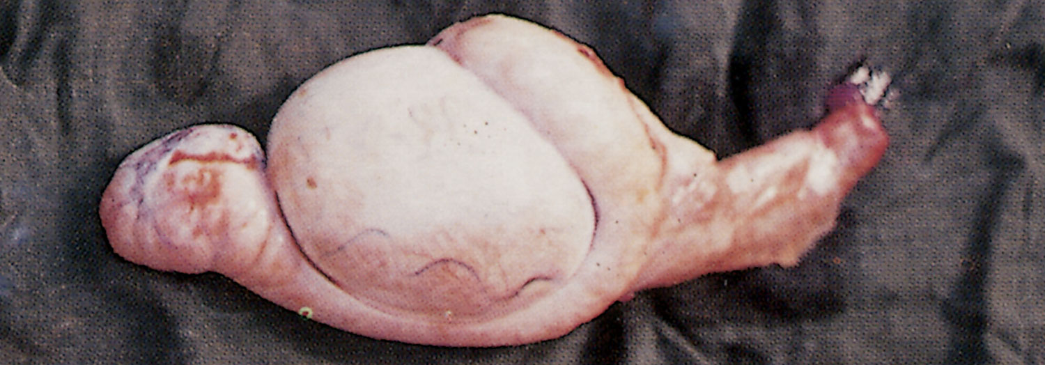
Peracute disease is characterized by severe, rapidly developing uni- or bilateral swelling and pain of the scrotum, high fever, listlessness, loss of appetite and body weight, and a stiff and straddled gait.6, 8, 17, 28, 46, 47, 50, 52, 60 On incision, the cavity of the tunica vaginalis contains copious amounts of a fibrinous to fibrino-purulent exudate (Figure 160.1). The swelling usually subsides after four days to a week and the enlarged epididymis (usually the tail) can then be palpated. 8, 17, 36, 46, 52, 60 In some animals scrotal fistulae, which discharge a slimy, greyish-white to greenish-yellow exudate to the externum, develop.28, 29, 47, 52, 60
Acute epididymitis is characterized by rapid enlargement of the tail of the epididymis without apparent concomitant swelling and pain of the scrotum, although, if the cavity of the tunica vaginalis is exposed by incision,8, 46, 52, 53, 58, 60, 67 variable amounts of fibrinous exudate are usually found to be adherent to the surface of the thickened tunica albuginea and tunica vaginalis.1, 28, 47
Microscopically, acute lesions in the affected part of the epididymis are characterized by necrosis, desquamation of the epithelium and cystic changes of the tubules, infiltration of numerous neutrophils and some macrophages and lymphocytes, and degeneration and necrosis of neutrophils and spermatozoa.8, 28, 29
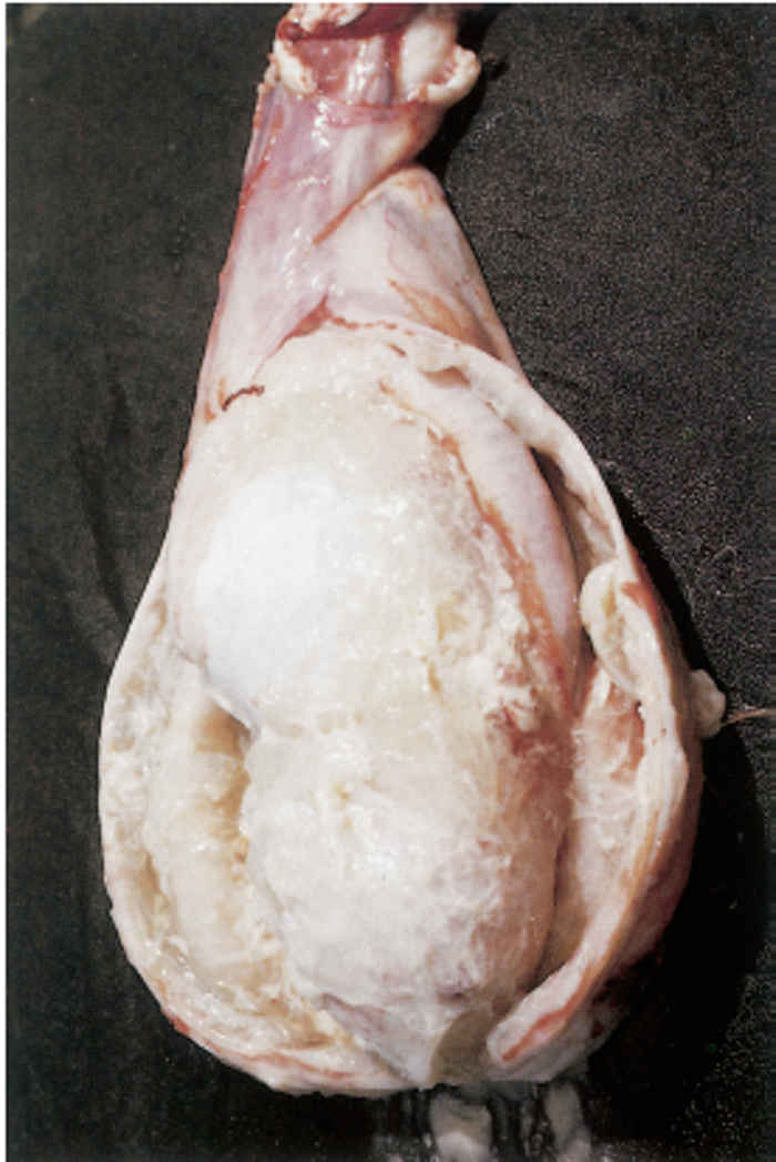
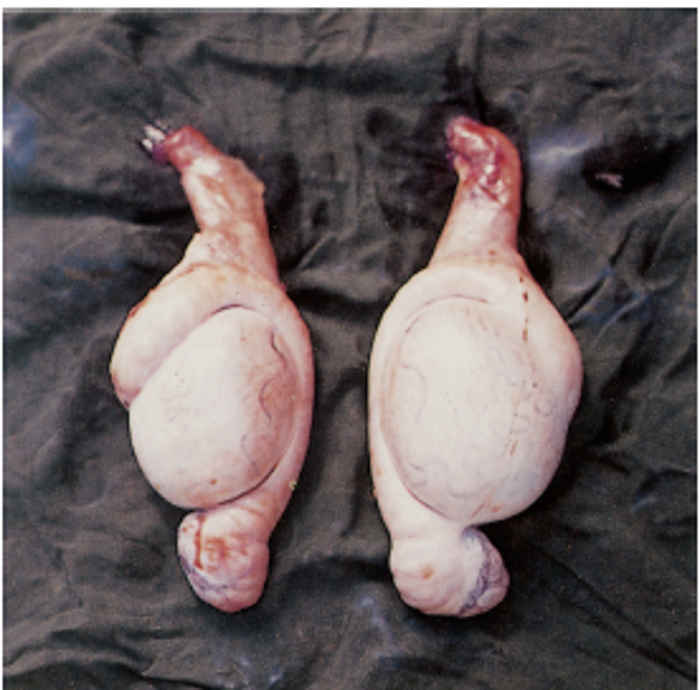
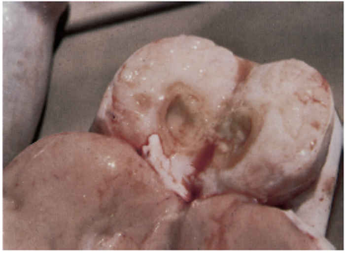
A subacute to chronic epididymitis is characterized by a slow, progressive enlargement and induration of the epididymis which is seldom detected clinically unless the part concerned is palpated. The tail of the epididymis of one or both testes is most commonly enlarged, sometimes by as much as five times, but the head and body of the epididymis may also be affected (Figure 160.2). The affected epididymis is not attached as there are generally no adhesions between the visceral and parietal layers of the tunica vaginalis.6, 8, 11, 17, 18, 28, 29, 46, 52, 53, 60 On incision of the affected part, the tunica albuginea is found to be thickened, the epididymal tissue is fibrotic and enlarged, and a greyish-white, purulent exudate or a granular, cheesy or calcified material is liberated (Figure 160.3). Microscopic lesions are characterized by fibrosis and infiltration of lymphocytes and plasma cells into the interstitium. The ductular epithelium may be hyperplastic and focally infiltrated by neutrophils. Spermatic granulomas, often present in the affected part of the epididymis, are characterized by central masses of sperm cells surrounded by foreign body giant cells, neutrophils, macrophages, lymphocytes, plasma cells, fibroblasts and fibrous connective tissue.8, 28, 29 Testicular atrophy may occur in rams with chronic epididymitis. Increased fibrous connective tissue, infiltration of lymphocytes and plasma cells in the interstitium and amorphous mineralized material in some of the seminiferous tubules may also be evident in the testis.28, 29 The epididymitis may be accompanied by orchitis, ampullitis, seminal vesiculitis, prostatitis, bulbo-urethritis, urethritis, balanoposthitis, chronic pyelonephritis and hydronephrosis.1, 8, 28, 63
Microscopic examination of stained semen smears of a clinically or subclinically affected ram generally shows varying numbers of bacteria, neutrophils, and desquamated epithelial cells.28, 29, 52–54
Apart from lesions in the urogenital system of rams, Gram-negative pleomorphic bacteria have also been associated with epididymitis in ennobled goats52 and cattle,19, 30, 58 acute purulent anterior vaginitis and cervicitis in ewes within two days of mating with infected rams,53 and acute purulent polyarthritis in young lambs. Experimentally, an acute gangrenous mastitis has been produced in ewes by infection of the mammary gland.2, 3, 63
Histophilus ovis has also been isolated from outbreaks of acute highly fatal septicaemia and meningitis in four- to six-month-old lambs. Up to 10 per cent of lambs may die over a period of weeks. Affected lambs are found dead, in recumbency showing nervous signs, or able to stand but severely depressed. Affected lambs usually die within 24 to 48 hours of first showing signs. The necropsy findings are variable and include a severe diffuse fibrinopurulent meningoencephalitis and micro-abscesses in the kidneys, heart, lungs and liver from which the organism has been isolated.23, 34 It has been suggested that the meningoencephalitis in sheep is similar to thrombo-embolic meningoencephalitis in cattle caused by the related organism, H. somni.41, 14
Diagnosis
The diagnosis of epididymitis as a result of Gram-negative pleomorphs is based on epidemiological data, clinical signs and pathology, examination of stained semen smears, and bacteriological examinations.53, 54
Semen smears stained with Giemsa and the modified Ziehl-Neelsen technique (to exclude Brucella ovis as a cause of epididymitis),48, 63 should be examined for the presence of neutrophils, epithelial cells and bacteria, the latter usually being found in close proximity to neutrophils. The examination of smears is useful in the diagnosis of both the clinical and subclinical disease, provided the epididymal ducts are patent.
The isolation of Gram-negative pleomorphs from infected semen samples presents no difficulties. However, the Gram-negative pleomorphic bacteria isolated from the genital tract of rams may be opportunistic pathogens and part of the normal flora,26 and the presence of such bacteria, with or without neutrophils, in semen samples does not signify that they are, in fact, the actual cause of the lesions.
At present there are no reliable serological tests that can be used to confirm a diagnosis of disease or infection caused by Gram-negative pleomorphs.51
Differential diagnosis
Epididymitis in rams caused by Gram-negative pleomorphs should be distinguished from those caused by B. ovis. The latter generally affects older rams and those used for flock breeding purposes, whereas epididymitis caused by Gram-negative pleomorphs occurs more frequently in virgin rams on a high plain of nutrition.12, 16, 37 Brucella ovis can be distinguished from the Gram-negative pleomorphs in semen smears by the fact that it stains positively with the modified Ziehl-Neelsen technique. It also has specific growth requirements and other bacteriological characteristics; and serological tests, such as the complement fixation test, can be used to identify B. ovis-infected rams.
In addition to A. seminis18, 22, 36, 37, 47, 58–60, 67 and H. ovis,53 other Gram-negative pleomorphs such as A. actinomycetemcomitans, 12, 16 Actinobacillus spp.,37 Actinobacillus-like organisms,53, 55, 60 Haemophilus spp., Moraxella spp.,12 Pasteurella spp.28 and pyogenic bacteria such as Trueperella (Corynebacterium) pyogenes and Corynebacterium pseudotuberculosis may be involved in epididymitis. Differentiation of these organisms can only be done by specific laboratory methods.
Control
The treatment of rams suffering from clinical or subclinicial epididymitis is of limited value.12, 17 As the majority of latently infected young rams would seem to clear themselves of infection between weaning and the late two-tooth stage, they should not be culled from the flock at too early an age.54 Final judgement as to the status of infection should preferably only be made when the rams are older than 15 months and after at least three examinations at monthly intervals have been conducted.53, 54 It is suggested that rams subclinically or latently infected at this age should undergo a course of parenterally administered tetracycline therapy, but the results are often disappointing.13, 25
It is well known that young rams may excrete neutrophils in their semen for some time, and that the semen may clear spontaneously without any form of treatment being applied. For this reason, in a treated animal, it is often difficult to decide whether such a positive response is the result of therapy or of self-cure.
It is advised that the breeding rams be tested for infection before the onset of the mating season. Similarly, rams should be tested before they are purchased or introduced to the flock for the first time.
The maintenance after weaning of young ram lambs in open range systems with minimal supplementary feeding has proved effective in the control of epididymitis caused by Gram-negative pleomorphs, but in the highly competitive field of ram production, this method is not very attractive.
At present there is no commercial vaccine available against A. seminis infection.
References
- AL-KATIB, W.A-K.H., 1981. Experimental Actinobacillus seminis infection of rams. Dissertation Abstracts International, 41, No.07, 2415–B. Ph.D. Thesis Kansas State University, Manhattan, U.S.A. (1980). Veterinary Bulletin Abstracts, 50, 944.
- ALESENOY, A.M., 1984. Comparative light and electron microscopic study of experimental Actinobacillus seminis in ewes. Veterinary Bulletin Abstracts, 54, 914.
- ALSENOY, A.M & DENNIS, S.M., 1985. Pathology of acute experimental Actinobacillus seminis mastitis in ewes. Australian Veterinary Journal, 62, 234–237.
- APPUHANY, S., LOW, J.C., COOTE, J.G. & PARTON, R., 1998. PCR methods and plasmid profile analysis for characterisation of Histophilus ovis strains. Journal of Medical Microbiology,47, 11, 987–992.
- BAGLEY, C.V., PASKETT, M.E., MATTHEWS, N.J. & STENQUIST, N.J., 1985. Prevalence and causes of ram epididymitis in Utah. Journal of the American Veterinary Medical Association, 186, 798–801.
- BAYNES, I.D. & SIMMONS, G.C., 1960. Ovine epididymitis caused by Actinobacillus seminis. Australian Veterinary Journal, 36, 454–459.
- BAYNES, I.D. & SIMMONS, G.C., 1966. Actinobacillus seminisin sheep. Australian Veterinary Journal, 42, 225.
- BAYNES, I.D. & SIMMONS, G.C., 1968. Clinical and pathological studies of Border Leicester rams naturally infected with Actinobacillus seminis. Australian Veterinary Journal, 44, 339–343.
- BEAYREGARD, M. & HIGGINS, R., 1983. Ovine mastitis due to Histophilus ovis. Canadian Veterinary Journal, 34, 284–286.
- BELONJE, P.C., 1965. Observations on the post-natal development of the penis in Merino ram lambs and wethers: The possible relationship to the passage of urinary calculi. Journal of the South African Veterinary Medical Association, 36, 381–383.
- BRUERE, A.N., WEST, D.M., MACLACHLAN, N.J., EDWARDS, J.D. & CHAPMAN,H.M., 1977. Genital infection of ram hoggets associated with a Gram-negative pleomorphic organism. New Zealand Veterinary Journal, 25, 191–193.
- BULGIN, M.S. & ANDERSON, B.C., 1983. Association of sexual experience with isolation of various bacteria in cases of ovine epididymitis. Journal of the American Veterinary Medical Association, 372–374.
- BULGIN, M.S., BRUSS, M.L. & ANDERSON, B.C., 1990. Methods for control of lamb epididymitis in large purebred flocks. Journal of the American Veterinary Medical Association, 196, 1110–1115.
- CASSIDY, J.P., MCDOWELL, S.W.J., REILLY, G.A.C., MCCONNEL, W.J., FORSTER, F. & LAWLER, D., 1977. Thrombotic meningoencephalitis associated with Histophilus ovisinfection in lambs in Europe. The Veterinary Record, 140: 8, 193–195.
- COUSINS, D.V. & LLOYD, J.M., 1988. Rapid identification of Haemophilus somnus, Histophilus ovis and Actinobacillus seminis using the APIZYM system. Veterinary Microbiology, 17, 1, 75–81.
- DE LONG, W.J., WALDHALM, D.G. & HALL, R.F., 1979. Bacteria isolates associated with epididymitis in rams from Idaho and Eastern Oregon flocks. American Journal of Veterinary Research, 40, 101–102.
- DE WET, J.A.L., 1985. Siektes by kleinvee. 6. Bybalontsteking (2). Landbouweekblad, 12 April 1985, 38–41.
- DE WET, J.A.L. & ERASMUS, J.A., 1984. Epididymitis of rams in the central and southern districts of the Orange Free State. Journal of the South African Veterinary Association, 55, 173–179.
- DIXON, R.J., STEVENSON, B.J. & SIMS, K.R., 1983. Actinobacillus seminis isolated from cattle. New Zealand Veterinary Journal, 31, 122–123.
- DODD, D.C. & HARTLEY, W.J., 1955. Specific suppurative epididymitis in rams. New Zealand Veterinary Journal, 3, 105–110.
- EKDAHL, M.O., MONEY, D.F.L. & MARTIN, C.A., 1968. Some aspects of epididymitis of rams in New Zealand. New Zealand Veterinary Journal, 16, 81–82.
- ERASMUS, J.A., DE WET, J.A.L. & PROZESKY, L., 1982. Actinobacillus seminis infection in a Walrich ram. Journal of the South African Veterinary Association, 53,129.
- GILL, J., 1992. Haemophilus agni (Histophilus ovis) as a cause of mortality in lambs. Surveillance (Wellington), 19, 2, 13.
- HAJTOS, I., FODOR, L., GLAVITIS, R. & VARGA, J., 1987. Isolation and characterization of Actinobacillus seminis strains from ovine semen samples and epididymitis. Journal of Veterinary Medicine, B (Infectious Diseases, Immunology, Food Hygiene, Veterinary Public Health). 34, 138–147.
- HAJTOS, I. & KIRALY, L., 1990. Effect of oxytetracycline treatment of subclinical genital infections by Histophilus ovis and Actinobacillus seminis in growing rams. (Hungarian) [Az xitetraciklin-kezeles hatasa novendek kosok Histophilus ovis es Actinobacillus seminis okozta szubkliniai genitalis fertozottsegere.] Magyar Allatorvosok Lapja, 45, 15–20.
- HEALY, M.C., HWANG, H.H., KLEINSCHUSTER, S.J., JOHNSON, A.V. & SYMONS, K.S., 1988. Comparison and partial characterization of the protein profiles and other membrane antigens of Actinobacillus species isolated from ram lambs with epididymitis. American Journal of Veterinary Research, 49, 1824–1832.
- HUMPHREY, J.D. & STEPHENS, L.R., 1983. ‘Histophilus somni’: a review. Veterinary Bulletin, 53, 987–1004.
- JANSEN, B.C., 1980. The aetiology of ram epididymitis. Ondersrstepoort Journal of Veterinary Research, 47, 101–107.
- JANSEN, B.C.,1980. A surgical technique for the experimental reproduction of epididymitis in rams. Onderstepoort Journal of Veterinary Research, 47, 281–283.
- JANSEN, B.C.,1981. The pathogenicity of Actinobacillus seminis in cattle. In: Landbounavorsing (Agricultural Research). Pretoria, South Africa: Department of Agriculture.
- JANSEN, B.C.,1983. The epidemiology of bacterial infection of the genitalia in rams. Onderstepoort Journal of Veterinary Research, 50, 275–282.
- JOHNSTONE, I.L., 1948. The growth and development of the penis in sheep: Their possible relationship to posthitis. Australian Veterinary Journal, 24, 86–88.
- KATER, J.C., MARSHAL S.C. & HARTLEY W.J., 1962. A specific suppurative synovitis and pyaemia in ewes. New Zealand Veterinary Journal, 10, 143–144.
- KEARNEY, K.P. & ORR, M.B., 1993. An outbreak of Haemophilus agni-Histophilus ovis septicaemia in lambs. New Zealand Veterinary Journal, 41, 149–150.
- LEES, W.V., MEEK, A.H. & ROSENDAL, S., 1990. Epidemiology of Histophilus somni in young rams. Canadian Journal of Veterinary Research, 54, 331–336.
- LIVINGSTON, C.W. & HARDY, W.J., 1964. Isolation of Actinobacillus seminis from ovine epididymitis. American Journal of Veterinary Research, 25, 660–663.
- LOZANO, E.A., 1986. Etiological significance of bacterial isolates from rams with palpable epididymitis. American Journal of Veterinary Research, 47, 1153–1156.
- MCDOWELL, S.W.J., CASSIDY, J.P. & MCCONNELL, W., 1994. A case of ovine abortion associated with Histophilus ovis Infection. The Veterinary Record, 134, 504.
- MCGILLIVERY, D.J., WEBBER, J.J. & DEAN, H.F., 1986. Characterisation of Histophilus ovis and related organisms by restriction endonuclease analysis. Australian Veterinary Journal, 63, 389–393.
- OSTER, G., 1978. Research review: The blood testis barrier in rats and monkeys. Research in Reproduction, 10, 4.
- PHILBEY, A.W., GLASTONBURG, J.R.W., ROTHWELL, J.T., LINKS, I.J. & SEARSON, J.E., 1991. Meningoencephalitis and other conditions associated with Histophilus ovis infection in sheep. Australian Veterinary Journal, 68, 387–390.
- PIECHULA, K., MUTTERS, R., BURBACH,S., KLUSSMEIER, R., POHL, S. & MANNHEIM, W., 1986. Deoxyribonucleic acid relationships of ‘Histophilus ovis/Histophilus somni’, Haemophilus haemoglobinophilus and Actinobacillus seminis. International Journal of Systematic Bacteriology, 1–7.
- PRETORIUS, P.S. & MARINCOWITZ, G., 1968. Post-natal penis development, testes descent and puberty in Merino lambs on different planes of nutrition. South African Journal of Agricultural Science, 11, 319–334.
- RAHALEY, R.S., 1978. Serological comparison between Histophilus ovis, Actinobacillus seminis and Brucella ovis. Australian Veterinary Journal, 54, 423–425.
- RAHALEY, R.S. & WHITE, W.E., 1977. Histophilus ovis infection in sheep in Western Victoria. Australian Veterinary Journal, 53, 124–127.
- SIMMONS, G.C, BAYNES, I.D. & LUDFORD, C.G., 1966. Epidemiology of Actinobacillus seminis in a flock of Border Leicester sheep. Australian Veterinary Journal, 42, 183–187.
- SPONENBERG, D.P., CARTER, M.E., CARTER, G.R., CORDES, D.O., STEVENS, S.E. & VEIT, H.P., 1983. Suppurative epididymitis in a ram infected with Actinobacillus seminis. Journal of the American Veterinary Medical Association, 182, 990–991.
- STAMP, J.T., MCEWEN, A.D., WATT, J.A.A. & NISBET, D.I., 1950. Enzootic abortion in ewes. 1. Transmission of the disease. The Veterinary Record, 62, 251–254.
- SWANEPOEL, M.L., 1984. A study for the differentiation of Actinobacillus seminis, A. actinomycetemcomitans, Histophilus ovis and Pasteurella haemolytica. Onderstepoort Journal of Veterinary Research, 51, 41–46.
- SWIFT, B.L., CRADDOCK, F., HANCOCK, R.J., THOMAS, G.M., TRUEBLOOD, M.S. & WEIBEL, J., 1982. Ram epididymitis: A clinical report. Theriogenology, 17, 344–347.
- TEKES, L. & HAJTOS, I., 1990. Trials with an enzyme-linked immunosorbent assay (ELISA) for the diagnosis of subclinical genital infections in rams caused by Histophilus ovis and Actinobacillus seminis. Journal of Veterinary Medicine Series B. 37, 549–555.
- VAN TONDER, E.M., 1973. Infection of rams with Actinobacillus seminis. Journal of the South African Veterinary Association, 44, 235–240.
- VAN TONDER, E.M., 1977. Actinobacillus seminis infection in sheep in South Africa. DVSc thesis, University of Pretoria, Pretoria.
- VAN TONDER, E.M., 1977. Examination of rams for genital soundness. Journal of the South African Veterinary Association, 48, 267–272.
- VAN TONDER, E.M., 1979. Actinobacillus seminis infection in sheep in the Republic of South Africa. I. Identifiction of the problem. Onderstepoort Journal of Veterinary Research, 46, 129–133.
- VAN TONDER, E.M., 1979. Actinobacillus seminis infection in sheep in the Republic of South Africa. II. Incidence and geographical distribution. Onderstepoort Journal of Veterinary Research, 46, 135–140.
- VAN TONDER, E.M., 1979. Actinobacillus seminis infection in sheep in the Republic of South Africa. III. Growth and cultural characteristics of A. seminis. Onderstepoort Journal of Veterinary Research, 46, 141–148.
- VAN TONDER, E.M., & BOULTON, T.F.W., 1970. The isolation of Actinobacillus seminis from bovine semen: A preliminary report. Journal of the South African Veterinary Medical Associatoin, 41, 287–288.
- WALKER, R.L., BIBERSTEIN, E.L., PRITCHETT, R.F. & KIRKHAM, C. 1985. Deoxyribonucleic acid relatedness among Histophilus somni, Haemophilus agni, Histophilus ovis, Actinobacillus seminis and Haemophilus influenza. International Journal of Systematic Bacteriology, 35, 46–49.
- WALKER, R.L. & LEAMASTER, B.R., 1986. Prevalence of Histophilus ovis and Actinobacillus seminis in the genital tract of sheep. American Journal of Veterinary Research, 47, 1928–1980.
- WALKER, R.L. & LEAMASTER, B.R., STELLFLUG, J.N., & BIBERSTEIN, E.L., 1986. Association of age of rams with distribution of epididymal lesions and etiologic agent. Journal of the American Veterinary Medical Association, 188, 393–396.
- WATSON, R.H., SAPSFORD, C.S. & MCCANE, I., 1956. The development of the testis, epididymis and penis in the young Merino ram. Australian Journal of Agricultural Research, 7, 574–590.
- WATT, D.A., 1970. Investigations of ovine brucellosis in Merino rams of Western Australia. Australian Veterinary Journal, 46, 506–508.
- WEBB, D.A., BAMFORD, V. & NAIRN, M.E., 1970. Actinobacillus seminis as a cause of polyarthritis and posthitis in sheep. Australian Veterinary Journal, 46, 516.
- WEBB, R.F., 1983. Bacteriological characteristics of Histophilus ovis and its relationship to similar bacteria. Research in Veterinary Science, 35, 25–29.
- WIGGINS E.L. & TERRILL, C.E., 1953. Variation in penis development in ram lambs. Journal of Animal Science, 12, 524–535.
- WORTHINGTON, R.W. & BOSMAN, P.P., 1968. Isolation of Acintobacillus semini in South Africa. Journal of the South African Veterinary Medical Association, 39, 81–85.


