- Infectious Diseases of Livestock
- Part 3
- Klebsiella spp. infections
- GENERAL INTRODUCTION: SPIROCHAETES
- Swine dysentery
- Borrelia theileri infection
- Borrelia suilla infection
- Lyme disease in livestock
- Leptospirosis
- GENERAL INTRODUCTION: AEROBIC ⁄ MICRO-AEROPHILIC, MOTILE, HELICAL ⁄ VIBROID GRAM-NEGATIVE BACTERIA
- Genital campylobacteriosis in cattle
- Proliferative enteropathies of pigs
- Campylobacter jejuni infection
- GENERAL INTRODUCTION: GRAM-NEGATIVE AEROBIC OR CAPNOPHILIC RODS AND COCCI
- Moraxella spp. infections
- Bordetella bronchiseptica infections
- Pseudomonas spp. infections
- Glanders
- Melioidosis
- Brucella spp. infections
- Bovine brucellosis
- Brucella ovis infection
- Brucella melitensis infection
- Brucella suis infection
- Brucella infections in terrestrial wildlife
- GENERAL INTRODUCTION: FACULTATIVELY ANAEROBIC GRAM NEGATIVE RODS
- Klebsiella spp. infections
- Escherichia coli infections
- Salmonella spp. infections
- Bovine salmonellosis
- Ovine and caprine salmonellosis
- Porcine salmonellosis
- Equine salmonellosis
- Yersinia spp. infections
- Haemophilus and Histophilus spp. infections
- Haemophilus parasuis infection
- Histophilus somni disease complex in cattle
- Actinobacillus spp. infections
- Actinobacillus equuli infections
- Gram-negative pleomorphic infections: Actinobacillus seminis, Histophilus ovis and Histophilus somni
- Porcine pleuropneumonia
- Actinobacillus suis infections
- Pasteurella and Mannheimia spp. infections
- Pneumonic mannheimiosis and pasteurellosis of cattle
- Haemorrhagic septicaemia
- Pasteurellosis in sheep and goats
- Porcine pasteurellosis
- Progressive atrophic rhinitis
- GENERAL INTRODUCTION: ANAEROBIC GRAM-NEGATIVE, IRREGULAR RODS
- Fusobacterium necrophorum, Dichelobacter (Bacteroides) nodosus and Bacteroides spp. infections
- GENERAL INTRODUCTION: GRAM-POSITIVE COCCI
- Staphylococcus spp. infections
- Staphylococcus aureus infections
- Exudative epidermitis
- Other Staphylococcus spp. infections
- Streptococcus spp. infections
- Strangles
- Streptococcus suis infections
- Streptococcus porcinus infections
- Other Streptococcus spp. infections
- GENERAL INTRODUCTION: ENDOSPORE-FORMING GRAM-POSITIVE RODS AND COCCI
- Anthrax
- Clostridium perfringens group infections
- Clostridium perfringens type A infections
- Clostridium perfringens type B infections
- Clostridium perfringens type C infections
- Clostridium perfringens type D infections
- Malignant oedema⁄gas gangrene group of Clostridium spp.
- Clostridium chauvoei infections
- Clostridium novyi infections
- Clostridium septicum infections
- Other clostridial infections
- Tetanus
- Botulism
- GENERAL INTRODUCTION: REGULAR, NON-SPORING, GRAM-POSITIVE RODS
- Listeriosis
- Erysipelothrix rhusiopathiae infections
- GENERAL INTRODUCTION: IRREGULAR, NON-SPORING, GRAM-POSITIVE RODS
- Corynebacterium pseudotuberculosis infections
- Corynebacterium renale group infections
- Bolo disease
- Actinomyces bovis infections
- Trueperella pyogenes infections
- Actinobaculum suis infections
- Actinomyces hyovaginalis infections
- GENERAL INTRODUCTION: MYCOBACTERIA
- Tuberculosis
- Paratuberculosis
- GENERAL INTRODUCTION: ACTINOMYCETES
- Nocardiosis
- Rhodococcus equi infections
- Dermatophilosis
- GENERAL INTRODUCTION: MOLLICUTES
- Contagious bovine pleuropneumonia
- Contagious caprine pleuropneumonia
- Mycoplasmal pneumonia of pigs
- Mycoplasmal polyserositis and arthritis of pigs
- Mycoplasmal arthritis of pigs
- Bovine genital mycoplasmosis
- Neurotoxin-producing group of Clostridium spp.
- Contagious equine metritis
- Tyzzer's disease
- MYCOTIC AND ALGAL DISEASES: Mycoses
- MYCOTIC AND ALGAL DISEASES: Pneumocystosis
- MYCOTIC AND ALGAL DISEASES: Protothecosis and other algal diseases
- DISEASE COMPLEXES / UNKNOWN AETIOLOGY: Epivag
- DISEASE COMPLEXES / UNKNOWN AETIOLOGY: Ulcerative balanoposthitis and vulvovaginitis of sheep
- DISEASE COMPLEXES / UNKNOWN AETIOLOGY: Ill thrift
- Eperythrozoonosis
- Bovine haemobartonellosis
Klebsiella spp. infections
This content is distributed under the following licence: Attribution-NonCommercial CC BY-NC  View Creative Commons Licence details here
Klebsiella spp. infections
M M HENTON
Introduction
Infections with Klebsiella spp. cause a variety of syndromes in livestock, the most important being endometritis, infertility and abortion in mares, mastitis in cows and sows, and neonatal septicaemia, particularly in foals. In southern Africa, the majority of infections are associated with the genital tract of Thoroughbred mares. Antibiotic resistant strains of Klebsiella commonly cause nosocomial infections in humans.28
Klebsiella was first described by Escherich in 1885 as Bakterium lactis aerogenes but as this name was later also used for Enterobacter, it was changed to Klebsiella, after the German bacteriologist, Edwin Klebs.28
The pathogenic capsular types found in South Africa are similar to those in other parts of the world (Table 149.1).4, 10, 22, 29
Aetiology and epidemiology
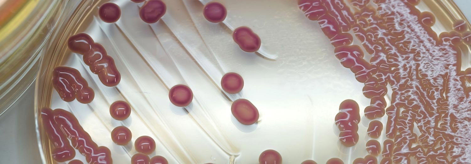
The genus Klebsiella belongs to the family Enterobacteriaceae. Klebsiella spp. are facultative anaerobes and are large, non-motile, Gram-negative rods (0,3 to 1,0 μm wide and 0,6 to 6,0 μm long) usually containing a thick, mucoid capsule.
In the laboratory they grow well on most growth media, forming large, moist and viscid colonies. Klebsiella spp. are inositol-positive, and most pathogenic strains are rapidly urease-positive. They are classified biochemically into K. pneumoniae, K. oxytoca, K. terrigena, and K. planticola. 28 Klebsiella pneumoniae is subdivided into three subspecies: pneumoniae, ozaenae and rhinoscleromatis. Klebsiella pneumoniae and K. oxytoca are isolated from various pathological processes in humans and animals, while K. terrigena and K. planticola are mainly found in soil and water.28
The biochemically defined species of Klebsiella are serotyped with one set of capsular typing antisera.9 Currently, all strains of Klebsiella are serologically classified into 77 currently recognized capsular types,28 and numbered from 1 to 82, as five of them (capsular types 73, 75, 76, 77 and 78) were subsequently found to be invalid.12 The lipopolysaccharide O-antigens are not usually determined, as the test methods (either unencapsulated mutants have to be selected, or an inhibition enzyme-linked immunosorbent assay performed) are cumbersome, and not as discriminatory, as there are only nine currently recognized O-groups.12 The serological classification gives a much clearer indication of pathogenicity than does biochemical typing.17, 26, 27 Plasmid profiles of isolates from horses and bovine mastitis show promise as indicators of virulence.16, 18 Fimbrial adhesins have been described, but their role in the pathogenesis of disease in animals is still unclear.31
Serotyping is done by means of the quelling (Neufeld) reaction; 9 live bacteria are mixed with capsular antisera, and the reaction indicated by refractile, precipitin patterns which are discernible microscopically on the surface of the bacterial capsule. The O-types of Klebsiella are usually not characterized, as they are few in number, and typing of the O-antigens is hampered by the large capsule.
Table 149.1 Number of common capsular types of Klebsiella spp. isolated from horses at the Veterinary Research Institute, Onderstepoort, from 1972 to 1999
| CAPSULAR TYPES | SUBCLINICAL INFECTIONS | DISEASED CASES | DISEASED CASES | ||
|---|---|---|---|---|---|
| Mares | NAMIBIA | RSA* HIGHVELD | RSA* BUSHVELD | ||
| 1 | 5 | 9 | 20 | - | 34 |
| 2 | 3 | 1 | 20 | - | 24 |
| 3 | 10 | 25 | 5 | - | 40 |
| 6 | 14 | 9 | 30 | 1 | 54 |
| 7 | 28 | 19 | 13 | - | 60 |
| 20 | 1 | - | 10 | - | 11 |
| 21 | 19 | 15 | 17 | - | 51 |
| 22 | 1 | 1 | 5 | - | 7 |
| 27 | 4 | 2 | 3 | - | 9 |
| 30 | 13 | 11 | 14 | - | 38 |
| 31 | 8 | - | - | - | 8 |
| 33 | 5 | 4 | 6 | - | 15 |
| 64 | 6 | 0 | 1 | - | 7 |
Klebsiella spp. are ubiquitous and occur in soil and water, or are part of the normal flora in the gastrointestinal tract of animals, albeit often in low numbers. They are particularly common in contaminated wood shavings and soil-containing shavings.25 Wood shavings used as bedding are therefore an important source of infection, especially when severely contaminated with faeces.27
Klebsiellae are opportunistic pathogens. The pathogenicity of specific capsular types for livestock species other than horses is largely unknown. In South Africa, capsular types 2, 6, 21, 30 and 33 are most commonly isolated from cattle. The types that are usually associated with mastitis in cows are 2, 19, 21, 30, 33 and 53. Opportunistic infections in pigs are more commonly caused by capsular types 22, 30, 31 and 33, and in sheep and goats types 2, 6, 21 and 33 predominate.13
Identification of the capsular types of Klebsiella isolated from the genital tract of horses from 1972 to 1999 at the Veterinary Research Institute, Onderstepoort, showed that certain types are more common than others (Table 149.1). Stallions may be subclinical carriers of these types and may infect mares venereally.5, 17, 19 It was found that capsular types 1, 2 and 6 were associated with endometritis in mares. Types such as 3, 31 and 64 occurred only as transient colonists of the uterus, whereas types 6, 7, 21 and 30 that commonly infect the immunocompromised uterus, were also regularly isolated from the cervix of healthy horses.
Endometritis in mares
Flaccid, gaping vulvar lips, a vulva which slants dorso-cranially, or pneumovagina as a result of poor conformation in which the dorsal commissure of the vulva is displaced to a position dorsally of the ischeal arch, foaling injuries to the genitalia (e.g. recto-vaginal lacerations or fistulas), urovagina and/or urouterus, ageing of the mare, and poor condition may predispose to Klebsiella endometritis. Endometritis usually results from an ascending infection after faecal contamination of the vulval lips, vestibulum and clitoral fossa.30
As Klebsiella is relatively resistant to most disinfectants, the injudicious use of disinfectants to wash the genitalia of mares and stallions may inhibit the normal genital flora and stimulate overgrowth of Klebsiella. Similarly, treatment of mares suffering from endometritis with antimicrobials not selected on the basis of an antibiogram may result in overgrowth of Klebsiella.
The strains of K. pneumoniae isolated from mares with endometritis are divided into:29
- Venereal strains (capsular types 1, 2 and 5): These strains are mostly transmitted venereally, but may also be transmitted mechanically by stallions that are not carriers of the infection. Environmental spread through contamination of pastures or bedding is also possible.
- Strains which infect the immunocompromised uterus (capsular types 6, 7, 21 and 30): The term ‘immunocompromised uterus’, as defined by Ricketts,29 embraces any malfunction of the uterus, be it structural or hormonal. Colonization of the external genitalia by these strains is common but transient in healthy animals.29 Stallions may transmit these organisms mechanically.
- Opportunists: Any of the other 82 capsular types may cause opportunistic venereal infections in horses.
Klebsiella is transmitted venereally or mechanically to mares. Transmission of Klebsiella may also occur by using improperly disinfected specula in mares.29 The bacteria may be isolated from the semen as well as the sheath of stallions that are carriers.15 The infection rate in mares served by a carrier stallion may be as low as 25 per cent, even with the most common pathogenic type (capsular type 1).13 Of those infected, more than 50 per cent recover spontaneously or after treatment. Mares may, however, remain infected for prolonged periods and shed Klebsiella type 1 for 1 to 17 months.13 On rare occasions, mares may remain infected and barren for up to five years.13
As the breeding of Thoroughbreds is seasonal, more cases of Klebsiella infections are detected in the spring than at any other time of the year.
Pastures may be contaminated by vaginal discharges and faeces containing Klebsiella.23 Horses grazing on these pastures may become infected and the bacteria may become part of their normal gastrointestinal tract flora.17
Mastitis
Mastitis caused by Klebsiella in cows is most common in countries with a cold climate where cows are kept indoors, especially on wood shavings.25 In South Africa, however, there is a marked preponderance of cases during the hot summer months of December to February.13 This may be related to calving, as Klebsiella mastitis is more common during early lactation when the udder is oedematous and milk production is at its highest.26, 27 Severe toxaemia, due to absorption of endotoxin, may occur in animals with peracute mastitis.24
Pathogenesis
In mares predisposed to endometritis, infection and colonization of Klebsiella usually results from contamination of the vulvar, clitoral and/or vestibular areas. From these areas the infection ascends to involve the uterus. Depending on the strain of Klebsiella involved and the susceptibility of the mare, the genital tract may become infected for a variable period. Semen seems to promote Klebsiella infections; mares that are repeatedly served carry the infection longer than those that have sexual rest.30
Clinical signs
Endometritis in mares
Mares predisposed to endometritis may become infected and show clinical signs at any stage of the oestrous cycle or during pregnancy. Venereally infected mares usually develop a vaginal discharge within three to five days after being served by a shedder stallion and are usually temporarily barren.
Endometritis in mares is generally manifested clinically by the presence of a scant or profuse greyish, viscid vaginal discharge which may contain floccules of pus. In mares with a profuse vaginal discharge, the hair on the buttocks and the tail may be matted by the exudate. The mucous membrane of the vagina and cervix is often dull and reddish-brown to purplish-red. In advanced cases, pyometra may result.30 On rectal palpation, the uterus is enlarged and atonic. The oestrous cycle in affected mares may be irregular, especially if the infection is secondary to a hormonal imbalance. Generally, mares and stallions with genital infection do not show systemic illness.
If a foetus becomes infected, abortion may occur, a weak, septicaemic foal may be born, or the foal may be normal at birth and develop septicaemia soon after birth.33, 34 Septicaemic foals show anorexia, depression, high fever, and usually develop severe polyarthritis, pneumonia and/or meningoencephalitis. If left untreated such foals die within a few days of birth.
Stallions rarely show any clinical signs but are commonly subclinically infected with Klebsiella.The prepuce is frequently colonized but Klebsiella may also infect the accessory sexual glands, kidneys or urinary bladder of stallions.5, 17, 19
Mastitis
The mastitis-metritis-agalactiae syndrome in sows is only rarely associated with Klebsiella infections, and when it occurs, it usually occurs during the immediate post-partum period.2
Klebsiella generally causes peracute mastitis in sows and cows, characterized by severe systemic signs which include fever, anorexia, and increased heart and respiratory rates. The milk is yellowish and watery and contains small floccules of pus. In animals with acute to chronic mastitis, systemic signs are mild or inapparent. The udder is usually not markedly swollen nor painful in animals with peracute, acute or chronic mastitis.2, 4
Other syndromes
Other syndromes that are caused by Klebsiella, such as septicaemia and pneumonia, occur sporadically. Young calves and piglets drinking milk from cows or sows with Klebsiella mastitis may die from septicaemia. Sporadic cases of septicaemia or pneumonia in livestock as a result of Klebsiella may occur. Polyarthritis in lambs has been reported.1
Pathology
Endometritis in mares
The lesions in mares suffering from endometritis vary according to the stage of development of the lesions, stage of the oestrous cycle, type of Klebsiella involved and which predisposing factors (vide supra) are present. In mild cases, only the superficial layers of the endometrium are affected, but in severe cases the endometrium may be focally denuded and have a dull, red, granular appearance.30 On histopathology, small abscesses may be present in the uterine glands and the luminal epithelium may be covered with small or copious amounts of pus, which is usually mucoid. As with other bacterial infections of the endometrium, a diffuse or focal neutrophilic infiltrate is present in one or more layers of the endometrium. In chronic infections, atrophy and fibrosis of the endometrium may be evident. Bacteria cannot always be found in histopathological sections of endometrial biopsies taken from Klebsiella-infected mares.
Endometrial or cervical cytology usually reveals a large number of neutrophils and increased numbers of epithelial cells, while bacteria may or may not be present, but are generally more plentiful than in histopathological sections.
Mastitis
Apart from the usual inflammatory changes associated with mastitis, severe cases may also develop thrombosis and infarction of the parenchyma.
Diagnosis and differential diagnosis
Endometritis in mares
Endometritis due to infection by Klebsiella spp. in mares is very similar clinically and pathologically to that caused by other bacteria, including Streptococcus spp., Pseudomonas aeruginosa and Taylorella equigenitalis.20, 21 Endometrial bacterial culture and cytology are therefore necessary to confirm the diagnosis. Swabs for bacterial isolation should be taken from the urethral fossa and sheath in stallions and from the endometrium of mares in oestrus. The bacteriological examination of swabs taken from the urethra, cervix and clitoral fossa of mares is a useful means of establishing persistent carriers.
Mastitis
Klebsiella mastitis may be confused with other causes of mastitis, particularly Escherichia coli infection, and can only be distinguished on bacterial culture.
Other syndromes
Apart from Klebsiella, septicaemia in foals is frequently caused by infection of E. coli, Actinobacillus equuli, A. suis or Streptococcus spp. acquired in utero, during parturition or during the early neonatal period.34
Control
Endometritis in mares
As Klebsiella is common in the intestinal tract of healthy animals and is also commonly found in the environment,28 methods to control Klebsiella infections should be aimed at those strains residing in or infecting the genital tract.
To prevent spread of infection on a stud farm, it is important to take swabs from mares that are possibly infected before they are covered. Stallions should also be swabbed two or three times during the breeding season.
Structural defects of the genital tract which predispose to infection, should be rectified surgically before any antimicrobial therapy is started. Mares and stallions carrying Klebsiella types 1, 2 and 5 should preferably be given sexual rest for a year. Mares should be treated as soon as possible with intrauterine antimicrobials (chosen with due regard to the antibiotic sensitivity of the isolate) to prevent endometrial fibrosis and should only be bred again after they have been shown to have recovered completely from the infection. Carrier stallions may be treated with disinfectants such as chlorhexidine or antimicrobials instilled into the sheath, but as most stallions are transient carriers and self-cure will result, it is preferable not to treat them. The injudicious use of antimicrobials and disinfectants can diminish the resident flora of the sheath and actually promote the multiplication of Klebsiella. If such stallions are to be used for breeding, artificial insemination with semen incubated in a diluent containing an appropriate antibiotic is effective in preventing the transmission of the infection to mares. Where artificial insemination is not allowed or practically impossible to perform, the semen diluent can be instilled into the mare’s uterus immediately prior to natural service. Such mares should then be treated with an appropriate antibiotic for at least three days after service.
Mares and stallions should be swabbed at least three times at 7- to 14-day intervals with at least one swab taken during oestrus, after treatment or sexual rest to ensure that they are free of infection. During this time infected mares should be kept in isolation and the swabbing repeated if necessary until all three swabs are found to be free of Klebsiella.
Strict hygiene should be applied at service and during any veterinary examination. Recovered mares should preferably only be mated once (as close as possible to ovulation), and antibiotic therapy as indicated by an antibiogram is usually necessary.32
Faeces should be removed from the stables at least once a day to minimize faecal contamination, and straw should be used instead of wood shavings. Efforts should be made to keep the bedding as dry as possible. All tail bandages and reproductive instruments should be disposable or sterilized after each use.
Mastitis
General mastitis therapy as well as supportive therapy using corticosteroids and intravenous fluids is necessary in Klebsiella mastitis. Drugs such as phenylbutazone and flunixin meglumine appear to have no beneficial effect.6 The addition of lime to sawdust bedding (1 kg lime to 10 kg sawdust) may be of value in the initial control of an outbreak. Lime decreases general bacterial counts in sawdust as well as decreasing the levels of Klebsiella on the teat skin, but this effect only lasts for two days, before bacterial levels rise again.14
References
- BERNABE, A., CONTRERAS, A., GOMEZ, M.A., SANCHEZ, A., CORRALES, J.C. & GOMEZ, A., 1998. Polyarthritis in kids associated with Klebsiella pneumoniae. The Veterinary Record, 142, 64–66.
- BERTSCHINGER, H.U., POHLENZ, J. & ROSS, R.F., 1986. Coliform mastitis. In: leman, a.d., straw, b., glock, r.d., mengeling, w.l., penny, r.h.c. & scholl, e., (eds). Diseases of Swine, 6th edn. Ames: Iowa State University Press.
- BLANCHARD, T.L., VARNER, D.D., LOVE, C.C., HURTGEN, J.P., CUMMINGS, N.R. & KENNEY, R.M., 1987. Use of a semen extender containing antibiotic to improve the fertility of a stallion with seminal vesiculitis due to Pseudomonas aeruginosa. Theriogenology, 28, 541–546.
- BRAMAN, S.K., EBERHART, R.J., ASBURY, M.A. & HERMANN, G.J., 1973. Capsular types of Klebsiella pneumoniae associated with bovine mastitis. Journal of the American Veterinary Medical Association, 162, 109–111.
- CROUCH, J.R.F., ATHERTON, J.C. & PLATT, H., 1972. Venereal transmission of Klebsiella aerogenes in a Thoroughbred stud from a persistently infected stallion. The Veterinary Record, 90, 21–28.
- DASCANIO, J.J., MECHOR, G.D., GROHN, Y.T., KENNEY, D.G., BOOKER, C.A., THOMSON, P., CHIFELLE, C.L., MUSSER, J.M. & WARNICK, L.D., 1995. Effect of phenylbutazone and flunixin meglumine on acute toxic mastitis in dairy cows. American Journal of Veterinary Research, 56, 1213–1218.
- DUGUID, J.P., 1959. Fimbriae and adhesive properties in Klebsiella strains. Journal of General Microbiology, 21, 271–286.
- ESCHERICH, T., 1885. Die Darmbacterien des Neugeborenen und Säuglings. Fortschrift Medicine, 3, 515–522 and 547–554.
- EWING, W.H., 1986. Edwards and Ewing’s Identification of Enterobacteriaceae. 4th edn. New York: Elsevier Science Publishing Company.
- FLOER, W., 1974. Klebsiella organisms in the genital organs and foetuses of Thoroughbred horses. Deutsche Tierärztliche Wochenschrift, 81, 20–22.
- GERLACH, G.F., 1989. Genetics and function of Klebsiella pneumoniae adhesins associated with fimbriae. Habilitations-schrift, Tierärztliche Hochschule, Hannover, Germany 140 pp.
- HANSEN, D.S., MESTRE, F., ALBERTI, S., HERNANDEZ-ALLES, S., ALVAREZ, D., DOMENECH-SANCHEZ, A., GIL, J., MERINO, S., TOMAS, J.M. & BENEDI, V.J., 1999. Klebsiella pneumoniae Lipopolysaccharide O Typing: Revision of Prototype Strains and O-Group Distribution among Clinical Isolates from Different Sources and Countries. Journal of Clinical Microbiology, 37, 56–62.
- HENTON, M.M., 1999. Onderstepoort Veterinary Institute, South Africa. Unpublished data.
- HOGAN, J.S. & SMITH, K.L., 1997. Bacteria counts in sawdust bedding. Journal of Dairy Science, 80, 1600–1605.
- KENNEY, R.M., BERGMAN, R.V., COOPER, W.L. & MORSE, G.W., 1975. Minimal contamination techniques for breeding mares: Technique and preliminary findings. Proceedings of the American Association of Equine Practitioners, S8–S15.
- KIKUCHI, N., BLAKESLEE, J.R. & HIRAMUNE, T., 1995. Plasmid profiles of Klebsiella pneumoniae isolated from horses. Journal of Veterinary Medicine and Science, 57, 113–115.
- KIKUCHI, N., IGUCHI, I. & HIRAMUNE, T., 1987. Capsule types of Klebsiella pneumoniae isolated from the genital tract of mares with metritis, extragenital sites of healthy mares and the genital tract of stallions. Veterinary Microbiology, 15, 219–228.
- KIKUCHI, N., KAGOTA, C., NOMURA, T., HIRAMUNE, T., TAKAHASHI, T. & YANAGAWA, R., 1995. Plasmid profiles of Klebsiella pneumoniae isolated from bovine mastitis. Veterinary Microbiology, 47, 9–15.
- KIKUCHI, N., TAKAYANAGI, N., KOSAKA, T., HIRAMUNE, T. & YANAGAWA, R., 1988. Presence of less heavily encapsulated Klebsiella pneumoniae capsular type 1 in semen of healthy stallions and cervical swabs of mares suffering from metritis and comparison of virulence between heavily and less heavily encapsulated strains. Japanese Journal of Veterinary Science, 50, 313–323.
- LE BLANC, M.M., 1989. Treatment protocols and preventive practices for mares with endometritis. Veterinary Medicine, 84, 906–911.
- LIU, I.K.M., 1989. Uterine infections in the mare. Equine Practice, 11, 26–32.
- MERKT, H., KLUG, E. BÖHM, K.H. & WEISS, R., 1974. Klebsiella organisms as agents of genital infection in horses. Berliner und Muünchener Tierärtztliche Wochenschrift, 87, 405–409.
- MERKT, H., KLUG, E., BÖHM, K.H. & WEISS, R., 1975. Recent observations concerning Klebsiella infections in stallions. Journal of Reproduction and Fertility, suppl. 23, 143–145.
- NEUMANN, P. & WENDT, K., 1985. Aetiopathogenesis of bovine mastitis due to Klebsiella pneumoniae. Monatschrifte für Veterinärmedizin, 40, 120–123.
- NEWMAN, L.E. & KOWALSKI, J., 1973. Fresh sawdust bedding as a potential source of Klebsiella organisms. American Journal of Veterinary Research, 34, 979–980.
- NOMURA, T., SUGIURA, H., MORIYA, H., KIKUCHI, N., HIRAMUNE, T. & YANAGAWA, R., 1989. Species and capsular types of Klebsiella isolated from cows and their environment. Journal of Rakuno Gakuen University, Natural Science, 14, 57–61.
- NONNECKE, B.J. & NEWBOULD, F.H., 1984. Biochemical and serologic characterization of Klebsiella strains from bovine mastitis and the environment of the dairy cow. American Journal of Veterinary Research, 45, 2451–2454.
- ORSKOV, I., 1984. Genus V. Klebsiella. In: KRIEG, N.R. & HOLT, J.G., (eds). Bergey’s Manual of Systematic Bacteriology. Baltimore, London: Williams and Wilkins.
- RICKETTS, S.W., 1981. Bacteriological examinations of the mare’s cervix: Techniques and interpretation of results. The Veterinary Record, 108, 46–51.
- ROBERTS, S.J., 1971. Veterinary Obstetrics and Genital Diseases. Ithaca, New York: Edwards Brothers.
- SEBGHATI, T.A.S., KORHONEN, T.K., HORNICK, D.B. & CLEGG, S., 1998. Characterization of the Type 3 Fimbrial Ashesins of Klebsiella strains. Infection and Immunity, 66, 2887–2894.
- VOLKMANN, D.H., 1992. Minimal contamination breeding techniques. Proceedings of Equine Practitioners Group Congress, 24–27 February 1992, Badplaas: 117–119
- WHITWELL, K.E., 1988. Infective placentitis in the mare. Equine Infectious Diseases, Proceedings of the Fifth International Conference. University of Kentucky, USA
- WILSON, W.D. & MADIGAN, J.E., 1989. Comparison of bacteriologic culture of blood and necropsy specimens for determining the cause of foal septicaemia: 47 cases (1978–1987). Journal of the American Veterinary Medical Association, 195, 1759–1763


