- Infectious Diseases of Livestock
- Part 3
- Yersinia spp. infections
- GENERAL INTRODUCTION: SPIROCHAETES
- Swine dysentery
- Borrelia theileri infection
- Borrelia suilla infection
- Lyme disease in livestock
- Leptospirosis
- GENERAL INTRODUCTION: AEROBIC ⁄ MICRO-AEROPHILIC, MOTILE, HELICAL ⁄ VIBROID GRAM-NEGATIVE BACTERIA
- Genital campylobacteriosis in cattle
- Proliferative enteropathies of pigs
- Campylobacter jejuni infection
- GENERAL INTRODUCTION: GRAM-NEGATIVE AEROBIC OR CAPNOPHILIC RODS AND COCCI
- Moraxella spp. infections
- Bordetella bronchiseptica infections
- Pseudomonas spp. infections
- Glanders
- Melioidosis
- Brucella spp. infections
- Bovine brucellosis
- Brucella ovis infection
- Brucella melitensis infection
- Brucella suis infection
- Brucella infections in terrestrial wildlife
- GENERAL INTRODUCTION: FACULTATIVELY ANAEROBIC GRAM NEGATIVE RODS
- Klebsiella spp. infections
- Escherichia coli infections
- Salmonella spp. infections
- Bovine salmonellosis
- Ovine and caprine salmonellosis
- Porcine salmonellosis
- Equine salmonellosis
- Yersinia spp. infections
- Haemophilus and Histophilus spp. infections
- Haemophilus parasuis infection
- Histophilus somni disease complex in cattle
- Actinobacillus spp. infections
- Actinobacillus equuli infections
- Gram-negative pleomorphic infections: Actinobacillus seminis, Histophilus ovis and Histophilus somni
- Porcine pleuropneumonia
- Actinobacillus suis infections
- Pasteurella and Mannheimia spp. infections
- Pneumonic mannheimiosis and pasteurellosis of cattle
- Haemorrhagic septicaemia
- Pasteurellosis in sheep and goats
- Porcine pasteurellosis
- Progressive atrophic rhinitis
- GENERAL INTRODUCTION: ANAEROBIC GRAM-NEGATIVE, IRREGULAR RODS
- Fusobacterium necrophorum, Dichelobacter (Bacteroides) nodosus and Bacteroides spp. infections
- GENERAL INTRODUCTION: GRAM-POSITIVE COCCI
- Staphylococcus spp. infections
- Staphylococcus aureus infections
- Exudative epidermitis
- Other Staphylococcus spp. infections
- Streptococcus spp. infections
- Strangles
- Streptococcus suis infections
- Streptococcus porcinus infections
- Other Streptococcus spp. infections
- GENERAL INTRODUCTION: ENDOSPORE-FORMING GRAM-POSITIVE RODS AND COCCI
- Anthrax
- Clostridium perfringens group infections
- Clostridium perfringens type A infections
- Clostridium perfringens type B infections
- Clostridium perfringens type C infections
- Clostridium perfringens type D infections
- Malignant oedema⁄gas gangrene group of Clostridium spp.
- Clostridium chauvoei infections
- Clostridium novyi infections
- Clostridium septicum infections
- Other clostridial infections
- Tetanus
- Botulism
- GENERAL INTRODUCTION: REGULAR, NON-SPORING, GRAM-POSITIVE RODS
- Listeriosis
- Erysipelothrix rhusiopathiae infections
- GENERAL INTRODUCTION: IRREGULAR, NON-SPORING, GRAM-POSITIVE RODS
- Corynebacterium pseudotuberculosis infections
- Corynebacterium renale group infections
- Bolo disease
- Actinomyces bovis infections
- Trueperella pyogenes infections
- Actinobaculum suis infections
- Actinomyces hyovaginalis infections
- GENERAL INTRODUCTION: MYCOBACTERIA
- Tuberculosis
- Paratuberculosis
- GENERAL INTRODUCTION: ACTINOMYCETES
- Nocardiosis
- Rhodococcus equi infections
- Dermatophilosis
- GENERAL INTRODUCTION: MOLLICUTES
- Contagious bovine pleuropneumonia
- Contagious caprine pleuropneumonia
- Mycoplasmal pneumonia of pigs
- Mycoplasmal polyserositis and arthritis of pigs
- Mycoplasmal arthritis of pigs
- Bovine genital mycoplasmosis
- Neurotoxin-producing group of Clostridium spp.
- Contagious equine metritis
- Tyzzer's disease
- MYCOTIC AND ALGAL DISEASES: Mycoses
- MYCOTIC AND ALGAL DISEASES: Pneumocystosis
- MYCOTIC AND ALGAL DISEASES: Protothecosis and other algal diseases
- DISEASE COMPLEXES / UNKNOWN AETIOLOGY: Epivag
- DISEASE COMPLEXES / UNKNOWN AETIOLOGY: Ulcerative balanoposthitis and vulvovaginitis of sheep
- DISEASE COMPLEXES / UNKNOWN AETIOLOGY: Ill thrift
- Eperythrozoonosis
- Bovine haemobartonellosis
Yersinia spp. infections
This content is distributed under the following licence: Attribution-NonCommercial CC BY-NC  View Creative Commons Licence details here
Yersinia spp. infections
S G FENWICK AND M G COLLETT
Introduction
The genus Yersinia is named after Alexandre Yersin, a French bacteriologist, who first isolated the plague bacillus in Hong Kong in 1894 while investigating a catastrophic epidemic of bubonic plague that killed an estimated 60 000 people. He named the bacterium Pasteurella pestis. Taxonomical revision now places this organism in the Yersinia genus, the oldest member of which is Y. pseudotuberculosis, so named because in 1883 Malassez and Vignal described a disease in guinea pigs characterized by nodules in internal organs resembling tuberculosis (cited by Schiemann, 1989).107 Yersinia enterocolitica has undergone a number of name changes since it was first isolated by McIvor and Pike in 1934, and called Flavobacterium pseudomallei. Since then it has been named Bacterium enterocoliticum, Pasteurella pseudotuberculosis X, Pasteurella pseudotuberculosis atypique, Pasteurella pseudotuberculosistype b and Les germes X. In 1964, Frederiksen demonstrated that these strains constituted a separate species which he named Yersinia enterocolitica, and at the same time proposed that the genus Yersinia be included in the family Enterobacteriaceae (cited by Bottone, 1981).11 The relationship between Y. pestis, Y. pseudotuberculosis and other Yersinia spp. was defined by DNA hybridization studies in 1980.7
Yersinia pseudotuberculosis infections occur in a wide range of mammalian and avian species and epidemics in laboratory animals and captive birds are common, particularly in the northern hemisphere.89 There is an hypothesis that the emergence of Y. pseudotuberculosis, which has a very close antigenic relationship to Y. pestis, may have limited the spread of plague in the last pandemic in Europe, and that the spread of infection to other parts of the world is due to shipments of livestock and/or bird migration.77, 113
Yersinia spp. pathogenic to livestock include Y. pseudotuberculosis7 and Y. enterocolitica. 6 Yersinia pseudotuberculosis causes sporadic outbreaks of gastroenterocolitis and/or abortion in cattle, sheep, goats and farmed deer, while Y. enterocolitica, which is generally less virulent than Y. pseudotuberculosis, has been incriminated in outbreaks of enterocolitis in sheep, goats and pigs. Strains of these organisms also cause enteric infections in humans and non-human primates.
The first descriptions of disease in sheep due to Y. pseudotuberculosis were from Australia, by Gilruth in 1909 and Hindmarsh in 1929.99 The incrimination of Y. pseudotuberculosis as a possible cause of bovine abortion was first reported by Mair and Harbourne in 1963.79 Livestock infections with Y. enterocolitica and Y. pseudotuberculosis have been reported principally from New Zealand and Australia37 but also from many other regions including Canada, the UK, Nigeria, China, Russia, India and Brazil. Infections with these organisms have not been recorded from South Africa. Various other Yersinia spp., considered non-pathogenic, are periodically isolated; they are, however, rarely believed to be significant and are found as commensals in the intestine, soil and water.117
Aetiology
Members of the genus Yersinia are Gram-negative coccobacilli, short rods or ovoid in form, ranging in size from 0,5–1 × 1–3 µm. Ovoid forms tend to be bipolar. The organisms are aerobic and facultatively anaerobic. They do not form endospores or true capsules. The production of flagella depends on the temperature of incubation, being absent at 37 °C but occurring at 22 °C, except in the case of Y. pestis which is non-motile at both temperatures. Using conventional media, Yersinia spp. are infrequently isolated because the colonies they produce after 48 hours of incubation are very small and easily overgrown by normal flora. In addition, rapid production of acid by bacteria such as Escherichia coli causes precipitation of bile salts in enteric media such as MacConkey agar, and this inhibits the growth of Yersinia spp.71
However, Y. enterocolitica and Y. pseudotuberculosis outgrow competitive microflora at 4 °C,11 and this feature is embodied in a technique known as cold-enrichment.95 A two-step enrichment procedure is superior to other methods for the recovery of pathogenic Y. enterocolitica serotypes.87 Plating onto a Yersinia selective medium, such as CIN agar (cefsulodin, irgasan, novobiocin), which contains a Yersinia antimicrobic supplement (Difco, USA), is used routinely after enrichment.16, 48, 52, 71, 96, 107, 110, 112 Growth of some strains of Y. enterocolitica and Y. pseudotuberculosis have been shown to be inhibited by CIN agar.39 The optimum incubation temperature for Yersinia is 28 to 30 °C. After 40 hours of incubation on selective media, characteristic deep-mauve colonies 1,5 mm in diameter are readily identifiable as Yersinia sp.110
The ability of Y. enterocolitica and Y. pseudotuberculosis, some strains of which are human pathogens, to survive and multiply at 4 °C, the temperature used for the preservation of chilled food, makes them of concern to the human food industry.56, 64, 94, 108
Yersinia spp. are catalase- and oxidase-negative. Acid is produced by fermentation of various carbohydrate substrates. Proteolytic activity is variable, as is production of urease. Nitrates are usually reduced.6, 8 The DNA base composition is within the range 46 to 49 per cent GC.13
Biochemical tests are used to distinguish Yersinia spp. from other bacterial genera and to differentiate the various species.17 Yersinia enterocolitica is a heterogeneous species, and a number of biotyping schemes have been proposed for subtyping.6, 88, 126 The biotyping scheme proposed by Wauters, Kandolo & Janssens in 1987 is the most commonly used and differentiates the species into five biotypes.126 All biotypes have been associated with livestock infections, but biotype 5 is the most frequently recovered.37
In addition to biotyping, the other phenotypic method used to differentiate strains of Y. enterocolitica is serotyping, using antibodies raised in rabbits to lipopolysaccharide Oantigens in a slide agglutination test.3, 124, 125, 129 Although over 30 O-factors have been described, only a few are found in pathogenic strains. Commercial antisera, including O:3, O:5,27 and O:9, are available for use in slide agglutination tests (Denka Seiken, Japan; Eco-Bio, Belgium).
A combination of biotyping and serotyping (bioserotyping) is usually used to characterize Y. enterocolitica strains for epidemiological purposes.38 Animal infections are predominantly associated with bioserotypes 2/O:9; 2/O:5,27; 3/O:5,27; 4/O:3 and 5/O:2,3. All except bioserotype 5/O:2,3 are also frequently recovered from human infections.37
Serotyping of Y. pseudotuberculosis is similarly performed to differentiate strains of the organism; 14 serotypes have been identified.118 Although all serotypes are considered pathogenic, animal infections are commonly associated with either serotypes I, II or III and commercial antisera are available for these.37 Yersinia pseudotuberculosis is a homogeneous species, with biochemical differences only seen between strains in serotype III, where melibiose-positive and melibiose-negative strains are recognized. Melibiose-negative strains have been reported to be less pathogenic than melibiose-positive ones.78, 120
Epidemiology
Yersinia spp. have been isolated from the faeces of healthy cattle,17, 40, 52 sheep,17 goats69, 91 and feral and farmed deer.50, 54 In surveys of faecal samples from healthy cattle, sheep, goats and farmed deer, the prevalence rate of Yersinia spp. is highest in young animals, and ranges from 0 to 50 per cent.17, 30, 52, 54, 69, 91 Experimental challenge of recently weaned calves or sheep with Y. pseudotuberculosis can lead to the establishment of enteric colonization and microscopic lesions in the lamina propria, as well as seroconversionin the absence of clinical disease.110, 111 Consequently, apparently healthy animals may be subclinically infected and faecal isolates from such animals should be interpreted with caution.52 Older animals are likely to be immune.110
Strains of Y. enterocolitica are frequently found in the throats of healthy pigs.4, 34, 55, 57, 87, 108, 123 Pigs may be the natural reservoir of human pathogenic Y. enterocolitica strains, particularly bioserotypes 2/O:9 and 4/O:3.117 While most reports are from temperate regions, carriage of Y. enterocolitica 1B/O:8 and 4/O:3 has been recorded in healthy pigs in Nigeria and Trinidad.1, 2 Although rare, reports of Y. enterocolitica infections in pigs have been encountered.115, 134, 135 Pigs are not prone to develop serious disease as they are capable of restricting the colonization of Y. enterocolitica to the throat and intestinal tract.106 Similarly, Y. pseudotuberculosis, which can be readily isolated from healthy pigs,119, 133 can also occasionally be recovered from diseased animals.46, 86 Pigs are believed to be the natural reservoir of Y. pseudotuberculosis serotype III.78, 81, 120
Strains of Y. pseudotuberculosis can cause disease (in decreasing order of prevalence) in farmed deer, cattle, goats, sheep and pigs.53 Serotype III is the most prevalent strain in cattle, sheep, goats and pigs, followed by serotypes I and II.10, 53, 69, 110, 113, 120 Serotypes I and II, on the other hand, are more prevalent in farmed deer. In New Zealand, red deer (Cervus elaphus) are the most commonly affected farm animal.49, 52 Birds and rodents are believed to be reservoirs of infection.5 Disease mainly involves young animals after weaning.49, 61, 76, 110, 111, 112 Maternally derived antibodies probably protect animals during the first weeks and months of life.110 The major method of transmission is the faecaloral route.100
Yersinia enterocolitica infections are reported sporadically from livestock in many countries, but are most common in sheep and goats in New Zealand and Australia, with bioserotype 5/O:2,3 being the predominant strain isolated.16, 37, 112 Results of surveys of goats in New Zealand have revealed that healthy animals commonly carry Y.enterocolitica bioserotype 5/O:2,3 in their gastrointestinal tracts.69, 91
Bioserotype 5/O:2,3 has been recovered less frequently from infections in deer, cattle and farmed alpaca in New Zealand. Other bioserotypes occasionally recovered from animal infections include 2/O:9; 2/O:5,27; 3/O:1,2,3 and 3/O:5,27. Bioserotype 4/O:3 is rarely recovered from livestock other than pigs.37
Both Y. enterocolitica and Y. pseudotuberculosis grow well in cold, wet environments such as found in New Zealand during winter and spring.8, 73 These conditions may enhance the pathogenicity of yersinias as enterotoxin production is increased from room temperature down to 4 °C.63 In Australia, Y. enterocolitica infections in sheep occur throughout the year.113 Yersinia pseudotuberculosis infections in sheep and cattle, on the other hand, occur mainly in the cooler months, i.e. winter and spring,96, 99, 110, 111, 113 and this seasonality possibly reflects the enhanced survival of the bacteria during that time of the year.110
Predisposing factors for yersiniosis outbreaks include heavy rainfall or flooding, cold weather, high summer temperatures, recent transportation, introduction of new animals, intensive stocking rates, nutritional stress, changes in husbandry, weaning, shearing and concurrent parasitism.16, 19, 49, 73, 76, 89, 96, 112 Piggery effluent or the contamination of feed with the faeces or carcasses of infected rodents or birds may be a source of infection for sheep and other animals.89, 95, 96, 99 Infected animals with diarrhoea are likely to cause heavy environmental contamination with rapid exposure of susceptible animals.110
Pathogenesis
Yersinia enterocolitica and Y. pseudotuberculosis are facultative intracellular pathogens that multiply primarily in the mononuclear phagocytic system of the host animal.8, 83 Organisms gain entry to the body via the oral route. Virulent strains invade the intestinal mucosa by means of ‘invasion’ genes such as inv and ail (= attachment invasion locus).27, 58, 83, 84, 132 These genes are lacking in non-pathogenic strains that consequently lack the ability to invade host cells.
The pathogenicity of Y. pseudotuberculosis and Y. enterocolitica strains also depends on a virulence-associated 40 to 48 MDa plasmid.41, 98 Non-pathogenic yersinias lack these plasmids.97 Several properties associated with the plasmid that enhance the virulence of the organism have been identified. For example, proteins are encoded for that inhibit phagocytosis and bacterial killing by host cells and which determine calcium and temperature dependency.5, 98 Strains harbouring the plasmid are calcium-dependent when grown at 37 °C, but not at 25 °C.21, 27, 41, 116 Both species also synthesize virulence-associated V and W antigens, as does Y. pestis. 15, 18, 22
Pathogenic strains of Y. enterocolitica produce a heatstable enterotoxin8, 32, 93 and some strains of Y. pseudotuberculosis (serotype III) produce an exotoxin.8, 44 chromosomally encoded features of virulent Y. enterocolitica include fimbriae, iron-uptake systems and urease activity.21 A number of phenotypic tests have been developed to identify virulent strains of Yersinia. 23, 36
Virulent strains of both Y. pseudotuberculosis and Y. enterocolitica produce typical microabscesses in the intestinal mucosa of susceptible animals, whereas avirulent strains do not.113 From the intestinal tract, organisms spread via the lymphatics to cause a mesenteric lymphadenitis and can then disseminate further through the portal veins to cause a bacteraemia and necrotic foci in the liver, spleen, lungs and/or other organs.89, 121
In endemic areas, most animals will acquire infection in the first two years of life. Variations in the prevalence of infection could be due to differences in the persistence of maternally derived immunity, the magnitude of challenge or the level of acquired immunity from a previous subclinical infection.113 Infection of sheep with Y. pseudotuberculosis does not induce cross-immunity to Y. enterocolitica. 113
Pre-existing gastrointestinal mucosal lesions, such as those caused by parasitism,may aid infection by yersiniae.89Disease may also result when cell-mediated immunity is compromised or when a latent infection is reactivated.5
Clinical signs and pathology
Cattle
Yersinia infections are restricted to the gastrointestinal tract in most cases. Yersinia pseudotuberculosis is more commonly isolated from cattle than Y. enterocolitica, which is rarely isolated from diarrhoeic animals. The course of the disease is usually acute or subacute.2, 35, 37 Affected animals show depression, diarrhoea, dehydration, pyrexia and recumbency or may be found dead. The faeces are foetid, watery and profuse and may be blood-flecked. More than one animal is usually affected. In some cases, however, the course may be more chronic with affected animals being in poor condition. Haematological examination may reveal a neutrophilia with a left shift and an increased fibrinogen concentration.110 Sporadic outbreaks of enterocolitis in older calves, yearlings and young adult cattle are characterized by hyperaemia of the abomasal and intestinal mucosa with occasional small ulcers present in the abomasum. The small and large intestines are generally dilated and filled with fluid, light green to tan contents, which may be frothy. Raised nodules with depressed centres may be present in the mucosa of the large intestine and the mesenteric lymph nodes are enlarged, congested and oedematous.5 Histologically, sections of the intestine show numerous, scattered, multifocal microabscesses in the superficial lamina propria and Peyer’s patches. These lesions comprise a dense aggregation of neutrophils and necrotic tissue surrounding colonies of Gram-negative coccobacilli. They may be present in both the small and large intestines, but are often confined to the former.
Sometimes the lesions are more serofibrinous with multifocal superficial suppuration and necrosis of the intestinal epithelium, together with numerous bacterial colonies. Microabscesses or small pyogranulomas, which may contain bacterial colonies, may be evident in the subcapsular and medullary sinuses of mesenteric lymph nodes.5 Foci of necrosis may also be seen in the liver.14, 19, 110 The pathology may be more striking in cases where the Yersinia infection is more fulminant. Lesions in subacute or chronic cases are generally less conspicuous.5
Yersinia pseudotuberculosis septicaemia occurs in cattle with lodgement of organisms in the gravid uterus, leading to abortion and, in some cases, pneumonia.45, 60, 70, 79 Yersinia pseudotuberculosis serotype I and Y. enterocolitica bioserotype 3/O:5,27 have been associated with bovine abortion, while Y. enterocolitica bioserotype 2/O:9 has been associated with water buffalo (Bubalus bubalis) abortions.12, 29, 79 Aborted bovine foetuses show minimal autolysis, and have excessive amber fluid, which may contain fibrin strands, in body cavities. Pale tan to white foci of necrosis, ranging from 0,1 to 10 mm in diameter, may be seen in the foetal liver or lungs.45, 67, 127Histologically, necrotic foci associated with bacteria may be evident in the liver and hepatic lymph nodes, while the foetal lung shows a diffuse bronchopneumonia with mixed inflammatory cells, including multinucleate giant cells, in airways. The lung lesions may be multifocal and fibrinopurulent to necrotizing and associated with bacteria.45, 67, 127 In the conjunctiva, infiltrations of mononuclear cells, plasma cells and a few neutrophils may be seenin the lamina propria. In the foetal colon, the lamina propria and submucosa contain large numbers of plasma cells and mononuclear cells and colonies of bacteria and inflammatory cells may be visible in the meconium.67 Cotyledons are red or tan and show varying degrees of necrosis, the intercotyledonary region is translucent or oedematous and there may be some pericotyledonary fibrin. Portions of the maternal caruncle may remain attached.60, 67 Histologically, the placenta shows necrosis, fibrinoid necrotic vasculitis and haemorrhage with suppuration and mononuclear cell infiltration in the cotyledons. Nutrients in the caruncle may favour colonization of Yersinia leading to villus infarction and chorionic invasion.67 Thrombosis, haemorrhage and necrosis are present in caruncle remnants.60, 67
Yersinia pseudotuberculosis serotype II is a rare cause of mastitis.82
Sheep
Outbreaks of enterocolitis due to Y. pseudotuberculosis (serotype III)96, 112 and Y. enterocolitica (bioserotypes 3/O:5,27 and 5/O:2,3)9, 37, 76, 96, 111 have been reported. Faecal excretion occurs two to seven days after experimental oral exposure with either organism.111, 112 Both organisms cause similar lesions, but Y. pseudotuberculosis is reported to be the more virulent of the two.96, 112 As with cattle, disease is usually acute or subacute. Affected animals are usually depressed, anorexic and in poor condition, and may have a watery, non-bloody and foul-smelling diarrhoea.
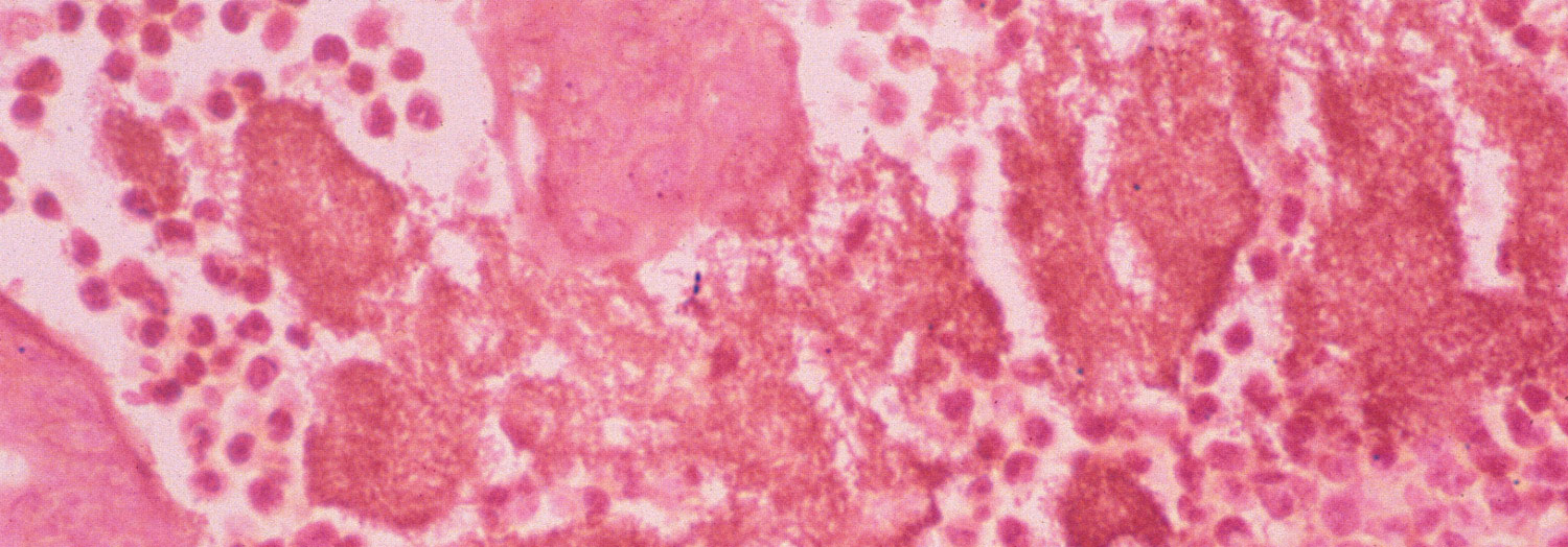
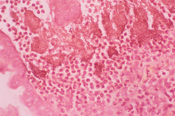
Affected animals frequently develop a marked neutrophilia and low serum albumin levels.111, 112 Morbidity rate varies from 1,0 to 90 per cent and mortality rate ranges from 0 to 6,7 per cent.76, 96 The abomasum and colon are reddened, the wall of the latter may be thickened and the contents are fluid and green or blood-stained. The lesions are more prevalent in the jejunum and ileum (Figure 155.1 and 155.2). Mesenteric lymph nodes are enlarged, congested and oedematous.96 The microscopic lesions in the intestine resemble those seen in cattle, i.e. a suppurative enterocolitis, characterized by microabscesses, which surround colonies of Gram-negative bipolar organisms in the lamina propria (Figure 155.3 and 155.4).76, 96, 111, 112 Leakage of albumin into the intestinal lumen probably leads to the hypoalbuminaemia and contributes to the diarrhoea.96 Occasional microscopic necrotic foci may be seen in the liver and mesenteric lymph nodes of sheep and goats infected with Y. pseudotuberculosis. 96, 111 Rare findings in sheep include epididymo-orchitis in rams,59, 80 caseous pseudotubercles in the spleen and liver,99 multifocal suppurative nephritis76 and erosive cholecystitis.96, 99 Yersinia intermedia and Y. frederiksenii, usually considered to be environmental and nonpathogenic, have occasionally been isolated from cases of diarrhoea and ill-thrift in sheep.96
Yersinia pseudotuberculosis (serotype III) and, less commonly, Y. enterocolitica (bioserotypes 1A/O:6,30; 3/O:5,27) can cause placentitis, abortion, stillbirth and neonatal death.12, 25, 26, 33, 65, 74, 92, 122 Ewes that abort appear clinically normal.92 Affected foetuses have subcutaneous oedema, serosanguinous fluid (sometimes containing fibrin strands) in body cavities and small (2 mm diameter or less) yellowish, necrotic or pyogranulomatous foci in the liver, and sometimes in the spleen, kidneys and mesenteric lymph nodes, with multiple haemorrhages in the lungs, kidneys and intestines. Necrogranulomatous focimay also be present in the heart. The lungs may reveal a suppurative bronchiolitis and alveolitis and necrotic arteritis. Gram-negative coccobacilli are present in many of the necrotic foci.26, 47, 65, 74, 121 An acute purulent leptomeningitis and ependymitis may also be present. The intercotyledonary chorio-allantois of the placenta may be thickened and ulcerated and covered with a yellowish, necrotic exudate, and some cotyledons may be necrotic. Diffuse inflammation, vasculitis and thrombosis are discernible histologically.26, 47, 74, 92 In experimentally infected pregnant ewes, the uterine and placental lesions include thrombosis, necrosis and severe suppuration in the caruncles and similar changes in the cotyledons. Judging by the severity of lesions and the concentration of bacteria, the placenta and gravid uterus are very susceptible to Y. pseudotuberculosis infection. In contrast, experimental infection of pregnant ewes with Y. enterocolitica, which is of low natural pathogenicity for the uterus, results in an infection which evolves slowly and only culminates in placental failure and foetal death when the effects of infection are superimposed on the normal decline in placental function at the end of gestation.26 Septicaemia or endotoxaemia may kill the ewe as well.65
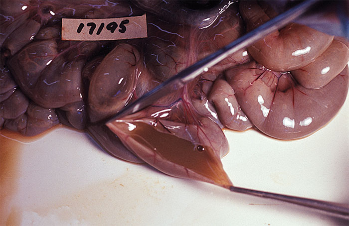
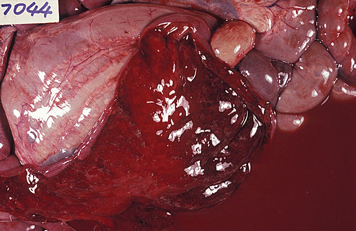
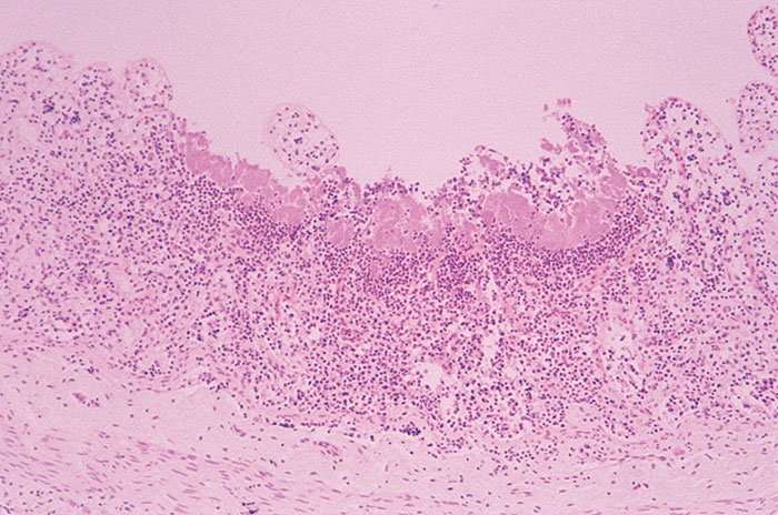
Figure 155.3 Microabscessation and necrosis of the mucosa and lamina propria of the jejunum of a sheep infected with Yersinia pseudotuberculosis. (By courtesy of Massey University, Palmerston North, New Zealand)
Goats
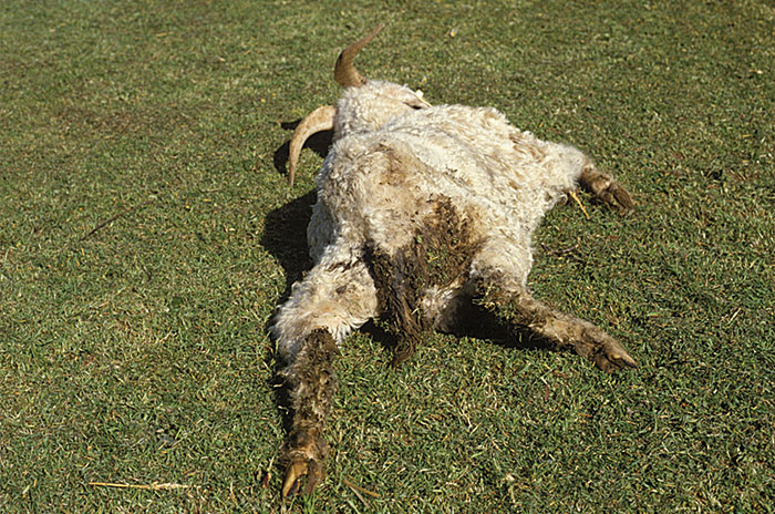
In goats, Y. enterocolitica (bioserotype 5/0:2,3) causes a similar enterocolitis to that seen in sheep.68, 112 This disease is the most common infectious cause of death in Angora (Figure 155.5) and other fibre-producing goat breeds less than 20 months old in New Zealand.16, 90 Yersinia pseudotuberculosis (serotypes I and III) can also cause enterocolitis as in sheep111 and this organism has occasionally been isolated from cases of abortion and early neonatal death of goat kids.130 Foetuses may be autolysed or fresh, and the placenta may reveal whitish foci of necrosis in the cotyledons. Histologically, there is suppuration in the placenta and in the foetal lung.130 Yersinia pseudotuberculosis (serotype III) has also been associated with mastitis in milking goats,20, 62 and enteritis76 and acute ulcerative conjunctivitis with swelling of the parotid gland in kids.75
Deer
In farmed deer, Y. pseudotuberculosis (serotypes I, II and III) is the most common infectious cause of death in New Zealand and Australia. Outbreaks of yersiniosis in farmed cervids have also been reported from Canada.104 Clinical signs, gross pathology and histopathology closely resemble those seen in domestic ruminants.49, 61 Infection of deer with Y. enterocolitica is usually asymptomatic.49
Pigs
Pigs are usually asymptomatic carriers of yersinias, but there are rare clinical reports of diarrhoeal disease associated with Y. enterocolitica (bioserotypes 2/O:9 and 4/O:3) originating from Australia and China.115, 134, 135 Diarrhoea and enterocolitis31, 46, 86, 111 ormultiple systemic abscesses and pseudotubercles85 have been associated with Y. pseudotuberculosis(serotypes IIa and III). In outbreaks of diarrhoea, enterocolitis and ill-thrift due to Y. pseudotuberculosis(serotype III) in 8- to 18-week-old pigs, the faeces ranged from unformed and grey to a watery, non-bloody diarrhoea. Multifocal haemorrhages were present in the ileal mucosa. Histopathologically, the lesion was a subacute enterocolitis with multiple microabscesses and colonies of Gram-negative coccobacilli in the intestinal lamina propria, and an associated locally extensive polymorphonuclear leukocyte infiltration.46, 111 Experimental infections using Y. enterocolitica bioserotypes 1B/O:8, 1B/O:21, 1B/O:13 and 4/O:3 have produced mild gastroenteritis in new-born piglets.101, 106, 109
Horses
Yersinia pseudotuberculosis is a rare cause of disease in horses. Serotype IIa has been isolated from a ten-week-old foal with numerous small abscesses in the liver, spleen and lungs.80 More recently, an outbreak of diarrhoea and pneumonia in foals caused by Y. pseudotuberculosis was reported from a stud farm in Poland. Ten per cent of affected foals died.28 One case of diarrhoea in an adult horse caused by Y. enterocolitica (bioserotype 3/O:5,27) occurred in New Zealand.37
Diagnosis
The clinical signs and gross pathology are not sufficiently distinctive to be diagnostic. The characteristic microscopic enteric lesions, however, can be useful in diagnosing infections in livestock.111 Consequently, bacterial isolation, identification and serotyping are essential. Fresh faecal samples, intestinal contents and mesenteric lymph nodes are the most appropriate samples for bacteriology. In cases of abortion, organisms can be cultured from the foetal abomasal contents and a wide variety of tissues, including liver, lungs and kidneys, and placenta and vaginal discharge.12, 26, 65, 67
Serological diagnosis of subclinical Yersinia infections in lambs has been demonstrated using an enzyme immunoassay that measured antibodies to outer membrane proteins.102 Serological cross-reactions occur between Y. enterocolitica bioserotype 2/O:9 and Brucella abortus and these have been shown to be due to the sugar moiety of the lipopolysaccharide in the cell wall.24, 43, 51, 117, 128 In some cases these cross-reactions have prompted the search for the presence of Y. enterocolitica 2/O:9 in the faeces of cattle and pigs presenting false-positive reactions in brucellosis screening tests.128, 131 Recently, a skin test has been developed that clearly eliminates brucellosis from false-positive serological reactions due to infection with Y. enterocolitica O:9.103
Differential diagnosis
In cattle, sheep and goats, diarrhoea due to yersiniosis should be differentiated from that caused by salmonellosis, campylobacteriosis (weaner colitis in sheep), coccidiosis,helminthosis and, in the case of young cattle, bovine virus diarrhoea. The microscopic enteric lesions due to yersiniosis, however, are distinctive and they are not a feature of either coccidiosis or salmonellosis. The latter is generally more severe, with erosion, ulceration and fibrinous effusion occurring.110 In sheep and cattle, abortion caused by Y. pseudotuberculosis may resemble that caused by Campylobacter spp., Salmonella serovars, Listeria monocytogenes, Pasteurella multocida and Toxoplasma gondii, and B. abortus, Campylobacter fetus, Salmonella serovars, Bacillus licheniformis, Aspergillus spp. and Mortierella wolfii, respectively.60 The placentitis due to Y. pseudotuberculosis closely resembles that due to Chlamydophila abortus, the cause of enzootic abortion of ewes.74, 92
Swine dysentery, salmonellosis, intestinal adenomatosis and post-weaning colibacillosis should be considered as differential diagnoses for diarrhoea due to yersiniosis in pigs.5
Control
Known predisposing factors, such as parasitism, nutritional stress, lack of shelter, and the presence of rodents, should be addressed (see Epidemiology). Good nutrition is important in avoiding clinical disease.100
Isolates of Y. pseudotuberculosis are sensitive in vitro to benzyl penicillin, ampicillin, tetracycline, streptomycin, neomycin, gentamycin, lincospectin, chloramphenicol and sulphamethoxazole/trimethoprim and variably sensitive to furazolidone and sulphonamides.8, 19, 46, 62, 66, 96, 110, 120, 127 Yersinia enterocolitica is sensitive in vitro to neomycin, streptomycin, lincospectin, tetracycline, and trimethoprim/ sulphonamide but resistant to ampicillin and furazolidone.8, 96, 112 Treatment with long-acting tetracyclines is recommended in the treatment of both Y. pseudotuberculosis and Y. enterocolitica infections.19, 76, 110, 114
Diarrhoea increases the susceptibility of sheep to flystrike.111, 112
Acquired immunity follows natural infection.89 Since antibodies to outer membrane proteins are found in animals and appear to be protective, vaccination may be useful.101 Vaccination of mares and their offspring with a formalin-inactivated culture of Y. pseudotuberculosis was employed following an outbreak of yersiniosis in Poland.28 A commercial vaccine (Yersiniavax, Agvax Developments Ltd, New Zealand) has been developed in New Zealand for use in farmed deer.72 It is a formalin-inactivated bacterin with an oil adjuvant containing Y. pseudotuberculosis serotypes I, II and III, and a two dose regime, given three to six weeks apart, has been shown to give significant protection against heavy experimental and field challenge. It is recommended for use in young deer, and should be administered at least four to seven weeks before the period of maximum stress, i.e. in autumn.72 Vaccination should be repeated annually.
References
- ADESIYUN, A.A., AGBONLAHOR, D.E., LOMBIN, L.H. & KWAGA, J.K.P., 1986. Occurrence of virulence markers in species of Yersinia isolated from animals in Nigeria. Veterinary Microbiology, 12, 289–294.
- ADESIYUN, A.A., KAMINJOLO, J.S., LOREGNARD, R. & KITSON-PIGGOTT, W., 1992. Frequency of isolation of Yersinia enterocolitica from livestock in Trinidad. The Veterinary Record, 131, 516.
- ALEKSIC, S. & BOCKEMUHL, J., 1984. Proposed revision of the Wauters et al. antigenic scheme for serotyping Yersinia enterocolitica. Journal of Clinical Microbiology, 20, 99–102.
- ANDERSEN, J.K., SORENSEN, R. & GLENSBJERG, M., 1991. Aspects of the epidemiology of Yersinia enterocolitica: a review. International Journal of Food Microbiology, 13, 231–238.
- BARKER, I.K., VAN DREUMEL, A.A. & PALMER, N., 1993. The alimentary system. In: JUBB, K.V.F., KENNEDY, P.C. & PALMER, N., (eds). Pathology of Domestic Animals, Vol. II, 4th edn. New York, London: Academic Press.
- BERCOVIER, H., BRENNER, D.J., URSING, J., STEIGERWALT, A.G., FANNING, G.R., ALONSO, J.M., CARTER, G.P. & MOLLARET, H.H., 1980. Characterisation of Yersinia enterocolitica sensu stricto. Current Microbiology, 4, 201–206.
- BERCOVIER, H., MOLLARET, H.H., ALONSO, J.M., BRAULT, J., FANNING, G.R., STEIGERWALT, A.G. & BRENNER, D.J., 1980. Intra- and interspecies relatedness of Yersinia pestis by DNA hybridisation and its relationship to Yersinia pseudotuberculosis. Current Microbiology, 4, 225–229.
- BERCOVIER, H. & MOLLARET, H.H., 1984. Genus XIV. Yersinia Van Loghem 1944. In: KRIEG, N.R. & HOLT, J.G., (eds.). Bergey’s Manual of Systematic Bacteriology, Vol I. Baltimore, London: Williams & Wilkins
- BIN-KUN, H., DE-SHENG, X., HONG-BI, O., SHI-XIANG, Z. & SLEE, K.J., 1994. Yersiniosis in sheep due to Yersinia enterocolitica. British Veterinary Journal, 150, 473–479.
- BLACKALL, P., 1977. Survey of the prevalence of Yersinia species in swine. Australian Veterinary Journal, 53, 407.
- BOTTONE, E.J., 1981. Yersinia enterocolitica. 1st edn. Boca Raton, Florida: CRC Press
- BREWER, R.A. & CORBEL, M.J., 1983. Characterisation of Yersinia enterocolitica strains isolated from cattle, sheep and pigs in the United Kingdom. Journal of Hygiene, Cambridge, 90, 425–433.
- BREWER, R.A. & CORBEL, M.J., 1986. Methods for the isolation and identification of Yersinia. Booklet 2521, Weybridge, Surrey: MAFF Publications.
- BROWN, C.C. & DAVIS, F.N., 1989. Yersinia pseudotuberculosis enteritis in four calves. Journal of Comparative Pathology, 101, 463–466.
- BRUBAKER, R.R., 1972. The genus Yersinia: Biochemistry and genetics of virulence. Current Topics in Microbiology, 37, 111–158.
- BUDDLE, B.M., HERCEG, M., RALSTON, M.J., PULFORD, H.D., MILLAR, K.R. & ELLIOTT, D.C., 1988. A goat mortality study in the southern North Island. New Zealand Veterinary Journal, 36, 167–170.
- BULLIANS, J.A., 1987. Yersinia species infection of lambs and cull cows at an abattoir. New Zealand Veterinary Journal, 35, 65–67
- BURROWS, T.W. & BACON, G.A., 1960. V and W antigens in strains of Pasteurella pseudotuberculosis. British Journal of Experimental Pathology, 41, 38–44.
- CALLINAN, R.B., COOK, R.W., BOULTON, J.G., FRASER, G.C. & UNGER, D.B., 1988. Enterocolitis in cattle associated with Yersinia pseudotuberculosis infection. Australian Veterinary Journal, 65, 8–11.
- CAPPUCCI, D.T., DANIELS, R.B., PERELLI-MINETTI, J.E., FURLONG, H.J. & WILCOX, M.A., 1978. Caprine mastitis associated with Yersinia pseudotuberculosis. Journal of the American Veterinary Medical Association, 173, 1589–1590
- CARNIEL, E., 1995. Chromosomal virulence factors of Yersinia. Contributions to Microbiology and Immunology, 13, 218–224.
- CARTER, P.B., ZAHORCHAK, R.J. & BRUBAKER, R.R., 1980. Plague virulence antigens from Yersinia enterocolitica. Infection and Immunity, 28, 638–640
- CHIESA, C., PACIFICO, L. & RAVAGNAN, G., 1993. Identification of pathogenic serotypes of Yersinia enterocolitica. Journal of Clinical Microbiology, 31, 2248–2249.
- CORBEL, M.J. & CULLEN, G.A., 1970. Differentiation of the serological response to Yersinia enterocolitica and Brucella abortus in cattle. Journal of Hygiene, Cambridge, 68, 519–529
- CORBEL, M.J., BREWER, R.A. & HUNTER, D., 1990. Characterization of Yersinia enterocolitica strains associated with ovine abortion. The Veterinary Record, 127, 526–527.
- CORBEL, M.J., ELLIS, B., RICHARDSON, C. & BRADLEY, R., 1992. Experimental Yersinia enterocolitica placentitis in sheep. British Veterinary Journal, 148, 339–349.
- CORNELIS, G., LAROCHE, Y., BALLIGAND, G. & SORY, M.P., 1987. Yersinia enterocolitica, a primary model for bacterial invasiveness. Reviews of Infectious Diseases, 9, 64–87.
- CZERNOMYSY-FUROWICZ, D., 1997. An outbreak of foal yersiniosis in Poland: Pathological and bacteriological examination. Zentralblatt für Bakteriologie, 286, 542–546.
- DAS, A.M., PARANJAPE, V.L. & WINBLAD, S., 1986. Yersinia enterocolitica associated with third tremester abortion in buffaloes. Tropical Animal Health and Production, 18, 109–112.
- DAVEY, G.M., BRUCE, J. & DRYSDALE, E.M., 1983. Isolation of Yersinia enterocolitica and related species from the faeces of cows. Journal of Applied Bacteriology, 55, 439–443.
- DE BARCELLOS, D.E.S.N. & DE CASTRO, A.F.P., 1981. Isolation of Yersinia pseudotuberculosis from diarrhoea in pigs. British Veterinary Journal, 137, 95–96
- DELOR, I., KAECKENBEECK, A., WAUTERS, G. & CORNELIS, G.R., 1990. Nucleotide sequence of yst, the Yersinia enterocolitica gene encoding the heat-stable enterotoxin, and prevalence of the gene among pathogenic and nonpathogenic yersiniae. Infection and Immunity, 58, 2983–2988.
- DENNIS, S.M., 1966. Recovery of Pasteurella pseudotuberculosis from a premature Merino lamb. The Veterinary Record, 79, 273–274
- DICKINSON, A.B. & MOCQUOT, G., 1961. Studies on the bacterial flora of the alimentary tract of pigs. I. Enterobacteriaceae and other Gram-negative bacteria. Journal of Applied Bacteriology, 24, 252–284.
- FALCAO, D.P., 1987. Yersiniosis in Brazil. Contributions to Microbiology and Immunology, 9, 68–75.
- FARMER, J.J., CARTER, G.P., MILLER, V.L., FALKOW, S. & WACHSMUTH, I.K., 1992. Pyrazinamidase, CR-MOX agar, salicin fermentation-esculin hydrolysis, and d-xylose fermentation for identifying pathogenic serotypes of Yersinia enterocolitica. Journal of Clinical Microbiology, 30, 2589–2594
- FENWICK, S.G., 1997. Yersinia enterocolitica infections in people and other animals — a New Zealand study. Ph.D. thesis, Massey University, Palmerston North, New Zealand.
- FUKUSHIMA, H., HOSHINA, K., NAKAMURA, R., ITO, Y. & GOMYODA, M., 1987. Epidemiological studies of Yersinia enterocolitica and Yersinia pseudotuberculosis in Shimane Prefecture, Japan. Contributions to Microbiology and Immunology, 9, 103–110.
- FUKUSHIMA, H. & GOMYODA, M., 1986. Growth of Yersinia pseudotuberculosis and Yersinia enterocolitica Biotype 3B serotype O3 inhibited on cefsulodin-irgasan-novobiocin agar. Journal of Clinical Microbiology, 24, 116–120.
- FUKUSHIMA, H., KOICHI, S., MISAO, T., KOICHI, O. & YOSHIHIRO, K., 1983. Isolation of Yersinia species from bovine faeces. Journal of Clinical Microbiology, 18, 981–982.
- GEMSKI, P., LAZERE, J.R. & CASEY, T., 1980. Plasmid associated with pathogenicity and calcium dependency of Yersinia enterocolitica. Infection and Immunity, 27, 682–685.
- GEMSKI, P., LAZERE, J.R., CASEY, T. & WOHLHIETER, J.A., 1980. Presence of a virulence-associated plasmid in Yersinia pseudotuberculosis. Infection and Immunity, 28, 1044–1047.
- GERBIER, G., GARIN-BASTUJI, B., POUILLOT, R., VERY, P., CAU, C., BERR, V., DUFOUR, B. & MOUTOU, F., 1997. False positive serological reactions in bovine brucellosis: evidence of the role of Yersinia enterocolitica serotype O:9 in a field trial. Veterinary Research, 28, 375–383.
- HAAGSMA, J., 1970. Enzootic death in mink caused by an exotoxin-producing strain of Yersinia pseudotuberculosis, type III. Netherlands Journal of Veterinary Science, 3, 77–84.
- HANNAM, D.A.R., 1993. Bovine abortion associated with Yersinia pseudotuberculosis. The Veterinary Record, 133, 372.
- HARPER, P.A.W., HORNITZKY, M.A.Z. & RAYWARD, D.G., 1990. Enterocolitis in pigs associated with Yersinia pseudotuberculosis infection. Australian Veterinary Journal, 67, 418–419.
- HARTLEY, W.J. & KATER, J.C., 1964. Perinatal disease conditions of sheep in New Zealand. New Zealand Veterinary Journal, 12, 49–57.
- HEAD, C.B., WHITTY, D.A. & RATNAM, S., 1982. Comparative study of selective media for recovery of Yersinia enterocolitica. Journal of Clinical Microbiology, 16, 615–621.
- HENDERSON, T.G., 1983. Yersiniosis in deer from the Otago-Southland region of New Zealand. New Zealand Veterinary Journal, 31, 221–224.
- HENDERSON, T.G., 1984. The isolation of Yersinia species from feral and farmed deer faeces. New Zealand Veterinary Journal, 32, 88–90.
- HILBINK, F., FENWICK, S.G., THOMPSON, E.J., PENROSE, M. & ROSS, G.P., 1995. Non-specific seroreactions against Brucella abortus in ruminants in New Zealand and the presence of Yersinia enterocolitica O:9. New Zealand Veterinary Journal, 43, 175–178.
- HODGES, R.T. & CARMAN, M.G., 1985. Recovery of Yersinia pseudotuberculosis from the faeces of healthy cattle. New Zealand Veterinary Journal, 33, 175–176.
- HODGES, R.T., CARMAN, M.G. & MORTIMER, W.J., 1984. Serotypes of Yersinia pseudotuberculosis recovered from domestic livestock. New Zealand Veterinary Journal, 32, 11–13.
- HODGES, R.T. & WOODS, E.P., 1984. Yersinia pseudotuberculosis recovered from the faeces of clinically healthy deer. New Zealand Veterinary Journal, 32, 79.
- HUNTER, D., HUGHES, S. & FOX, E., 1983. Isolation of Yersinia enterocolitica from pigs in the United Kingdom. The Veterinary Record, 112, 322–323.
- HURVELL, B., 1981. Zoonotic Yersinia enterocolitica infection: Host range, clinical manifestations and transmission between animals and man. In: BOTTONE, E.J., (ed.). Yersinia enterocolitica. Boca Raton, Florida: CRC Press.
- HURVELL, B., GLATTHARD, V. & THAL, E., 1979. Isolation of Yersinia enterocolitica from swine at an abattoir in Sweden. Contributions to Microbiology and Immunology, 5, 243–248.
- ISBERG, R.R., VOORHIS, D.L. & FALKOW, S., 1987. Identification of invasin: A protein that allows enteric bacteria to penetrate cultured mammalian cells. Cell, 50, 769–778.
- JAMIESON, S. & SOLTYS, M.A., 1947. Infectious epididymo-orchitis of rams associated with Pasteurella pseudotuberculosis. The Veterinary Record, 59, 351.
- JERRETT, I.V. & SLEE, K.J., 1989. Bovine abortion associated with Yersinia pseudotuberculosis infection. Veterinary Pathology, 26, 181–183.
- JERRETT, I.V., SLEE, K.J. & ROBERTSON, B.I., 1990. Yersiniosis in farmed deer. Australian Veterinary Journal, 67, 212–214.
- JONES, T.O., MAIR, N.S. & FOX., E., 1982. Caprine mastitis associated with Yersinia pseudotuberculosis infection. The Veterinary Record, 110, 231.
- KAPPERUD, G., 1982. Enterotoxin production at 4, 22, and 37 °C among Yersinia enterocolitica and Y. enterocolitica-like bacteria. Acta Pathologica Microbiologica et Immunologica Scandinavica. Sect. B., 90, 185–189.
- KAPPERUD, G., 1991. Yersinia enterocolitica in food hygiene. International Journal of Food Microbiology, 12, 53–66.
- KARBE, E. & ERICKSON, E.D., 1984. Ovine abortion and stillbirth due to purulent placentitis caused by Yersinia pseudotuberculosis. Veterinary Pathology, 21, 601–606.
- KANAZAWA, Y. & KURAMATA, T., 1974. Difference in susceptibility to benzylpenicillin between Yersinia enterocolitica and Yersinia pseudotuberculosis. Japanese Journal of Microbiology, 18, 483–485.
- KENNEDY, P.C. & MILLER, R.B., 1993. The female genital system. In: JUBB, K.V.F., KENNEDY, P.C. & PALMER, N., (eds). Pathology of Domestic Animals, Vol III, 4th edn. New York, London: Academic Press.
- KROGSTAD, O., TEIGE, J. & LASSEN, J., 1972. Yersinia enterocolitica type 2 associated with disease in goat. Acta Veterinaria Scandinavica, 13, 594–596
- LANADA, E.B., 1990. The epidemiology of Yersinia infections in goat flocks. M.Sc. thesis, Massey University, Palmerston North, New Zealand
- LANGFORD, E.V., 1969. Pasteurella pseudotuberculosis associated with abortion and pneumonia in the bovine. Canadian Veterinary Journal, 10, 208–211.
- LYNCH, J.A., 1986. Improved Yersinia isolation from enteric specimens. Canadian Veterinary Journal, 27, 154.
- MACKINTOSH, C.G., 1993. Vaccine against yersiniosis in deer. Surveillance, 20, 25..
- MACKINTOSH, C.G. & HENDERSON, T.G., 1984. The epidemiology of yersiniosis in deer. In: Proceedings No. 1, Deer Branch, New Zealand Veterinary Association, Wellington, New Zealand.
- MACLEOD, N.S.M., PATTERSON, I.A.P. & ROGERSON, F., 1992. Yersinia pseudotuberculosis and ovine abortion. The Veterinary Record, 131, 84.
- MCSPORRAN, K., 1983. Yersinia pseudotuberculosis in goat kids. Surveillance, 10, 22.
- MCSPORRAN, K.D., HANSEN, L.M., SAUNDERS, B.W. & DAMSTEEGT, A., 1984. An outbreak of diarrhoea in hoggets associated with infection by Yersinia enterocolitica. New Zealand Veterinary Journal, 32, 38–39.
- MAIR, N.S., 1973. Yersiniosis in wildlife and its public health implications. Journal of Wildlife Diseases, 9, 64–71..
- MAIR, N.S., FOX, E. & THAL, E., 1979. Biochemical, pathogenicity and toxicity studies of type III strains of Yersinia pseudotuberculosis isolated from the caecal contents of pigs. Contributions to Microbiology and Immunology, 5, 359–365.
- MAIR, N.S. & HARBOURNE, J.F., 1963. The isolation of Pasteurella pseudotuberculosis from a bovine foetus. The Veterinary Record, 75, 559–561.
- MAIR, N.S. & ZIFFO, G.S., 1974. Isolation of Y. pseudotuberculosis from a foal. The Veterinary Record, 94, 152–153.
- MERILAHTI-PALO, R., LAHESMAA, R., GRANFORS, K., GRIPPENBERG-LERCHE, C. & TOIVANEN, P., 1991. Risk of Yersinia infection among butchers. Scandinavian Journal of Infectious Diseases, 23, 55–61.
- MESSERLI, J., 1972. Yersinia pseudotuberculosis, erreger einer mastitis beim rind. Zentralblatt für Bakteriologie, Parasitenkunde, Infektionskrankheiten und Hygiene, Erste Abteilung Originale, Reihe A, 222, 280–282..
- MILLER, V.L., BLISKA, J.B. & FALKOW, S., 1990. Nucleotide sequence of the Yersinia enterocolitica ail gene and characterization of the ail protein product. Journal of Bacteriology, 172, 1062–1069.
- MILLER, V.L, FARMER, J.J., HILL, W.E. & FALKOW, S., 1989. The ail locus is found uniquely in Yersinia enterocolitica serotypes commonly associated with disease. Infection and Immunity, 57, 121–131.
- MORITA, M., NAKAMATSU, M. & GOTO, M., 1968. Pathological studies on pseudotuberculosis rodentium. III Spontaneous swine cases. Japanese Journal of Veterinary Science, 30, 233–239.
- NEEF, N.A. & LYSONS, R.J., 1994. Pathogenicity of a strain of Yersinia pseudotuberculosis isolated from a pig with porcine colitis syndrome. The Veterinary Record, 135, 58–63.
- NESBAKKEN, T. & KAPPERUD, G., 1985. Yersinia enterocolitica and Yersinia enterocolitica-like bacteria in Norwegian slaughter pigs. International Journal of Food Microbiology, 1, 301–309
- NILEHN, B., 1969. Studies on Yersinia enterocolitica with special reference to bacterial diagnosis and occurrence in human acute enteric disease. Acta Pathologica et Microbiologica Scandinavica, Supplement, 206, 1–48
- OBWOLO, M.J., 1976. A review of yersiniosis (Yersinia pseudotuberculosis infection). The Veterinary Bulletin, 46, 167–171.
- ORR, M., CRAIGHEAD, L., & CAMERON, S., 1987. Yersiniosis outbreak in goat weanlings. Surveillance, 14, 10–11.
- ORR, M., CRAIGHEAD, L., KYLE, B. & MACKINTOSH, C., 1990. The isolation of Yersinia species from the faeces of farmed goats. Surveillance, 17, 27–28
- OTTER, A., 1996. Ovine abortion caused by Yersinia pseudotuberculosis. The Veterinary Record, 138, 143–144.
- PAI, C.H. & MORS, V., 1978. Production of enterotoxin by Yersinia enterocolitica. Infection and Immunity, 22, 334–338.
- PALUMBO, S.A., 1986. Is refrigeration enough to restrain foodborne pathogens? Journal of Food Protection, 49, 1003–1009.
- PATERSON, J.S. & COOK, R., 1963. A method for the isolation of Pasteurella pseudotuberculosis from faeces. Journal of Pathology and Bacteriology, 85, 241–242.
- PHILBEY, A.W., GLASTONBURY, J.R.W., LINKS, I.J. & MATTHEWS L.M., 1991. Yersinia species isolated from sheep with enterocolitis. Australian Veterinary Journal, 68, 108–110.
- PORTNOY, D.A. & MARTINEZ, R.J., 1985. Role of a plasmid in the pathogenicity of yersinia species. Current Topics in Microbiology and Immunology, 118, 29–51.
- PORTNOY, D.A., MOSELY, S.L. & FALKOW, S., 1981. Characterization of plasmids and plasmid-associated determinants of Yersinia enterocolitica pathogenesis. Infection and Immunity, 31, 775–782.
- PULLAR, E.M., 1932. Pseudo-tuberculosis of sheep due to B. pseudotuberculosis rodentium (so-called ‘pyaemic hepatitis’). Australian Veterinary Journal, 8, 181–183.
- RADOSTITS, O.M., BLOOD, D.C. & GAY, C.C., 1994. (eds). Veterinary Medicine. 8th edn. London, Philadelphia: Baillière Tindall.
- ROBINS-BROWNE, R.M., BORDUN, A.M. & SLEE, K.J., 1993. Serological response of sheep to plasmid-encoded proteins of Yersinia species following natural infection with Y. enterocolitica and Y. pseudotuberculosis. Journal of Medical Microbiology, 39, 268–272
- ROBINS-BROWNE, R.M., TZIPORI, S., GONIS, G., HAYES, J., WITHERS, M. & PRPIC, J.K., 1985. The pathogenesis of Yersinia enterocolitica infection in gnotobiotic piglets. Journal of Medical Microbiology, 19, 297–308.
- SAEGERMAN, C., VO, T-K.O., DE WAELE, L., GILSON, D., BASTIN, A., DUBRAY, G., FLANAGAN, P., LIMET, J.N., LETESSON, J-J. & GODFROID, J., 1999. Diagnosis of bovine brucellosis by skin test: Conditions for the test and evaluation of its performance. The Veterinary Record, 145, 214–218.
- SANFORD, S.E., 1995. Outbreaks of yersiniosis caused by Yersinia pseudotuberculosis in farmed cervids. Journal of Veterinary Diagnostic Investigation, 7, 78–81.
- SCHIEMANN, D.A., 1979. Synthesis of a selective agar medium for Yersinia enterocolitica. Canadian Journal of Microbiology, 25, 1298–1304.
- SCHIEMANN, D.A., 1988. The pathogenicity of Yersinia enterocolitica for piglets. Canadian Journal of Veterinary Research, 52, 325–330.
- SCHIEMANN, D.A., 1989. Yersinia enterocolitica and Yersinia pseudotuberculosis. In: DOYLE, M.P., (ed.). Foodborne Bacterial Pathogens. New York: Marcel Dekker, Inc., pp. 601–672.
- SCHOFIELD, G.M., 1992. Emerging food-borne pathogens and their significance in chilled foods. Journal of Applied Bacteriology, 72, 267–273.
- SHU, D., SIMPSON, H., XU, R.J., MELLOR, D.J., REYNOLDS, G.W., ALLEY, M.R., FENWICK, S.G. & MARSHALL, R.M., 1995. Experimental infection of newborn piglets with Yersinia enterocolitica: An animal model of enteritis. New Zealand Veterinary Journal, 43, 5–56.
- SLEE, K.J., BRIGHTLING, P. & SEILER, R.J., 1988. Enteritis in cattle due to Yersinia pseudotuberculosis infection. Australian Veterinary Journal, 65, 271–275.
- SLEE, K.J. & BUTTON, C., 1990. Enteritis in sheep, goats and pigs due to Yersinia pseudotuberculosis infection. Australian Veterinary Journal, 67, 320–322..
- SLEE, K.J. & BUTTON, C., 1990. Enteritis in sheep and goats due to Yersinia enterocolitica infection. Australian Veterinary Journal, 67, 396–398.
- SLEE, K.J. & SKILBECK, N.W., 1992. Epidemiology of Yersinia pseudotuberculosis and Y. enterocolitica infections in sheep in Australia. Journal of Clinical Microbiology, 30, 712–715
- SPIER, C., SLEE, K.J. & BUTTON, C., 1990. Treatment of Yersinia infection with tetracyclines. Australian Veterinary Journal, 67, 471.
- STEPHENS, C.P., TAYLOR, J.D. & BATES, J.R., 1999. Enterocolitis in pigs in Southeast Queensland associated with Yersinia enterocolitica. In: Proceedings of the IXth International Congress of Bacteriology and Applied Microbiology, Sydney.
- STRALEY, S.C., SKRZYPEK, E., PLANO, G.V. & BLISKA, J.B., 1993. Yops of Yersinia spp. pathogenic for humans. Infection and Immunity, 61, 3105–3110.
- SWAMINATHAN, B., HARMON, M.C. & MEHLMAN, I.J., 1982. A review: Yersinia enterocolitica. Journal of Applied Bacteriology, 52, 151–183
- TSUBOKURA, M. & ALEKSIC, S., 1995. A simplified antigenic scheme for serotyping of Yersinia pseudotuberculosis: Phenotypic characterisation of reference strains and preparation of O and H factor sera. Contributions to Microbiology and Immunology, 13, 99–105
- TSUBOKURA, M., OTSUKI, K., FUKUDA, T., KUBOTA, M., IMAMURA, M., ITAGAKI, K., YAMAOKA, K. & WAKATSUKI, M., 1976. Studies on Yersinia pseudotuberculosis. IV. Isolation of Y. pseudotuberculosis from healthy swine. Japanese Journal of Veterinary Science, 38, 549–552.
- TSUBOKURA, M., OTSUKI, K., KAWAOKA, Y. & MARUYAMA, T., 1984. Characterization and pathogenicity of Yersinia pseudotuberculosis isolated from swine and other animals. Journal of Clinical Microbiology, 19, 754–756.
- VALLI, V.E.O., 1993. The hematopoietic system. In: JUBB, K.V.F., KENNEDY, P.C. & PALMER, N., (eds). Pathology of Domestic Animals, Vol. III, 4th edn. New York, London: Academic Press.
- WATSON, W.A. & HUNTER, D., 1960. The isolation of Pasteurella pseudotuberculosis from an ovine foetus. The Veterinary Record, 72, 770–772.
- WAUTERS, G., 1979. Carriage of Yersinia enterocolitica serotype 3 by pigs as a source of human infection. Contributions to Microbiology and Immunology, 5, 249–252.
- WAUTERS, G., 1981. Antigens of Yersinia enterocolitica. In: BOTTONE, E.J., (ed.). Yersinia enterocolitica, Boca Raton, Florida: CRC Press.
- WAUTERS, G., ALEKSIC, S., CHARLIER, J. & SCHULZ, G., 1991. Somatic and flagellar antigens of Yersinia enterocolitica and related species. Contributions to Microbiology and Immunology, 12, 239–243.
- WAUTERS, G., KANDOLO, K. & JANSSENS, M., 1987. Revised biogrouping scheme of Yersinia enterocolitica. Contributions to Microbiology and Immunology, 9, 14–21.
- WELSH, R.D. & STAIR, E.L., 1993. Yersinia pseudotuberculosis bovine abortion. Journal of Veterinary Diagnostic Investigation, 5, 109–111
- WEYNANTS, V., TIBOR, A., DENOEL, P.A., SAEGERMAN, C., GODFROID, J., THIANGE, P. & LETESSON, J-J., 1996. Infection of cattle with Yersinia enterocolitica O:9 a cause of the false positive serological reactions in bovine brucellosis diagnostic tests. Veterinary Microbiology, 48, 101– 112.
- WINBLAD, S., 1967. Studies on serological typing of Yersinia enterocolitica. Acta Pathologica et Microbiologica Scandinavica, Supplement, 187, 115.
- WITTE, S.T., SPONENBERG, D.P. & COLLINS, T.C., 1985. Abortion and early neonatal death of kids attributed to intrauterine Yersinia pseudotuberculosis infection. Journal of the American Veterinary Medical Association, 187, 834.
- WRATHALL, A.E., BROUGHTON, E.S., GILL, K.P.W. & GOLDSMITH, G.P., 1993. Serological reactions to Brucella species in British pigs. The Veterinary Record, 132, 449–454.
- YOUNG, V.B., MILLER, V.L., FALKOW, S. & SCHOOLNIK, G.K., 1990. Sequence, localization and function of the invasin protein of Yersinia enterocolitica. Molecular Microbiology, 4, 1119–1128.
- ZEN-YOJI, H., SAKAI, S., MARUYAMA, T. & YANAGAWA, Y., 1974. Isolation of Yersinia enterocolitica and Yersinia pseudotuberculosis from swine, cattle and rats at an abattoir. Japanese Journal of Microbiology, 18, 103–105.
- ZHENG, X.B., 1987. Isolation of Yersinia enterocolitica from the faeces of diarrhoeic swine. Journal of Applied Bacteriology, 62, 521–525.
- ZHENG, X.B. & XIE, C., 1996. Isolation, characterisation and epidemiology of Yersinia enterocolitica from humans and animals. Journal of Applied Bacteriology, 81, 681–684.


