- Infectious Diseases of Livestock
- Part 3
- Clostridium septicum infections
- GENERAL INTRODUCTION: SPIROCHAETES
- Swine dysentery
- Borrelia theileri infection
- Borrelia suilla infection
- Lyme disease in livestock
- Leptospirosis
- GENERAL INTRODUCTION: AEROBIC ⁄ MICRO-AEROPHILIC, MOTILE, HELICAL ⁄ VIBROID GRAM-NEGATIVE BACTERIA
- Genital campylobacteriosis in cattle
- Proliferative enteropathies of pigs
- Campylobacter jejuni infection
- GENERAL INTRODUCTION: GRAM-NEGATIVE AEROBIC OR CAPNOPHILIC RODS AND COCCI
- Moraxella spp. infections
- Bordetella bronchiseptica infections
- Pseudomonas spp. infections
- Glanders
- Melioidosis
- Brucella spp. infections
- Bovine brucellosis
- Brucella ovis infection
- Brucella melitensis infection
- Brucella suis infection
- Brucella infections in terrestrial wildlife
- GENERAL INTRODUCTION: FACULTATIVELY ANAEROBIC GRAM NEGATIVE RODS
- Klebsiella spp. infections
- Escherichia coli infections
- Salmonella spp. infections
- Bovine salmonellosis
- Ovine and caprine salmonellosis
- Porcine salmonellosis
- Equine salmonellosis
- Yersinia spp. infections
- Haemophilus and Histophilus spp. infections
- Haemophilus parasuis infection
- Histophilus somni disease complex in cattle
- Actinobacillus spp. infections
- Actinobacillus equuli infections
- Gram-negative pleomorphic infections: Actinobacillus seminis, Histophilus ovis and Histophilus somni
- Porcine pleuropneumonia
- Actinobacillus suis infections
- Pasteurella and Mannheimia spp. infections
- Pneumonic mannheimiosis and pasteurellosis of cattle
- Haemorrhagic septicaemia
- Pasteurellosis in sheep and goats
- Porcine pasteurellosis
- Progressive atrophic rhinitis
- GENERAL INTRODUCTION: ANAEROBIC GRAM-NEGATIVE, IRREGULAR RODS
- Fusobacterium necrophorum, Dichelobacter (Bacteroides) nodosus and Bacteroides spp. infections
- GENERAL INTRODUCTION: GRAM-POSITIVE COCCI
- Staphylococcus spp. infections
- Staphylococcus aureus infections
- Exudative epidermitis
- Other Staphylococcus spp. infections
- Streptococcus spp. infections
- Strangles
- Streptococcus suis infections
- Streptococcus porcinus infections
- Other Streptococcus spp. infections
- GENERAL INTRODUCTION: ENDOSPORE-FORMING GRAM-POSITIVE RODS AND COCCI
- Anthrax
- Clostridium perfringens group infections
- Clostridium perfringens type A infections
- Clostridium perfringens type B infections
- Clostridium perfringens type C infections
- Clostridium perfringens type D infections
- Malignant oedema⁄gas gangrene group of Clostridium spp.
- Clostridium chauvoei infections
- Clostridium novyi infections
- Clostridium septicum infections
- Other clostridial infections
- Tetanus
- Botulism
- GENERAL INTRODUCTION: REGULAR, NON-SPORING, GRAM-POSITIVE RODS
- Listeriosis
- Erysipelothrix rhusiopathiae infections
- GENERAL INTRODUCTION: IRREGULAR, NON-SPORING, GRAM-POSITIVE RODS
- Corynebacterium pseudotuberculosis infections
- Corynebacterium renale group infections
- Bolo disease
- Actinomyces bovis infections
- Trueperella pyogenes infections
- Actinobaculum suis infections
- Actinomyces hyovaginalis infections
- GENERAL INTRODUCTION: MYCOBACTERIA
- Tuberculosis
- Paratuberculosis
- GENERAL INTRODUCTION: ACTINOMYCETES
- Nocardiosis
- Rhodococcus equi infections
- Dermatophilosis
- GENERAL INTRODUCTION: MOLLICUTES
- Contagious bovine pleuropneumonia
- Contagious caprine pleuropneumonia
- Mycoplasmal pneumonia of pigs
- Mycoplasmal polyserositis and arthritis of pigs
- Mycoplasmal arthritis of pigs
- Bovine genital mycoplasmosis
- Neurotoxin-producing group of Clostridium spp.
- Contagious equine metritis
- Tyzzer's disease
- MYCOTIC AND ALGAL DISEASES: Mycoses
- MYCOTIC AND ALGAL DISEASES: Pneumocystosis
- MYCOTIC AND ALGAL DISEASES: Protothecosis and other algal diseases
- DISEASE COMPLEXES / UNKNOWN AETIOLOGY: Epivag
- DISEASE COMPLEXES / UNKNOWN AETIOLOGY: Ulcerative balanoposthitis and vulvovaginitis of sheep
- DISEASE COMPLEXES / UNKNOWN AETIOLOGY: Ill thrift
- Eperythrozoonosis
- Bovine haemobartonellosis
Clostridium septicum infections
This content is distributed under the following licence: Attribution-NonCommercial CC BY-NC  View Creative Commons Licence details here
Clostridium septicum infections
M W ODENDAAL AND N P J KRIEK
Introduction
Several disease syndromes are associated with Clostridium septicum infection in livestock: malignant oedema and gas gangrene in ruminants, pigs and horses, gangrenous abomasitis known as braxy or bradsot in sheep, gangrenous abomasitis in lambs and calves following infection of abomasal ulcers, and post-parturient gas gangrene of the perineum, vulva and genital tract particularly of ewes and does, but also of cows (in South Africa this is often referred to as ‘baarmoeder sponssiekte’, [literally ‘uterine gas gangrene’]).
Malignant oedema develops as a consequence of wound infections and is an acute, febrile, highly fatal condition characterized by the presence of an oedematous and gangrenous inflammation of the affected parts, as well as toxaemia. >1, 10, 12, 13, 15, 18, 19, 22, 30 Clostridium septicum may be a secondary invader, and often occurs in conjunction with other clostridia. It is considered to be the primary cause of malignant oedema in up to 30 per cent of cattle suffering from the disease.27 Other histotoxic clostridia associated with gas gangrene in animals are C. perfringens type A or C, C. novyi, C. chauvoei and C. sordellii.
Braxy is a highly fatal disease which has, for a very long time, been known to occur in north-western Europe, principally in Norway, Denmark, North Germany, Iceland and the Faroe Islands, where it is known as ‘bradsot’ (‘quick plague’). It is also well known in parts of the UK and Ireland, North America and Australia. Braxy is characterized by severe oedema and gangrenous inflammation of the mucous membrane of the abomasum and severe toxaemia. Some cases of braxy may be due to C. novyi and are indistinguishable from the condition caused by C. septicum.
Clostridium septicum is a normal inhabitant of the intestinal contents of animals and is a frequent and rapid postmortal tissue invader and common contaminant; the longer the interim period between death and collection of tissue specimens, the better the chances of isolating C. septicum from animal tissues.
In southern Africa, losses of livestock, particularly cattle and sheep, as a result of malignant oedema, post-parturient gas gangrene and gangrenous abomasitis are encountered sporadically but braxy has not been reported in the region.
Aetiology
Clostridium septicum is usually a straight or slightly curved, Gram-positive bacillus 3 to 6 μm long and 0,4 to 1,2μm wide. Filaments up to 35 μm long are formed on the peritoneal surface of organs (particularly the diaphragmatic surface of the liver) of infected animals. It often stains unevenly with Gram’s stain. The bacterial cells usually occur singly or in pairs, are motile due to the presence of peritrichous flagella and exhibit pleomorphism and citrons, spindles and barred rods under certain conditions. Spores which are oval, subterminal and distend the cell are formed abundantly.5, 25
Clostridium septicum is easily cultured on blood agar, and after two days’ incubation colonies are 1 to 8 mm in diameter, slightly raised, circular, semitranslucent, grey and glossy, with markedly irregular to rhizoid margins. A zone of complete haemolysis surrounds each colony. Due to the motility of the organisms, the colonies have a tendency to swarm on solid media which can be prevented by shortening the incubation period, increasing the agar content to 4 to 6 per cent, or by spreading specific antiserum over the surface of the agar plate.
Clostridium septicum is anaerobic. Optimum growth is attained at temperatures of between 37 and 40 °C, while most strains will also grow at 44 °C. Growth ceases at 46 °C. The presence of fermentable carbohydrate or serum in the culture medium stimulates its growth. Essential factors to sustain growth of the bacterium on artificial media include biotin, nicotinic acid, pyridoxine, thiamin, cysteine, tryptophan, adenine, arginine, aspartic acid, histidine, isoleucine, phenylalanine, serine, threonine, tyrosine and valine.25
Clostridium septicum has saccharolytic and nonproteolytic properties and does not produce lipase or lecithinase. Cellobiose, fructose, galactose, lactose, maltose,mannose and trehalose are fermented and esculin is hydrolysed by 90 to 100 per cent of strains. Arabinose, amygdalin, glycogen, inositol, inulin, mannitol, melezitose, melibiose, raffinose, rhamnose, sorbitol, starch, sucrose and xylose are not fermented by 90 to 100 per cent of strains.7, 20
Six different serological groups of C. septicum have been identified, based on combinations of two somatic (O) antigens and five types of flagellar (H) antigens that may be present. Serological typing is useful in epidemiological investigations of strains isolated from different sources.23, 25 Clostridium septicum and C. chauvoei share a common spore antigen which is responsible for cross-agglutination between them.
Clostridium septicum produces four different exotoxins. The alpha toxin is haemolytic, lethal and necrotizing, the beta toxin is a deoxyribonuclease, the gamma toxin is a hyaluronidase and the delta toxin is a thiol-activated cytolysin (an oxygen-labile haemolysin) which in the case of C. septicum is named septicolysin. The delta toxin is haemolytic, and considered lethal and cardiotoxic.26 The production of toxins in a culture medium is determined by the nutritional composition of the medium, strain of the bacterium, length of incubation, and presence of a haemolysin-inactivating factor.4
The alpha toxin is a protein with an isoelectric point of 8,4 and a molecular weight of 48 000 Daltons.2 Of the four toxins the alpha toxin is the only one that is lethal and in vitro appears during the first day of growth. It is labile, and haemolyses the red blood cells of humans, cattle, sheep, pigs and rabbits, but not those of dogs, horses, guinea pigs and chickens. This toxin also has a direct effect on cardiac muscle when injected into guinea pigs.25 The alpha toxin of C. septicum is serologically related to the alpha toxin of C. chauvoei.25 No specific information is available on the structure or the activity of the other three toxins.
Epidemiology
Clostridium septicum occurs in soil, water and the intestinal contents of animals. Clostridium septicum alone may be responsible for infecting a wound or it may be present in combination with one or more of the other histotoxic clostridia such as C. chauvoei, C. perfringens type A or C, C. sordellii and C. novyi, as well as other anaerobic and aerobic organisms.
All breeds, sexes and age groups of cattle, sheep and goats may contract malignant oedema. Pigs and horses are less susceptible than domestic ruminants, while dogs and cats are resistant or suffer at most from a slight local reaction at the site of infection. Most cases present after infection of wounds such as those caused by surgical procedures (castration, tail-docking, and ear-marking) in lambs, or contracted during shearing, parturition or vaccination.18 The failure to use aseptic techniques during surgical operations, sterile equipment when administering intramuscular injections or strict hygienic measures during shearing procedures, predisposes to the development of malignant oedema.14
Crow pick is a rapidly fatal form of gangrene in sheep, resulting from an infection of wounds caused by the pecking of crows of animals, such as parturient ewes, which are down and unable to escape from the birds’ attack. The birds are particularly attracted to the eyes, and in this way a sheep’s eye may eventually be torn from its socket, the resulting wound forming an ideal site for the proliferation of infecting bacteria such as C. septicum, C. novyi or C. perfringens. 9 Crows are carrion eaters and their beaks are often contaminated by clostridia.
Post-parturient gas gangrene of the perineum, vulva and genital tract has been reported in cows, ewes and does. In South Africa it occurs most frequently in Boer and Angora goats and Dorper and Merino sheep. Pluriparous ewes with twins or triplets are more commonly affected than those with single lambs or kids. A prevalence rate as high as 30 per cent has been reported in some Boer goat herds. It is assumed that the portals of entry of the bacteria are lacerations and contusions contracted during partus, although the presence of these cannot always be verified.31
Braxy is no longer considered a disease of major importance because of its present low prevalence, although at one time it was of some significance in the regions in which it occurred. It is a disease of sheep in countries with cold winters, and can be closely correlated with the type of husbandry practised, being particularly prevalent in animals that have to be overwintered on summer grazing.27 Cases usually occur in autumn and early to midwinter when heavy frost and snow are present. In the USA, however, it tends to occur in the spring.22 It usually affects animals in good condition of 6 to 18 months of age. The morbidity rate in an affected flockmaybe as high as 50 per cent. Isolated cases of a disease similar to braxy have been reported in calves in the USA.12, 22 Ulcers in the abomasum of calves may also become infected with C. septicum, resulting in gangrenous abomasitis.
Pathogenesis
The entrance of C. septicum into the tissues is favoured when a mucous membrane or the skin is injured. The presence of effusions, coagulated blood and necrotic tissue are necessary in most forms of C. septicum infections for the provision of the local anaerobic and other conditions required for the germination of spores of the organism and its vegetative growth and toxin production. Once it has become established and has elaborated sufficient amounts of its toxins, particularly alpha toxin, to cause tissue necrosis, the infection becomes self-generating.
The pathogenesis of braxy is not understood. In most cases, the organism invades the mucosa of the abomasum or, more rarely, that of the duodenum, but as there is no obvious predisposing lesion in the affected mucosa which would have allowed such invasion to occur, the precise circumstances under which it does take place remain obscure. As the disease is associated with cold weather, it has been hypothesized that the ingestion of frosted vegetation plays a role in some way, possibly by causing chilling with devitalization and tissue injury of the mucous membrane which, in turn, allows the penetration and proliferation of C. septicum.8 Absorption of toxins into the blood stream results in fatal toxaemia.
The fact that prior injury to the abomasal mucosa probably plays a role in the pathogenesis of braxy was exemplified, albeit inadvertantly, in an experiment in which a number of sheep were infused via an abomasal cannula with a solution containing glucose and volatile fatty acids.10 An acute haemorrhagic abomasitis resembling that of braxy and from which C. septicum was isolated, developed in some of the sheep, and the centrifugal distribution of the lesions around the abomasal cannula suggested that the infusion solution may have damaged the abomasal mucosa, thus facilitating infection with the bacterium.
A form of gangrenous abomasitis in lambs and calves is associated with infection by C. septicum of abomasal ulcers.22
Clinical signs and pathology
In animals that contract malignant oedema, clinical signs develop within four days of infection and are characterized by the development of rapidly expanding oedematous swellings, which may or may not be crepitous due to the presence of gas bubbles, at the site of infection. These swellings may be associated with lameness if the muscles of locomotion in the region are affected. Horses rapidly succumb to the infection after developing toxic shock and intravascular coagulation.21
At necropsy, affected areas are infiltrated with large amounts of inflammatory oedema which extend into the fasciae (Figure 183.1). The muscles in the area are either markedly congested or pale reddish or brownish and usually do not contain foci of necrosis. The exudate lacks the typical rancid odour that is so characteristic of C. chauvoei infections.
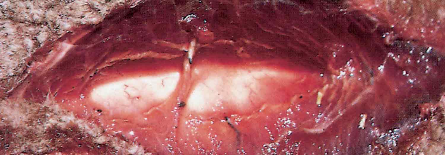
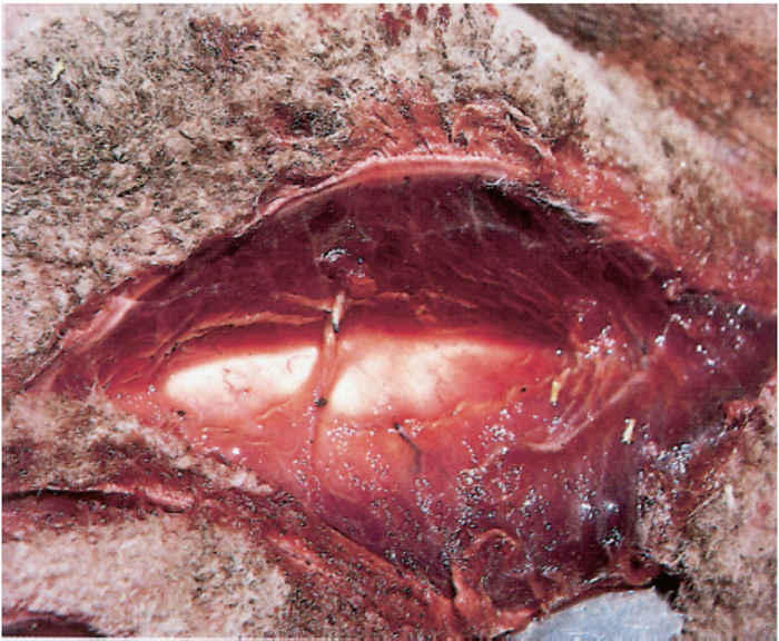
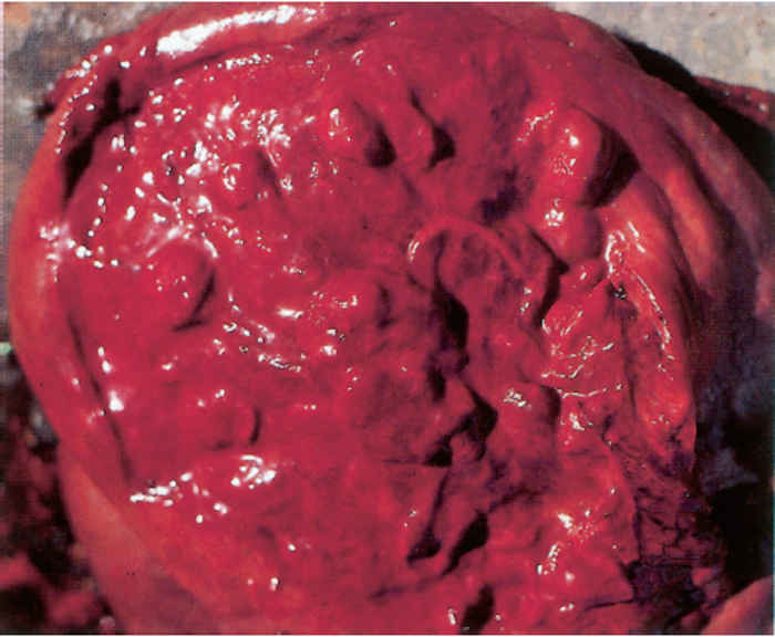
The incubation period of braxy is usually less than one day and the duration of the disease may be a matter of a few hours; death frequently supervenes before any clinical signs are evident. Animals that are seen to be ill usually manifest signs of abdominal pain and bloat, develop a high fever, and are inappetent and depressed. Death occurs within a few hours of the onset of clinical signs. Post-mortem changes develop rapidly.15 Necropsy reveals a marked, diffuse inflammation of the abomasal wall; the abomasal folds are intensely red, may be emphysematous, contain focal ulcers, and are several times their normal thickness due to severe oedema (Figure 183.2). Fibrin may cover the mucosal lesions and the serosa of the abomasum over the affected areas in the mucosa.12 The abomasal content is blood-stained, scant or voluminous and foul-smelling. There are severe degenerative changes in the liver and kidneys, a turbid, reddish-brown peritoneal exudate, and petechial haemorrhages in serosal membranes. Histologically, affected parts of the mucosa, submucosa and adjacent muscular layers of the abomasum manifest a pronounced inflammatory oedema, areas of necrosis associated with a neutrophil infiltrate, emphysema, and numerous Gram-positive, rod-shaped bacteria in clumps or short chains. Thrombosis may occur in the blood vessels in the submucosa.10, 12
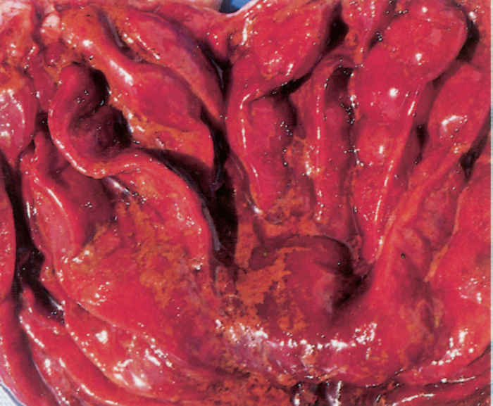
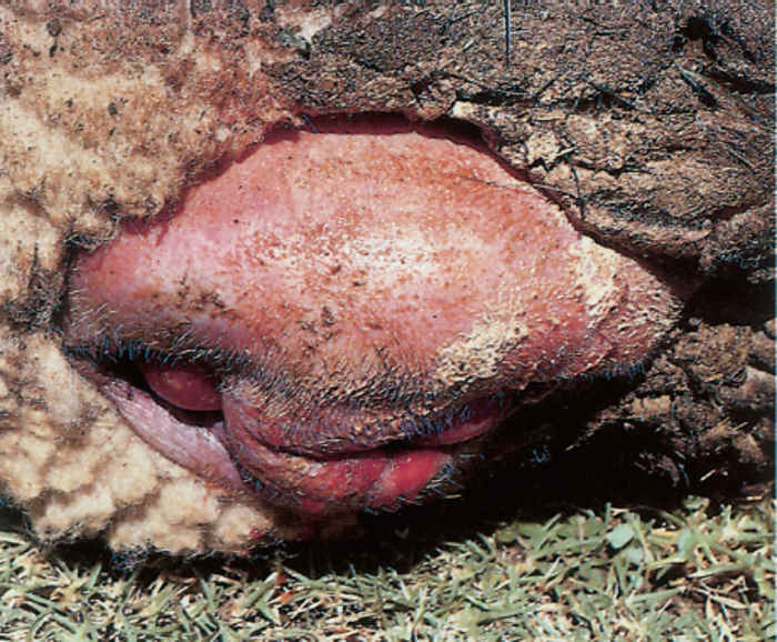
Clinical signs of post-parturient gas gangrene develop within 12 to 24 hours of partus and affected animals usually die within 12 to 24 hours of being noticeably ill. The signs include fever, lethargy, straining and in some a dark haemorrhagic discharge from the vulva. The vulva and the perineum are usually severely swollen due to the presence of a marked oedema and emphysema (Figure 183.3). Similar lesions occur in the genital tract, the uterus being atonic, red and emphysematous (Figure 183.4).
Lambs and calves suffering from the condition as a result of the infection of abomasal ulcers with C. septicum show signs of abdominal pain and are often bloated. At necropsy there are usually several ulcers in the abomasal mucosa which are associated with severe oedema and emphysema of the mucosa and submucosa, particularly in close proximity to the ulcers. The lesions may become more generalized and involve the mucosa and submucosa of the entire abomasum. Numerous petechial haemorrhages may be scattered in the abomasal mucosa. In some affected animals the ulcers penetrate the wall of the abomasum, resulting in peritonitis and sudden death.
Diagnosis
A presumptive diagnosis of the disease caused by C. septicum is based on the history, clinical signs and pathology. The diagnosis should, however, be confirmed by bacterial culture and identification of the organisms.
As C. septicum is a common post-mortem invader, specimens for the confirmation of the diagnosis should be collected as soon as possible after the death of the animal, chilled, and submitted at 4 to 8 °C to a diagnostic laboratory. Specimens required for culturing include blocks of the affected tissue, such as muscle, abomasum, and uterus. As in the case of blackquarter, six thin, unfixed and air-dried impression smears on microscope slides should be prepared from the lesions for Gram’s stain and the immunofluorescent test for the identification of C. septicum.3, 28
Specimens collected for bacterial isolation should be cultured on blood agar and incubated under anaerobic conditions for 18 to 24 hours at 35 to 37 °C. Typical colonies of C. septicum should be selected when the culture is contaminated and subcultured until a pure culture is obtained. The bacterium is then identified by biochemical techniques.7
Differential diagnosis
The clinical signs and lesions of the syndromes caused by C. septicum cannot be differentiated with certainty from those caused by the other histotoxic clostridia, including C. chauvoei, C. novyi, C. sordellii, or C. perfringens types A or C. The signs and lesions of malignant oedema may also be very similar to those of anthrax, and those caused by the bites of snakes such as puff adders (Bitis arietans arietans).
Control
Treatment of animals suffering from malignant oedema and braxy is rarely successful, but may be considered during the early course of the disease.19 The treatment includes high doses of antimicrobials, including penicillin, aminobenzylpenicillin and cephaloridine, and, if applicable, provision of adequate surgical drainage of the area and irrigation of the site with hydrogen peroxide. The administration of specific antitoxin may be considered.24 Antitoxin is not available in South Africa.
Immunoprophylaxis is the method of choice to control C. septicum infections. A toxoid vaccine is marketed by Onderstepoort Biological Products in South Africa;11 bacterin/ toxoid combinations or multicomponent vaccines are also commercially available under various trade names.6, 17, 29 Animals that are vaccinated for the first time should receive two injections of the vaccine at a four-week interval and an annual booster thereafter. Subsequently, animals at risk should be vaccinated three to four weeks before shearing or parturition.
Special precautions should be taken when sheep are sheared or animals are castrated or their tails docked. These procedures should be conducted as aseptically as possible. Shearing sheds and pens should be kept as clean as possible and, once cases have occurred, should be thoroughly cleansed and disinfected. Dead animals should be disposed of by incineration or deep burial to minimize the spread of spores.
In order to prevent braxy sheep should, if possible, not be overwintered on summer grazing.27 If an outbreak does occur, sheep should immediately be vaccinated and placed in another paddock or camp. The kraaling or yarding of sheep at night and the feeding of hay before allowing them to graze on frosted vegetation is said to be of value in preventing the disease.16
Vegetative organisms that contaminate instruments are easily killed by sterilization at high temperatures or by chemical disinfectants. The spores, however, are extremely resistant to most forms of sterilization. They are inactivated by exposure to 3 per cent formaldehyde for 30 minutes.
References
- BAINS, B.S. & MACKENZIE, M.A., 1975. An outbreak of gangrenous cellulitis caused by C. septicum in a broiler flock. Australian Veterinary Journal, 51, 106–107.
- BALLARD, J., BRYANT, A., STEVENS, D. & TWETEN, R.K., 1992. Purification and characterization of the lethal toxin (alpha-toxin) of C. septicum. Infection and Immunity, 60, 784–790.
- BATTY, I. & WALKER, P.D., 1963. Differentiation of Clostridium septicum and Clostridium chauvoei by the use of fluorescent labelled antibodies. Journal of Pathology and Bacteriology, 85, 517–521.
- BERNHEIMER, A.W., 1944. Nutritional requirements and factors affecting the production of toxin of C. septicum. Journal of Experimental Pathology, 80, 321–331.
- BISPING, W. & AMTSBERG, G., 1988. Colour Atlas for the Diagnosis of Bacterial Pathogens in Animals. Berlin & Hamburg: Paul Parey Scientific Publishers.
- BROWN, K.K., PARIZEK, R.E. & STEWART, R.C., 1976. Prevention of clostridial disease in cattle and sheep by vaccination with a multivalent bacterin-toxoid. Veterinary Medicine/Small Animal Clinician, 71, 1717–1721.
- CATO, E.P., GEORGE, W.L. & FINEGOLD, S.M., 1986. Genus Clostridium Prazmowski 1880. In: sneath, p.h.a., mair, n.s., sharpe, m.e. & holt, j.g., (eds). Bergey’s Manual of Systematic Bacteriology. Vol. II. Baltimore & London: Williams & Wilkens.
- DAVIES, G.O., 1947. Gaiger and Davies’ Veterinary Pathology and Bacteriology. 3rd edn. London: Baillière, Tindall & Cox.
- EDGAR, G., 1931. Anaerobic infection in sheep brought about by the attack of crows (Corvus spp.) (‘crow pick’). Australian Veterinary Journal, 7, 64–68.
- ELLIS, T.M., ROWE, J.B. & LLOYD, J.M., 1983. Acute abomasitis due to C. septicum infection in experimental sheep. Australian Veterinary Journal, 60, 308–309.
- ERASMUS, B.J., CAMERON, C.M., HUNTER, P., CILLIERS, J.A., OBEREM, P.T., STOLTSZ, W.H. & DE WAAL, D.T., 1990. Onderstepoort Vaccines. Booklet issued by the Department of Agriculture and Development and obtained from the Directorate Agricultural Information, Private Bag X144, Pretoria 0001.
- EUSTIS, S.L. & BERGELAND, M.E., 1981. Suppurative abomasitis associated with C. septicum infection. Journal of the American Veterinary Medical Association, 178, 732–734.
- FOWLER, N.G. & HUSSAINI, S.N., 1975. Clostridium septicum infection and antibiotic treatment in broiler chickens. The Veterinary Record, 107, 14–15.
- HARWOOD, D.G., 1984. Apparent iatrogenic clostridial myositis in cattle. The Veterinary Record, 115, 412.
- HENNING, M.W., 1956. Animal Diseases in South Africa. 3rd edn. South Africa: Central News Agency.
- HUNGERFORD, T.G., 1990. Diseases of Livestock. 9th edn. Sydney: McGraw-Hill Book Company.
- KNOTT, G.K.L., ERWIN, B.G. & CLASSICK, L.G., 1985. Benefits of a clostridial vaccination program in feedlot cattle. Veterinary Medicine, 79, 95–97.
- MACHEAK, M.E., 1978. Clostridial diseases of cattle. Veterinary Medicine/ Small Animal Clinician, February, 195–200.
- PERDRIZET, J.A., CALLIHAN, D.R., REBHUN, W.C. & SHIN, S.J., 1987. Successful management of malignant edema caused by Clostridium septicum in a horse. Cornell Veterinarian, 77, 328–338.
- PHILLIPS, K.D., BRAZIER, J.S., LEVETT, P.N. & WILLIS, A.T., 1985. Clostridia. In: collins, c.h. & grange, j.m., (eds). Isolation and Identification of Micro-organisms of Medical and Veterinary Importance. London: Academic Press.
- REBHUN, W.C., SHIN, S.J., KING, J.M., BAUM, K.H. & PATTEN, V., 1985. Malignant edema in horses. Journal of the American Veterinary Medical Association, 187, 732–736.
- SAUNDERS, G., 1986. Diagnosing braxy in lambs and calves. Veterinary Medicine, 81, 1050–1051.
- SHIRASAKA, S., TERANISHI, H., BENNO, Y. & AZUMA, R., 1983. Serological characterization of C. septicum strains from clinical materials of chickens and cows. Japanese Journal of Veterinary Science, 45, 861–863.
- SHIRASAKA, S. & UMEKI, F., 1984. Antibiotic susceptibility of Clostridium septicum isolated from chickens and cattle. Japanese Journal of Veterinary Science, 46, 397–399.
- SMITH, L.D.S. & WILLIAMS, B.L., 1984. The Pathogenic Anaerobic Bacteria. 3rd edn. Springield, Illinois: Charles C. Thomas.
- STEPHEN, J. & PIETROWSKI, R.A., 1986. Bacterial Toxins. Washington, D.C.: American Society for Microbiology.
- STERNE, M., 1981. Clostridial infections. British Veterinary Journal, 137, 443–454.
- STERNE, M. & BATTY, I., 1975. Pathogenic Clostridia. Boston & London: Butterworths.
- STERNE, M., BATTY, I., THOMPSON, A. & ROBERTSON, J.M., 1962. Immunisation of sheep with multi-component clostridial vaccines. The Veterinary Record, 74, 909–913.
- VAN HEERDEN, J. & BOTHA, W.S., 1982. Clostridial myositis in a horse. Journal of the South African Veterinary Association, 53, 211.
- VAN TONDER, E.M., 1979. Regional Diagnostic Laboratory, Middelburg, CP. South Africa. Unpublished data.


