- Infectious Diseases of Livestock
- Part 3
- Staphylococcus aureus infections
- GENERAL INTRODUCTION: SPIROCHAETES
- Swine dysentery
- Borrelia theileri infection
- Borrelia suilla infection
- Lyme disease in livestock
- Leptospirosis
- GENERAL INTRODUCTION: AEROBIC ⁄ MICRO-AEROPHILIC, MOTILE, HELICAL ⁄ VIBROID GRAM-NEGATIVE BACTERIA
- Genital campylobacteriosis in cattle
- Proliferative enteropathies of pigs
- Campylobacter jejuni infection
- GENERAL INTRODUCTION: GRAM-NEGATIVE AEROBIC OR CAPNOPHILIC RODS AND COCCI
- Moraxella spp. infections
- Bordetella bronchiseptica infections
- Pseudomonas spp. infections
- Glanders
- Melioidosis
- Brucella spp. infections
- Bovine brucellosis
- Brucella ovis infection
- Brucella melitensis infection
- Brucella suis infection
- Brucella infections in terrestrial wildlife
- GENERAL INTRODUCTION: FACULTATIVELY ANAEROBIC GRAM NEGATIVE RODS
- Klebsiella spp. infections
- Escherichia coli infections
- Salmonella spp. infections
- Bovine salmonellosis
- Ovine and caprine salmonellosis
- Porcine salmonellosis
- Equine salmonellosis
- Yersinia spp. infections
- Haemophilus and Histophilus spp. infections
- Haemophilus parasuis infection
- Histophilus somni disease complex in cattle
- Actinobacillus spp. infections
- Actinobacillus equuli infections
- Gram-negative pleomorphic infections: Actinobacillus seminis, Histophilus ovis and Histophilus somni
- Porcine pleuropneumonia
- Actinobacillus suis infections
- Pasteurella and Mannheimia spp. infections
- Pneumonic mannheimiosis and pasteurellosis of cattle
- Haemorrhagic septicaemia
- Pasteurellosis in sheep and goats
- Porcine pasteurellosis
- Progressive atrophic rhinitis
- GENERAL INTRODUCTION: ANAEROBIC GRAM-NEGATIVE, IRREGULAR RODS
- Fusobacterium necrophorum, Dichelobacter (Bacteroides) nodosus and Bacteroides spp. infections
- GENERAL INTRODUCTION: GRAM-POSITIVE COCCI
- Staphylococcus spp. infections
- Staphylococcus aureus infections
- Exudative epidermitis
- Other Staphylococcus spp. infections
- Streptococcus spp. infections
- Strangles
- Streptococcus suis infections
- Streptococcus porcinus infections
- Other Streptococcus spp. infections
- GENERAL INTRODUCTION: ENDOSPORE-FORMING GRAM-POSITIVE RODS AND COCCI
- Anthrax
- Clostridium perfringens group infections
- Clostridium perfringens type A infections
- Clostridium perfringens type B infections
- Clostridium perfringens type C infections
- Clostridium perfringens type D infections
- Malignant oedema⁄gas gangrene group of Clostridium spp.
- Clostridium chauvoei infections
- Clostridium novyi infections
- Clostridium septicum infections
- Other clostridial infections
- Tetanus
- Botulism
- GENERAL INTRODUCTION: REGULAR, NON-SPORING, GRAM-POSITIVE RODS
- Listeriosis
- Erysipelothrix rhusiopathiae infections
- GENERAL INTRODUCTION: IRREGULAR, NON-SPORING, GRAM-POSITIVE RODS
- Corynebacterium pseudotuberculosis infections
- Corynebacterium renale group infections
- Bolo disease
- Actinomyces bovis infections
- Trueperella pyogenes infections
- Actinobaculum suis infections
- Actinomyces hyovaginalis infections
- GENERAL INTRODUCTION: MYCOBACTERIA
- Tuberculosis
- Paratuberculosis
- GENERAL INTRODUCTION: ACTINOMYCETES
- Nocardiosis
- Rhodococcus equi infections
- Dermatophilosis
- GENERAL INTRODUCTION: MOLLICUTES
- Contagious bovine pleuropneumonia
- Contagious caprine pleuropneumonia
- Mycoplasmal pneumonia of pigs
- Mycoplasmal polyserositis and arthritis of pigs
- Mycoplasmal arthritis of pigs
- Bovine genital mycoplasmosis
- Neurotoxin-producing group of Clostridium spp.
- Contagious equine metritis
- Tyzzer's disease
- MYCOTIC AND ALGAL DISEASES: Mycoses
- MYCOTIC AND ALGAL DISEASES: Pneumocystosis
- MYCOTIC AND ALGAL DISEASES: Protothecosis and other algal diseases
- DISEASE COMPLEXES / UNKNOWN AETIOLOGY: Epivag
- DISEASE COMPLEXES / UNKNOWN AETIOLOGY: Ulcerative balanoposthitis and vulvovaginitis of sheep
- DISEASE COMPLEXES / UNKNOWN AETIOLOGY: Ill thrift
- Eperythrozoonosis
- Bovine haemobartonellosis
Staphylococcus aureus infections
This content is distributed under the following licence: Attribution-NonCommercial CC BY-NC  View Creative Commons Licence details here

Staphylococcus aureus infections
M M HENTON
Introduction
Staphylococcus aureus is a commensal bacterium of animals and humans that most commonly occurs on the skin and in the nasopharynx, but may also be present in the alimentary and genital tracts.20 It is a potential pathogen and may cause a range of pyogenic conditions, the major one in livestock being mastitis in cattle, sheep and goats. Other conditions for which it is periodically responsible include tick pyaemia of lambs;13 chronic pyogranulomatous in- flammation known as ‘botryomycosis’ which may occur in horses, cattle and pigs; folliculitis and furunculosis in horses, goats and sheep;14 pyoderma in goats, piglets and cattle;11 staphylococcal dermatitis in sheep (also known as facial or periorbital eczema12); polyarthritis in young animals; impetigo or subcorneal pustular dermatitis of piglets; and dermatitis of the udder in goats.19 Wounds may become infected with S. aureus, and it may play a primary or secondary role in a great variety of infections. These include purulent bronchopneumonia and pyothorax, osteomyelitis, salpingitis and abscess formation. A variety of virulence determinants have been described,10 but the effects of these have only been studied in detail in human infections and bovine mastitis. In addition, many strains of S. aureus are an important cause of food poisoning in humans, but animals are resistant.10, 20
Staphylococcus aureus of cattle, pigs, horses, poultry and humans can be distinguished from each other, and may be regarded as representing different ecovars (or biovars).20 There is an association between biovars and their hosts; biovar A consists of human strains, B of pig and poultry strains, C of cattle and sheep strains, D of hare strains, and E, which is now called Staphylococcus intermedius, is commonly isolated from dogs, horses and pigeons.6 The association between biovars and their hosts is not absolute, e.g. human strains (A) can cause mastitis in animals.
Some of the conditions mentioned above, and of which S. aureus is the cause, are discussed below.
Tick pyaemia of lambs
Synonym: Cripples
Tick pyaemia of lambs is a pyogenic staphylococcal infection of two- to ten-week-old animals reported in the United Kingdom and Ireland, resulting from infestation with the Ixodes ricinus tick. Affected animals often suffer concurrent tick-borne fever caused by Ehrlichia (formerly Cytoecetes) phagocytophila (see Lesser-known rickettsias infecting livestock).2, 25 The syndrome has not been reported in southern Africa.
Staphylococcus aureus appears to be transmitted by I. ricinus, although the process would seem to be purely mechanical: the bacteria enter the skin of lambs via the wounds produced by the mouth parts of infected ticks. From these primary sites of infection the bacteria spread haematogenously to localize in many organs and tissues of the body, particularly the joints, where they produce a suppurative in- flammatory reaction.
As only young lambs between two and ten weeks of age are affected, it seems probable that an acquired immunity to S. aureus develops in older animals.13 It appears that tick-borne fever, though it is usually a mild disease, may predispose to the development of tick pyaemia by the suppression of the humoral and cell-mediated immune responses which occur during the course of the disease.1 Additionally, the parasites also infect neutrophils, thus further impairing the defence against infection with S. aureus. 13
The disease occurs most often on hill farms in Scotland, Wales, north-east England and Ireland, and can result in serious economic losses. Prevalence rates as high as 29 per cent have been reported on some farms, although they are generally less than this; a figure of about 5 per cent is usual on farms on which the disease occurs.23
The most commonly recognized forms of tick pyaemia are characterized by the development of suppurative polyarthritis that results in lameness (crippling) and multiple abscesses in other organs and tissues, or of vertebral abscessation with pressure on the spinal cord and consequent paresis or paralysis. Abscesses may, however, develop in virtually any organ or tissue, resulting in lambs which are thin, dull and unthrifty, or even in sudden death if one of the vital organs is involved.13
Conditions such as joint ill (both suppurative and nonsuppurative polyarthritides), lameness caused by foot rot, foot abscess or toe abscess (see Fusobacterium necrophorum, Dichelobacter (Bacteroides) nodosus and Bacteroides spp. infections), nutritional muscular dystrophy (white muscle disease), ill-thrift, and tick paralysis should be considered in the differential diagnosis of tick pyaemia.
The treatment of lambs suffering from tick pyaemia is often disappointing, as by the time the disease is noticed, lesions are well advanced and irreparable damage has occurred.13 The control of the disease is primarily dependent upon the control of the vector tick. Alternatively, holding ewes and their lambs in a fenced, tick-free or relatively tickfree-pasture until the lambs are about six weeks old is effective. In addition, suppression of either the S. aureus or E. phagocytophila infections by the administration of long-acting antibiotics to which the organisms are susceptible may reduce the prevalence of tick pyaemia.2, 13 Long-acting benzathine penicillin given to lambs at three weeks of age produced a marked decrease in the prevalence of tick pyaemia, while long-acting tetracycline protected them against experimental tick-borne fever for about three weeks.2
Staphylococcal dermatitis in sheep
Synonyms: Facial or periorbital eczema, facial dermatitis, necrotic or ulcerative dermatitis
An apparently contagious purulent dermatitis, frequently termed facial or periorbital eczema, which affects mainly the face of sheep and which is associated with infection by some strains of Staphylococcus aureus, has been described in the United Kingdom, particularly Scotland.15 The disease has been reviewed by Martin.12
The term ‘facial eczema’ used for this disease is a misnomer and should not be used, as this name is more generally reserved for the form of photosensitization of unpigmented skin caused by the hepatotoxic mycotoxin sporidesmin, which is elaborated by the fungus Pithomyces chartarum. 17 The term ‘periorbital eczema’ is also inappropriate, as the condition is not eczematous according to the strict definition of the word.17
A form of staphylococcal folliculitis and furunculosis has been encountered in sheep and goats in South Africa in association with the bites of Simulium flies (‘black flies’).21 Lesions occur on the parts of the body that are not covered by wool — chiefly the face and pinnae, but also the perineum. Papules develop which may give rise to pustules, ulceration and scab formation. Healing lesions may be confused with ringworm.
While staphylococci are commensal bacteria on the skin and in the nose, mouth and vagina of sheep,12, 24 it is not known whether dermatopathogenic strains are frequent or regular inhabitants of the skin.12
Numerous organisms are present in the exudate of the lesion in staphylococcal dermatitis, and it appears that strains of the staphylococci isolated from cases of the disease are more pathogenic for the skin than those that cause other conditions in sheep.12 Such dermatopathogenic strains are coagulase-positive, produce alpha and beta haemolysins, and most also elaborate delta haemolysin. However, no other characteristics which could specifically identify them have been established. Although it appears that the staphylococci are the primary pathogens, mixed infections with a variety of other bacteria are not uncommon.12
The pathogenesis of the condition is not known, but it is thought that the bacteria gain entrance to the skin through minor injuries; the skin of the head being possibly more subjected to infection because it is not protected by wool. Close contact during feeding seems to facilitate the spread of infection between sheep.17 The history is frequently that of sheep being fed from troughs, and it is possible that headto-head contact during feeding, particularly in overcrowded conditions, could cause skin abrasions which then become invaded by the staphylococci.
The disease is more prevalent in adult sheep, although younger animals may occasionally be affected. As many as 50 per cent of the animals in affected flocks may develop the disease over a period of several months.12 The lesions are generally confined to the woolless parts of the head, particularly the skin over bony prominences. Common sites are around the eyes and ears, the pinnae of the ears, over the nasal and maxillary bones, and at the base of the horns, where irregularly shaped, deep, suppurative ulcers, 10 to 60 mm in diameter, form at one or more of these sites. They are covered with blackish scabs and surrounded by in- flamed zones of alopecic and thickened skin. These lesions exude pus and bleed easily, the blood and exudate adhering to the surrounding skin. Some sheep have red, thickened areas of skin which exude a serous fluid. Severely affected sheep are said to present an ‘alarming and distressing’ appearance, but the disease does not appear to cause systemic disturbances.18 Infections that spread to involve an eye may result in blindness. On rare occasions, ulcerative dermatitis in sheep involves only the legs (particularly the skin adjacent to the coronary bands)22 or teats.5 Healing of the skin lesions is by scar tissue formation and in individual animals takes place slowly over a period of five to six weeks. It commences at the periphery and in the depth of the ulcers. Recovered sheep do not seem to develop immunity against reinfection,4 and the disease may reappear in affected flocks in consecutive years.12
Staphylococcal skin lesions of lesser severity may manifest only as folliculitis in early post-natal lambs.15 The lesions in these animals are confined to one or more of the hairless sites of the body, which include the lips, expecially the anterior aspect of the upper lips, around the nostrils, the ventral aspect of the tail, perineum, and near the teats.
In the differential diagnosis of staphylococcal dermatitis, orf (see Orf) and dematophilosis (see Dermatophilosis) should be considered. In the case of staphylococcal dermatitis, removal of a scab reveals a deep ulcer, whereas in orf the scabs cover areas of granulation tissue.18 Dermatophilosis can be distinguished from staphylococcal dermatitis by making a smear of the exudate below the crusts and by demonstrating Dermatophilus congolensis after the smear has been stained with Giemsa or other suitable stains.
In order to control the disease, affected sheep should be isolated and treated and, if trough feeding is practised, provision of adequate trough-space for each individual must be ensured so that head bumping is minimized.12 Isolates of the causative organism are sensitive to most antibiotics and affected sheep can be treated either by topical application of an antibiotic together with other accepted forms of wound therapy or, in serious cases, by topical therapy combined with parenteral antibiotic administration.12
Botryomycosis
Botryomycosis is a chronic, localized, pyogranulomatous condition of horses, cattle, pigs, dogs, cats and humans that is caused by several species of bacteria, most commonly staphylococci. The term ‘botryomycosis’ is technically erroneous.16 Etymologically it is derived from the Greek word botrys, meaning a bunch of grapes, and ‘mycosis’ because it was originally thought to be caused by a fungus. The use of the term has persisted because of the close clinical and histopathological resemblance of the disease to fungal granulomas.3
Most reported cases of botryomycosis are caused by Staphylococcus aureus, but Escherichia coli, Pseudomonas aeruginosa, Actinobacillus lignieresii, species of Bacteroides, Streptococcus and Proteus, and Actinobacillus equuli3, 16 have also been implicated. The lesion caused by A. lignieresii in the bovine tongue (‘wooden tongue’) (see Actinobacillus lignieresii infections) is actually a form of botryomycosis, as is the chronic granulomatous mastitis caused by S. aureus in cows, sows and mares.3, 11
Well-known botryomycotic conditions are ‘scirrhous cord’ (from scirrhus = hard, indurated growth with predominance of fibrous tissue), which is a chronic, persistent funiculitis resulting from the bacterial contamination of the incised end of the spermatic cord following castration, particularly of horses (Figure 169.1) and pigs; ‘breast boil’ in horses; and chronic staphylococcal mastitis in cows and sows.
Lesions of botryomycosis generally involve the skin and adjacent tissues, but also the viscera. Grossly botryomycotic lesions consist of single or multiple firm nodules or tumorous growths which contain pockets of pus of varying sizes, and suppurative tracts.
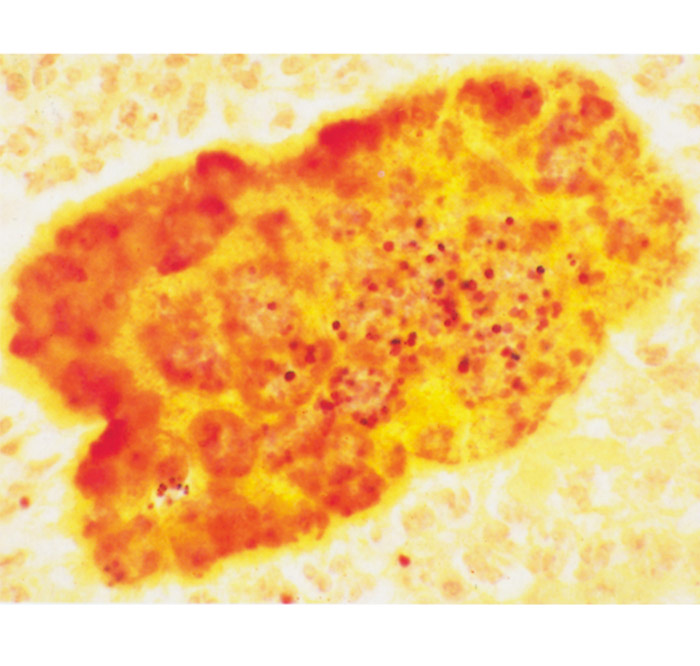
They frequently ulcerate and exude pus in which small granules, such as those found in certain mycoses, may be just visible to the naked eye as whitish or yellowish grains.11, 16 Some lesions, such as those of scirrhous cord in geldings, may become very large. Histologically the granules consist of masses of bacteria surrounded by eosinophilic material, known as Splendore-Hoeppli material, from which club-like bodies sometimes radiate at the periphery. This material probably represents glycoprotein antigen-antibody complexes and stains positively with the periodic-acid Schiff stain.11 The cell reaction generally consists of varying proportions of neutrophils, epithelioid cells, macrophages, lymphocytes, plasma cells and, in some, giant cells. There is often exuberant fibroplasia.
Predisposing causes of scirrhous cord in horses subsequent to castration are cutting the spermatic cord too close to the testicle, resulting in the protrusion of the cord through the scrotal wound after the operation; undue tension on the cord during the procedure; and adherence of the edge of the scrotal wound to the incised end of the cord.7, 8 The lesion, which consists of a firm, irregular, cauliflower- or mushroom-like (hence its French name of ‘champignon’) mass of connective tissue permeated by small abscesses and purulent sinus tracts, continues to expand slowly in size over many months or even years. In horses, it may attain the size of a human head. The lesion may extend into the inguinal canal, assuming the shape of a cone, and even into the abdominal cavity, or it may hang down from the scrotum, sometimes reaching the hocks. Alternatively, it may fill the scrotal sac, giving the impression that the animal still possesses a testicle. Pus discharges intermittently from the mass. Extensive adhesions develop between the growth and normal surrounding tissues and hamper surgical intervention, which is usually accompanied by severe haemorrhage. Well-developed lesions are associated with pain, fever and loss of condition.7, 8
The condition in horses known as ‘breast boil’ is encountered particularly in draught animals and is essentially a botryomycotic myositis of the neck and pectoral regions.11 The organisms gain entrance to the muscles through skin lesions caused by ill-fitting harnesses. This results in typical botryomycotic lesions, with swelling of the affected part, which slowly enlarges until it interferes with the horse’s work. The skin over the lesion becomes tense and eventually ruptures, with pus being discharged. The swelling may then subside to some extent and the skin wound heals. The swelling recurs and the whole process is periodically repeated, with the muscle lesion progressively increasing in size.9
The definitive diagnosis of botryomycosis rests on the results of bacteriological and histopathological examination of the lesions. In the latter case, the application of special staining techniques may be required (Figure 169.2). Botryomycotic granulomas have an identical macroscopic appearance to those of eumycotic mycetomas (see Mycoses) caused by true fungi, and granulomas caused by Actinomyces and Nocardia spp.3
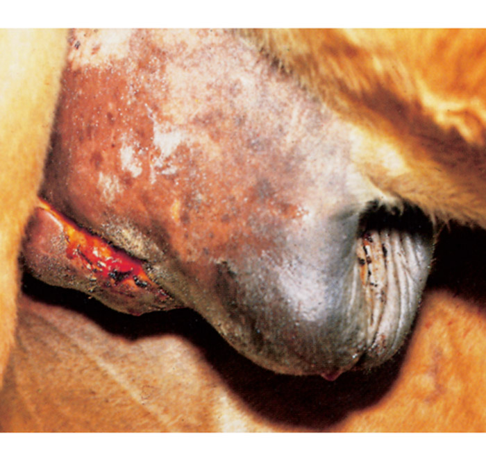
Figure 169.1 Scirrhous cord in a horse caused by Staphylococcus aureus infection following castration. Note swelling of the prepuce and protrusion of granulation tissue through the incision. (By courtesy of Prof. R.D. Gottschalk, Department of Surgery, Faculty of Veterinary Science, University of Pretoria, Onderstepoort)
The location of the lesions in botryomycosis often presents difficulties in its treatment, because simple surgical drainage of the lesions and the systemic administration of antimicrobial drugs are usually ineffective.16 The Splendore-Hoeppli material that surrounds the bacterial colonies and the granulomatous reaction with its large component of fi- brous connective tissue apparently impede the penetration of drugs and protect the organism. The best therapeutic results are obtained by complete surgical excision of the lesion, if this is possible, or by radical surgical debulking, followed by prolonged systemic antibiotic therapy, with the selection of the drug based on an antibiogram of the organism.16 Prevention lies in attention to aseptic surgical techniques and postoperative hygiene, or, in the case of botryomycosis developing secondarily to mechanical injury of the skin (such as that caused by the harness or plant thorns or bristles), in the removal of the cause if this is possible. Most cases of botryomycosis in horses involve wound infections of the skin, with or without the presence of a foreign body.14, 16
Folliculitis and furunculosis
Folliculitis and furunculosis due to staphylococcal infection are more common in horses, goats and sheep than in other livestock species. Most are caused by cutaneous trauma, and physiological stress is the predisposing factor.16
In horses there are three common forms, namely pastern folliculitis, truncal folliculitis and tail pyoderma. These conditions have been reviewed by Mullowney and Fadok.14 In pastern folliculitis, which occurs infrequently, lesions develop on the posterior aspect of the pastern and fetlock regions of one or more limbs. Initial lesions consist of papules and pustules which, if left untreated, coalesce to produce large, painful areas of ulceration and suppuration. Treatment consists of clipping the hairs from the affected area, washing the lesion, and the regular application of an antimicrobial ointment. The systemic administration of antibiotics is usually not necessary. The condition should be differentiated from greasy heel (pastern dermatitis).16
Most lesions in truncal folliculitis and furunculosis, which is also known by a variety of other names including saddle boil, saddle scab, acne, and summer rash, occur in the areas of the skin that are covered by saddles or harnesses. In temperate climates, it is encountered mainly in the spring and early summer. The condition is associated with sweating and friction, and is less common in wellgroomed horses. The lesions may persist for two or more weeks, and may be so painful that the animal is rendered unfit for work. Treatment consisting of the daily washing of the affected area with a povidone-iodine scrub and the intramuscular administration of penicillin for seven days is usually successful. However, the condition may recur.
Tail pyoderma of horses usually occurs as a result of skin trauma produced by tail-rubbing, which is provoked by insect bites, mange, lice or pinworm (Oxyuris equi) infestations, or as a vice. Small pustules form on the dorsal surface of the tail. The affected skin is pruritic, which compounds the self-mutilation. The disease progresses to a chronic pyoderma which responds poorly to treatment. Therapy includes the removal of predisposing causes, prevention of tail-rubbing, protection of the tail by bandaging, and topical and systemic application of antimicrobials.
Caprine staphylococcal folliculitis and furunculosis have been reviewed by Scott et al. 19 Moist and filthy environments, trauma, and the stress of parturition predispose to the condition, which is manifested by the development in the skin of superficial to deep papules, pustules, erosions, and ulcers which are covered by scabs. They may be painful and result in systemic disturbances. Lesions commonly commence on the udder and may spread to the ventral abdomen, medial thighs, perineum, distal limbs, face, and pinnae. Therapy consists of topical and systemic application of antimicrobial drugs to which the organism is susceptible, while the administration of an autogenous bacterin may be helpful. Infections by Corynebacterium pseudotuberculosis, Trueperella (Corynebacterium) pyogenes and Pseudomonas aeruginosa may produce asimilar disease but are much rarer than the staphylococcal disease.
References
- BATUNGBACAL, M.R. & SCOTT, G.R., 1982. Suppression of the immune response to clostridial vaccine by tick-borne fever. Journal of Comparative Pathology, 92, 409–413.
- BRODIE, T.A., HOLMES, P.H. & URQUHART, G.M., 1986. Some aspects of tick-borne disease of British sheep. The Veterinary Record, 118, 415–418.
- DONOVAN, G.A. & GROSS, T.L., 1984. Cutaneous botryomycosis (bacterial granulomas) in dairy cows caused by Pseudomonas aeruginosa. Journal of the American Veterinary Medical Association, 184, 197–198
- FRASER, J., SCOTT, F.M.M., ANGUS, K.W. & MARTIN, W.B., 1982. Experimental re-infection of the skin of sheep with Staphylococcal aureus. The Veterinary Record, 111, 485–486.
- GUNNING, R.F. & BOSWORTH, P.A., 1989. Staphylococcal dermatitis involving the teats of lactating ewes. The Veterinary Record, 124, 146–147.
- HAJEK, V., 1976. Staphylococcus intermedius, a new species isolated from animals. International Journal of Systematic Bacteriology, 26, 401–408.
- HAYES, M.H., 1952. Veterinary Notes for Horse Owners. A Manual of Horse Medicine and Surgery. London: Hurst & Blackett Ltd.
- HOBDAY, F.T.G., 1913. Manual of the Practice of Veterinary Medicine. London: Ballière, Tindall & Cox.
- HUNGEFORD, T.G., 1990. Diseases of Livestock. 9th edn. Sydney: McGraw-Hill Book Company.
- JONSSON, P. & WARDSTROM, T., 1993. Staphylococcus. In: GYLES, C.L. & THOEN, C.O., (eds). Pathogenesis of bacterial infections in animals. Ames: Iowa State University Press.
- JUBB, K.V.F., KENNEDY, P.C. & PALMER, N., 1985. Pathology of Domestic Animals. 3rd edn. New York: Academic Press.
- MARTIN, W.B., 1983. Staphylococcal dermatitis in sheep. The Veterinary Annual, 23, 104–108.
- MITCHELL, G.B.B., BRODIE, T.A. & WEBSTER, K.A., 1988. Tick pyaemia (enzootic staphylococcal infection): Recent developments. The Veterinary Annual, 28, 69–73. London: Butterworth & Co.
- MULLOWNEY, P.C. & FADOK, V.A., 1984. Dermatologic diseases of horses. Part II. Bacterial and viral skin diseases. The Compendium on Continuing Education for the Practising Veterinarian, 6, S16–S22.
- PARKER, B.N.J., BONSON, M.D. & CARROL, P.J., 1983. Staphylococcal dermatitis in unweaned lambs. The Veterinary Record, 113, 570–571.
- SCOTT, D.W., 1988. Large Animal Dermatology. Philadelphia: W.B. Saunders Company.
- SCOTT, F.M.M., FRASER, J. & MARTIN, W.B., 1980. Staphylococcal dermatitis in sheep. The Veterinary Record, 107, 572–574.
- SCOTT, F.M.M., FRASER, J. & SCOTT, G.R., 1991. Pyoderma. In: MARTIN, W.B. & AITKEN, I.D., (eds). Diseases of Sheep. 2nd edn. London: Blackwell Scientific Publications.
- SCOTT, D.W., SMITH, M.C. & MANNING, T.O., 1984. Caprine dermatology. Part I. Normal skin and bacterial and fungal disorders. The Compendium on Continuing Education for the Practising Veterinarian, 6, S190–221
- SCHLEIFER, K.H., 1986. Family 1. Micrococcaceae Prevot 1961. In: SNEATH, P.H.A., MAIR, N.S., SHARPE, M.E. & HOLT, J.G., (eds). Bergey’s Manual of Systematic Bacteriology.Vol. 2. Baltimore: Williams & Wilkins.
- SCHNEIDER, D.J., 1993. Veterinary Diagnostic Laboratory, Stellenbosch. Personal communication.
- SYNGE, B.A., SCOTT, F.M.M. & MACDOUGALL, D.C., 1985. Dermatitis of the legs of sheep associated with Staphylococcus aureus. The Veterinary Record, 116, 459–460.
- WATSON, W.A., 1964. Studies on the distribution of the sheep tick (Ixodes ricinus L.) and the occurrence of enzootic staphylococcal infection of lambs in North-West Yorkshire and North-East Lancashire. The Veterinary Record, 76, 743–747.
- WATSON, W.A., 1965. The carriage of pathogenic staphylococci by sheep. The Veterinary Record, 77, 477–478.
- WEBSTER, K.A. & MITCHELL, G.B.B., 1986. Experimental production of tick pyaemia. The Veterinary Record, 119, 186–187.


