- Infectious Diseases of Livestock
- Part 3
- Dermatophilosis
- GENERAL INTRODUCTION: SPIROCHAETES
- Swine dysentery
- Borrelia theileri infection
- Borrelia suilla infection
- Lyme disease in livestock
- Leptospirosis
- GENERAL INTRODUCTION: AEROBIC ⁄ MICRO-AEROPHILIC, MOTILE, HELICAL ⁄ VIBROID GRAM-NEGATIVE BACTERIA
- Genital campylobacteriosis in cattle
- Proliferative enteropathies of pigs
- Campylobacter jejuni infection
- GENERAL INTRODUCTION: GRAM-NEGATIVE AEROBIC OR CAPNOPHILIC RODS AND COCCI
- Moraxella spp. infections
- Bordetella bronchiseptica infections
- Pseudomonas spp. infections
- Glanders
- Melioidosis
- Brucella spp. infections
- Bovine brucellosis
- Brucella ovis infection
- Brucella melitensis infection
- Brucella suis infection
- Brucella infections in terrestrial wildlife
- GENERAL INTRODUCTION: FACULTATIVELY ANAEROBIC GRAM NEGATIVE RODS
- Klebsiella spp. infections
- Escherichia coli infections
- Salmonella spp. infections
- Bovine salmonellosis
- Ovine and caprine salmonellosis
- Porcine salmonellosis
- Equine salmonellosis
- Yersinia spp. infections
- Haemophilus and Histophilus spp. infections
- Haemophilus parasuis infection
- Histophilus somni disease complex in cattle
- Actinobacillus spp. infections
- Actinobacillus equuli infections
- Gram-negative pleomorphic infections: Actinobacillus seminis, Histophilus ovis and Histophilus somni
- Porcine pleuropneumonia
- Actinobacillus suis infections
- Pasteurella and Mannheimia spp. infections
- Pneumonic mannheimiosis and pasteurellosis of cattle
- Haemorrhagic septicaemia
- Pasteurellosis in sheep and goats
- Porcine pasteurellosis
- Progressive atrophic rhinitis
- GENERAL INTRODUCTION: ANAEROBIC GRAM-NEGATIVE, IRREGULAR RODS
- Fusobacterium necrophorum, Dichelobacter (Bacteroides) nodosus and Bacteroides spp. infections
- GENERAL INTRODUCTION: GRAM-POSITIVE COCCI
- Staphylococcus spp. infections
- Staphylococcus aureus infections
- Exudative epidermitis
- Other Staphylococcus spp. infections
- Streptococcus spp. infections
- Strangles
- Streptococcus suis infections
- Streptococcus porcinus infections
- Other Streptococcus spp. infections
- GENERAL INTRODUCTION: ENDOSPORE-FORMING GRAM-POSITIVE RODS AND COCCI
- Anthrax
- Clostridium perfringens group infections
- Clostridium perfringens type A infections
- Clostridium perfringens type B infections
- Clostridium perfringens type C infections
- Clostridium perfringens type D infections
- Malignant oedema⁄gas gangrene group of Clostridium spp.
- Clostridium chauvoei infections
- Clostridium novyi infections
- Clostridium septicum infections
- Other clostridial infections
- Tetanus
- Botulism
- GENERAL INTRODUCTION: REGULAR, NON-SPORING, GRAM-POSITIVE RODS
- Listeriosis
- Erysipelothrix rhusiopathiae infections
- GENERAL INTRODUCTION: IRREGULAR, NON-SPORING, GRAM-POSITIVE RODS
- Corynebacterium pseudotuberculosis infections
- Corynebacterium renale group infections
- Bolo disease
- Actinomyces bovis infections
- Trueperella pyogenes infections
- Actinobaculum suis infections
- Actinomyces hyovaginalis infections
- GENERAL INTRODUCTION: MYCOBACTERIA
- Tuberculosis
- Paratuberculosis
- GENERAL INTRODUCTION: ACTINOMYCETES
- Nocardiosis
- Rhodococcus equi infections
- Dermatophilosis
- GENERAL INTRODUCTION: MOLLICUTES
- Contagious bovine pleuropneumonia
- Contagious caprine pleuropneumonia
- Mycoplasmal pneumonia of pigs
- Mycoplasmal polyserositis and arthritis of pigs
- Mycoplasmal arthritis of pigs
- Bovine genital mycoplasmosis
- Neurotoxin-producing group of Clostridium spp.
- Contagious equine metritis
- Tyzzer's disease
- MYCOTIC AND ALGAL DISEASES: Mycoses
- MYCOTIC AND ALGAL DISEASES: Pneumocystosis
- MYCOTIC AND ALGAL DISEASES: Protothecosis and other algal diseases
- DISEASE COMPLEXES / UNKNOWN AETIOLOGY: Epivag
- DISEASE COMPLEXES / UNKNOWN AETIOLOGY: Ulcerative balanoposthitis and vulvovaginitis of sheep
- DISEASE COMPLEXES / UNKNOWN AETIOLOGY: Ill thrift
- Eperythrozoonosis
- Bovine haemobartonellosis
Dermatophilosis
This content is distributed under the following licence: Attribution-NonCommercial CC BY-NC  View Creative Commons Licence details here
Dermatophilosis
L T ZARIA AND J D AMIN
Introduction
Dermatophilosis is an acute, subacute or chronic skin disease that affects a wide range of domestic and wild animal species as well as humans and is characterized by the development of an exudative epidermitis followed by scab formation, alopecia and thickening of the skin. The disease is caused by Dermatophilus congolensis, a facultatively anaerobic, Gram-positive actinomycete that produces motile zoospores that invade the skin. The morbidity and mortality rates of the disease vary depending on the season; it constitutes an important economic problem to livestock production, particularly during rainy seasons.
The disease was first reported in 1915 in the annual report of the Veterinary Department of the then Belgian Congo (now Democratic Republic of Congo) by Van Saceghem.269 Since then, it has been diagnosed in many tropical, subtropical and temperate parts of the world. When the disease was first reported, the causative agent was named as Dermatophilus congolensis and subsequently as Actinomyces dermatonomus,33 Actinomyces congolensis 98 and Streptothrix bovis. It is now classified as Dermatophilus congolensis.75
The disease is also known by different names depending upon the hosts affected and the clinical picture. Initially, it was named ‘contagious dermatitis’, but this name is inappropriate due to the number of infective agents known to cause bovine dermatitis. The commonly used term ‘mycotic dermatitis’ in sheep and cattle is incorrect as it implies that the disease is caused by a fungus. The use of the term ‘cutaneous streptothricosis’ is also incorrect as it implies that the aetiological agent is a Streptothrix sp. Similarly, the terms ‘nocardial dermatitis’ and ‘cutaneous nocardiosis’ are also inappropriate.
Dermatophilosis is of economic importance because it can cause great losses to the livestock and leather industries. 3, 71, 84, 102, 124, 173, 176, 229, 260 These are mainly attributed to poor quality of hides and skin and to low meat production as affected animals may lose weight and become emaciated. Other losses are the result of decreased milk production, decreased working capacity of draught cattle,120 forced culling of infected animals before they reach market value, and death in severe cases. The disease in cows can cause as much as a 20 per cent drop in milk production.163, 178 However, the reduced growth rate of calves suckling affected cows is considered to be more the result of the lesions on the udder and teats than the decreased milk production.98 Severely emaciated cows may show anoestrus.
The fertility of terminally affected bulls may be reduced as a result of abnormalities in spermatozoa; sterility may even occur.188 In woolled sheep, economic losses result from damage to the fleece and hides.27, 259, 275 The scabs on severely affected sheep may also cause considerable inconvenience as a result of loss of time and breakage of machines during the shearing process. In Australia, the most important economic effect is as a consequence of secondary blowfly strike of the lesions in sheep.72
Dermatophilus congolensis infection may occur in humans, 10, 11, 46, 57, 108, 109, 130, 197, 255 and pitted keratolysis in humans is thought possibly to be associated with the organism. 174, 233, 277, 282
Aetiology
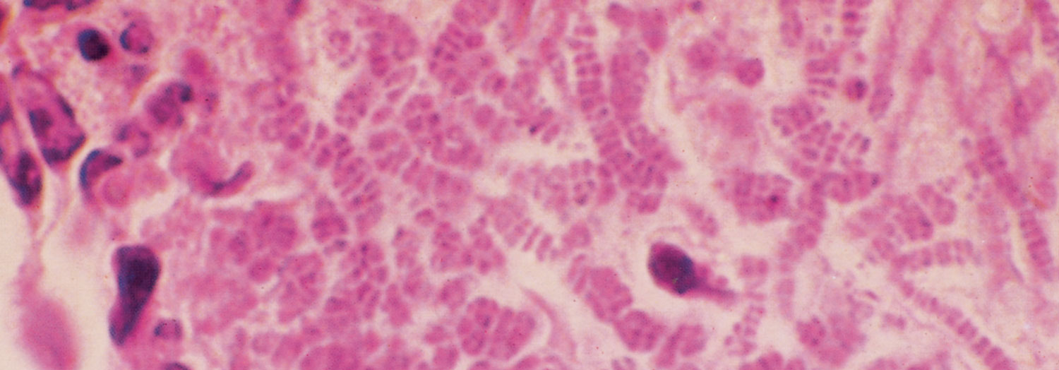
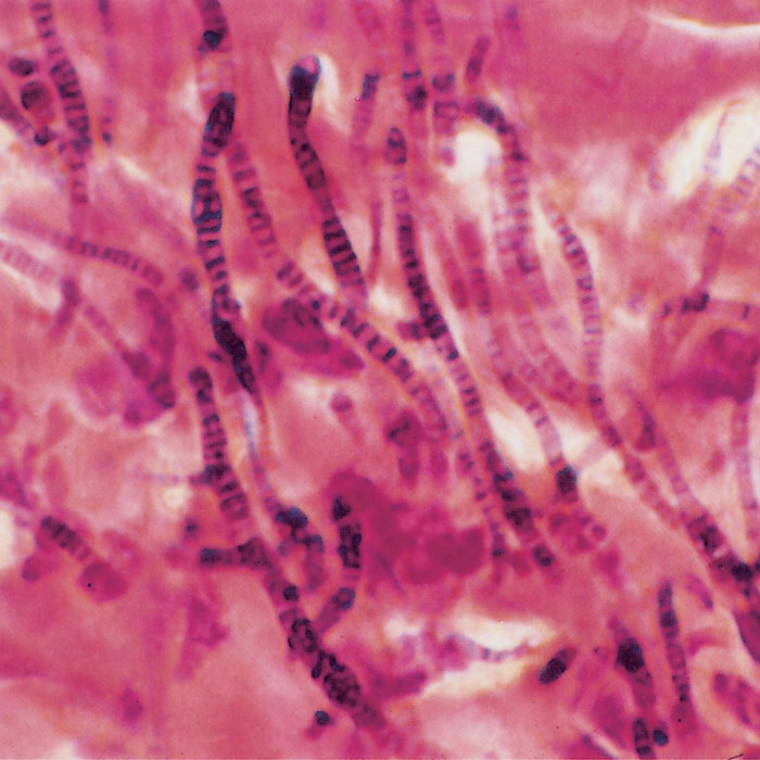
Dermatophilus congolensis is classified in the family Dermatophilaceae, order Actinomycetales. It is a pleomorphic, aerobic and facultatively anaerobic, Gram-positive bacterium which produces zoospores. On germinating, the zoospores bud to produce a filament that is less than 1 μm wide, which later forms right-angled lateral branching. Initially transverse and then longitudinal septa develop in the filaments, giving rise to parallel rows of encapsulated coccoid cells, 1,5 μm in diameter (zoospores) of equal size that can either be single-stranded or up to eight rows wide (Figures 200.1 and 200.2).77
All these structures are observed in smears made from lesions but may appear fragmented as a result of the smear making process.
The zoospores can remain dormant in the absence of any adequate stimulus, such as moisture, and, when in this state, are highly resistant to desiccation and may even remain viable after heating to 100 °C for up to 30 minutes. They lose their outer capsule and become motile by means of a tuft of flagella within 24 hours with the advent of warm, humid conditions, and can migrate from the moist scabs of the skin lesions that are induced by them.207, 212 Motile zoospores are not very resistant to physical and chemical changes such as pH, osmotic changes and desiccation; these result in decreased viability after a few hours outside the scab lesions, but in a favourable substrate, such as the persistently moistened skin of a suitable host, they lose their motility, and germinate.
Dermatophilus congolensis grows best in an atmosphere containing 10 per cent carbon dioxide.76, 137, 211 There is pronounced haemolysis on media containing sheep but not horse blood,77 and aerial filaments are more profuse. The bacterium can grow at a temperature of 22 to 37 °C, but the optimum growth temperature is 37 °C.137 It can be cultured on or in most ordinary laboratory media but optimal growth occurs on blood agar and brain heart infusion agar, and in tryptone broth; abundant growth can be achieved on Leoffler’s and Dorset egg media.137 It does not grow on media such as coagulated egg albumin, Sabouraud’s dextrose agar, potato dextrose agar, MacConkey’s agar, deoxychocolate citrate agar, Czapek’s agar, and Edward’s crystal violet agar. 7, 137, 162, 200, 268
There is a great variation in the colonial appearance of the organism; a single strain often gives rise to diverse forms on different media under varying conditions.76, 78 On blood agar, the colonies are very small, round to squarish, irregular, greyish-white, rough and granular, and are usually tough and adherent through invasion of the substrate by the filaments.77 There is beta-haemolysis and sometimes short, white aerial filaments may be observed. In older cultures the colonies become yellowish to orange and they are smooth, opaque and larger, and the zone of haemolysis also increases in extent. Strain variations in the rate of growth under microaerophilic condition occur,19, 76, 137 and pigment production also varies: some strains produce greyishyellow pigment, while some produce greyish-white to bright orange pigments.76, 130, 263
In liquid media, growth usually appears as a thick sediment, the supernatant broth being clear. The sediment is generally flocculent, granular or flaky but in some it is ropy or taffy-like.76, 137, 263
The bacterium is catalase positive, hydrolyses urea and starch and is proteolytic for gelatin. When grown in enriched media, it produces acid from glucose and fructose but not from dulcitol, lactose, mannitol, salicin, sorbitol, sucrose and xylose.76, 77, 137
Epidemiology
Dermatophilosis has a world-wide distribution but is most prevalent in humid, tropical and subtropical regions.21, 23, 34, 40, 44, 46, 48, 49, 51, 60, 80, 84, 88, 107, 113, 117, 129, 138, 154, 163, 172, 174, 178, 182, 189, 190, 195, 197, 203, 215, 231, 233, 237, 238, 240, 252, 253, 265, 266, 270 Predisposing factors such as rainfall, high humidity 64, 132 and ambient temperature, skin lesions (due to, for example, ectoparasites, thorns and other trauma), skin pigmentation, managemental practices, intercurrent diseases, malnutrition, stress and heredity,2, 123, 140 play an important role in the epidemiology and manifestations of the disease. The effect of climate is, however, one of the most prominent epidemiological features of dermatophilosis. Another important feature is that animals in which the disease has regressed often become reinfected repeatedly in successive wet seasons.
Dermatophilosis affects a wide range of domestic and wild animals, including terrestrial and aquatic mammals.65 Among domestic animals, it occurs most commonly in cattle, sheep, goats and horses,25, 31, 36, 51, 62, 86, 235, 246 but mules, donkeys,127 water buffalo (Bubalus bubalis)115, 174 and pigs may also be affected.115, 128, 165, 254, 267, 271 Lambs aged three to nine months are particularly susceptible.122 It rarely occurs in dogs, cats and camels (Camelus dromedarius). 30, 39, 63, 67, 73, 105, 248
Occasional infections have been reported in many wild animal species, including white-tailed deer (Odocoileus virginianus), woodchuck (Marmota monax), striped skunk (Mephitis mephitis), racoon (Pryocyon lotor), giraffe (Giraffa camelopardalis) and antelope,79, 221, 227–229, 257 chamois (Rupicapra sp.), zebra (Equus burchelli), small rodents and monkeys,63, 99 polar bears (Tholarctos maritimus),175, 250, 251 snakes and lizards.166, 244 Birds are highly resistant, but experimental infection of the disease in White Leghorn fowls has been induced.232
In subtropical areas of South America and Africa, sheep are most commonly affected,44, 109, 117, 138, 168, 178, 195 while in regions that have a monsoon climate, such as tropical regions, cattle and water buffalo are the main animal species affected.174, 248 In New Zealand, Australia and South Africa, the disease mainly occurs in sheep.
Dermatophilosis is transmitted by direct or indirect contact. Close contact between animals, such as rubbing against each other,190 favours mechanical transfer of the organism.122 Experimental infection of sheep with D. congolensis can be effected by intradermal inoculation of the organism or by application of cultures or infected-scab suspensions to the skin with or without wetting, depilation, scarification or removal of the sebaceous overlay.9, 14, 33, 46, 117, 255, 262, 270, 275, 279 Infection spreads readily between infected and susceptible sheep, particularly when both affected animals are wet.279 Indirect transference of the zoospores may occur mechanically through contact with plants, other objects or insects and arachnids (see below) harbouring the organism. The wetted skin and fleece, and particularly the wetted crusts of lesions, are attractive to flies which promote the transmission of infection.204, 219
In animals, the epidermis is protected against infection by three barriers, namely the hair coat or fleece, the sebum which impregnates the hair coat or wool and forms a layer on the skin, and the stratum corneum.213, 215, 219
The degree of development of each barrier, and hence the protection it provides, varies greatly between animals and at different sites on the body. In sheep, the thickness of the stratum corneum is inversely proportional to the density of the hair coat, and the thickness and completeness of the sebaceous film on the skin are directly related to the density of hair or wool-fibre population because of the association of sebaceous glands with hair or wool follicles. Resistance of the non-woolled areas to infection therefore depends mainly on the integrity of the stratum corneum, while the woolled areas of the skin, which have a very thin stratum corneum, are protected mainly by the greasy fleece and the sebaceous film on the skin surface.213 This sebaceous film on the woolled skin is more protective in adult Merino sheep than in young lambs or in coarser-woolled sheep, in which the film may be incomplete.213, 215, 219 The protectiveness of the sebaceous film apparently depends entirely on the mechanical properties of the sebum.213
Prolonged wetting of the skin causes emulsification and disruption of the sebaceous film and maceration of the stratum corneum, rendering the skin susceptible to infection by D. congolensis.47, 104, 213
The zoospores are found mostly in the scabs and dried exudate of the lesions,33, 206, 207, 209, 210, 212, 213, 215 from which, if thorough wetting occurs, they are released in large numbers. As virtually no viable zoospores are detectable at the surface of scabs and exudate before they have been soaked, affected animals are probably not an important source of infection unless their lesions are wet.173, 266 The number of viable zoospores in scabs decreases rapidly as scabs dry out, but small numbers may survive in them for several months.209, 212, 219, 240
Outbreaks of dermatophilosis in sheep are often associated with wet weather conditions,13, 26, 52, 89, 160, 207, 208, 215, 219, 256 dipping,26, 52, 121, 122, 131, 213, 270, 275 shearing,122 skin penetration sites of grass seeds or thorns of plants,26, 213, 219 ear-punching and insect bite sites.
Outbreaks of the disease may occur in both summer and winter.26, 89, 160, 219, 256 Under natural conditions, rain, misty weather and dew are important in the release of zoospores.6, 13, 26, 33, 52, 89, 160, 219, 256, 275 Continuous wetting of the feet, face and parts of the fleece in young lambs from grazing wet pastures plays a contributory role in outbreaks of dermatophilosis. 13 The disease has also been reported in lambs a few days after birth, possibly as a result of the wetness of the skin caused by foetal fluids,241 and around the mouth from suckling. 222
The association of dipping with outbreaks of dermatophilosis is generally attributed not only to the wetting of the skin and the subsequent release of zoospores, but also to the detrimental effect of the dip on the integrity of the sebaceous film of the skin. Dermatophilus congolensis is known to survive in dipping fluids containing certain acaricides and, where animals are regularly dipped to control ectoparasites, organisms in scabs and crusts of infected animals can contaminate the fluid.10, 93, 218, 237, 264, 278, 283
In addition to its role in the activation and release of zoospores, water or dipping fluids may also transport zoospores from existing lesions to non-infected areas on the same animal.121, 132, 219 That this happens is borne out by the development of acute lesions in sites lower down on the body to chronic ones, and the confluent lines or channels of affected skin down the sides of infected sheep exposed to prolonged heavy rain.219
Mechanical disruption of the protective barriers of the skin is caused by the shearing of sheep, which not only removes the protective fleece, but also frequently causes abrasions and wounds of the skin.191, 213, 217
Dermatophilosis in cattle occurs in all age groups,43, 107, 197, 198, 234, 237, 245, 253 and all age groups and sexes appear to be equally susceptible.135, 172, 189, 190, 225 There are, however, breed differences. In West Africa, N’dama and Mturu breeds are believed to possess a degree of natural resistance, while the White Tulani and Zebu breeds and some European breeds with relatively long hair coats are highly susceptible. 14, 42, 122, 123 The lighter skinned Wagumbe cattle in Nigeria would appear to be more severely affected than the variegated Wagumbe.50, 203 Paradoxically, in Guadeloupe, the Creole breed of cattle is highly resistant but the same breed and its crosses in Martinique are highly susceptible. 139, 140, 157, 158 The introduction of the highly susceptible and high producing dairy breeds, such as the Holstein and Jersey, into West Africa where the disease is hyperendemic, has been greatly hampered.
Higher serum levels of some minerals, such as zinc and iron, have been found in breeds resistant to dermatophilosis as compared to those in susceptible breeds,66, 68, 112, 243 but the role that these minerals play in the resistance of susceptibility of animals to the disease is not clear.69 Decreased blood levels of ascorbic acid and cholesterol have also been reported.58, 276
There is an increased prevalence of the disease in cattle in the rainy season compared to that in the dry season.49, 60, 253, 255 Severe wetting or saturation of the hair and skin for several days or weeks is often associated with a higher prevalence of dermatophilosis than is found in areas receiving a high but intermittent rainfall. During the rainy seasons, other predisposing factors, such as malnutrition, ectoparasites and intercurrent diseases, may also play a role in the epidemiology of the disease. 281
In Nigeria the prevalence of the dermatophilosis in cattle may be as high as 50 per cent during the rainy season. It has been estimated there that, based on a 10 to 12 per cent prevalence rate, about one million cattle are infected annually, and about 287 000 of the cases are likely to be chronically affected and consequently die or are culled, which result in severe economic losses.224
Infestations with arthropods,224 especially the hard tick Amblyomma variegatum, have long been associated with an increased prevalence of dermatophilosis,20, 35, 37, 133, 156, 161, 170, 171, 180, 182, 186, 190, 198, 226, 272, 273 the lesions occurring particularly in the most heavily infested areas of the body but there is no cogent explanation for this.180, 273 It has been postulated that skin lesions due to delayed hypersensitivity as a result of repeated feeding of the ticks provide a portal of entry to D. congolensis 217 and that the systemic effects on the immune system of the host of the immunomodulating factors, such as prostaglandin E2 (PGE2), present in the saliva of ticks, predispose to the disease. The nymphal stage of A. variegatum has been experimentally incriminated in transmitting the organism.225 It has also been observed in herds subjected to tick control measures that the disease either does not occur, or its prevalence and severity are diminished. 94, 116, 161, 198, 231
Dermatophilus congolensis has been isolated from certain ticks such as Hyalomma asticum, A. variegatum and Boophilus microplus.117, 167, 171, 190, 272, 280 Successful transmission of dermatophilosis has been achieved by feeding A. variegatum, collected from infected animals, on noninfected animals.133, 198, 225
Biting and non-biting flies, such as Musca domestica, Lyperosia spp., Stomoxys calcitrans, tabanids, Glossina morsitans, Lucilia cuprina and Calliphoria spp., and mosquitoes have been implicated in the transmission and spread of dermatophilosis,204 although the actual role they play in its epidemiology is not very clear. It has been suggested that they may cause a hypersensitivity type of reaction in the skin. 7, 134, 184
Other ectoparasites which can create a portal of entry for D. congolensis in animals, and which have been incriminated in the transmission of the disease, are Cochliomyia macelloma, lice, and mites such as Demodex spp. and Chorioptes spp. 204 Experimentally, the disease has been transmitted to rabbits by S. calcitrans and M. domestica, the flies remaining infective for up to 24 hours after feeding on infected rabbits. 204
The establishment of the infection is facilitated by damage of the skin barrier by various mechanical injuries such as those caused by the feeding of ox-pecker birds (Buphagus spp.), shearing, branches of trees and spines of thorny bushes and other sharp objects.107, 184 In draught oxen, lesions often occur in the skin of those areas of the neck which bear the yoke. 84 It has been suggested that oral lesions observed in some animals may have been secondary to abrasions caused by browsing on thorny bushes.100, 185, 247
In those cases of the disease which are associated with mechanical injuries, serum and/or blood oozing from the lesions may attract flies and other insects carrying D. congolensis, as well as provide nutrient and a favourable environment for the establishment of the organism with consequent development of the disease. 2
Dermatophilus congolensis infection has been associated with rinderpest,6 lumpy skin disease,103, 141 besnoitiosis and trypanosomiosis 27 in cattle, and with contagious pustular dermatitis (orf) in sheep. 5, 21
Pathogenesis
Following infection, the zoospores reach susceptible sites of the skin by their own motility 209, 215 and a positive chemotactic response to carbon dioxide which diffuses through the skin.209, 210
In contrast to dermatophytes, which are parasites of the stratum corneum, D. congolensis invades the living cell layers of the epidermis and the sheaths of the hair or wool follicles, in which extensive proliferation of the organism occurs; it does not invade the dermis or the hair or wool fibres.14, 15, 214, 215 The organisms induce increased keratinization of the epidermis and an acute inflammation characterized by the accumulation of an exudate rich in neutrophils beneath the invaded part of the epidermis; the exudate separates the infected from the non-infected epidermis.106 The organisms do not penetrate the layer of the exudate, which apparently acts as a barrier to invasion of the uninfected layers of the epidermis. The new epidermis that forms underneath the exudate is invaded from the side, usually by filamentous organisms from adjacent follicle sheaths.214 The infected epidermis is then again separated from the uninfected epidermis by a layer of exudate, and so the process continues in cycles until a thick, laminated crust is formed, composed of alternating layers of para- and orthokeratotic hyperkeratosis and exudate.106, 214 The new layers of epidermis are formed mainly as outgrowths from the sheaths of the follicles, and are also infected by bacterial filaments originating from these sites. The presence of the organism in the crusts acts as a source of infection to other animals and of re-infection in affected animals during rainy seasons.
The immunology of dermatophilosis is not well understood. 6, 146, 148, 151, 193 Antigens of D. congolensis consist of exoantigens which are mucoid and of the hexosaminegalactose, xylose peptide type, and there is an immunological cross-reactivity of several strains of D. congolensis with respect to somatic agglutinogens, haemolysins and precipitinogens, but flagellar agglutinogen displays no strain variation.219, 242 The antigenic structure of the organism has been established as complex and appears to depend partly on the phase of the life-cycle. Crude antigenic products of D. congolensis strains have been separated into ‘exoantigens’ and ‘endoplasm’ antigens which are derived from the cytoplasmic contents of the whole cell.118, 144, 219
Studies of the humoral immune response35 during natural and experimental infections of dermatophilosis in various hosts have established the presence of antibodies which do not afford any protection against re-infection nor do they have any influence on the course of the disease once it is established. However, successful immunization of cattle against D. congolensis infection by using a whole-cell antigen has been reported.29, 38, 56, 96, 97, 126, 145, 152, 153, 183, 196, 201, 217, 258, 259
In Nigeria, experiences in the field and during the performance of serological procedures for diagnostic purposes have shown that in dermatophilosis different levels and types of antibody occur in naturally infected cattle which depend on the stage and severity of the disease. For instance, antibody to extracellular antigen is in abundance in the acute as well as the mild disease, while chronically infected animals show higher levels of antibody to wholecell, cell wall and cytoplasmic antigens.149, 150, 152 During the early stages of the disease, infected animals show higher antibody responses to whole-cell associated antigen than to extracellular antigens which indicate that the immunocompetent cells of the host come in contact with the cell wall of D. congolensis before extracellular antigens have been produced.145
It is thought that D. congolensis and certain poxviruses, such as orf virus in sheep and lumpy skin disease virus in cattle, may have a synergistic action, as it is often present in lesions caused by these viruses.1, 179, 182, 299, 237 Severe lesions similar to those observed in field cases of stawberry footrot have been induced in lambs infected with the orf virus and D. congolensis together, but not with either organism alone.
Clinical signs and pathology
Generally, in all the affected animal species, the disease is characterized by the appearance, in the areas affected, of an exudative to proliferative epidermitis with subsequent formation of scabs and crusts under which the hair or fleece tend to break or matt together. The matted hair or fleece may sometimes become detached, leaving raw areas. The detached hair or wool and crusts during the early stages of the disease to some extent resemble camel hair paint brushes; this stage is often referred to as the ‘paint brush stage’ of the disease. In the chronic stage, the scabs are dry and when removed, leave only a soft pinkish area because of the healing that has already occurred.99, 177, 185
In flocks of sheep, the prevalence of lumpy wool may vary from individual cases to severe outbreaks where almost all the animals are affected.13, 26, 33, 53, 89, 160, 256 The mortality rate is usually very low, but up to 20 per cent of affected lambs may die.41, 236, 275
Mild lesions are inconspicuous and not easy to detect in sheep. Even advanced lesions can be concealed by the fleece and may only be detected by palpation of the animal or during shearing and classing of the wool.21, 89
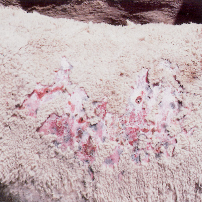
The size, severity and distribution of the lesions vary markedly among affected animals. In sheep, the lesions may occur in the woolled13, 17, 26, 33, 111, 192, 240 or the hairy parts of the body,41, 87, 89, 222, 236, 256 or both. Acute to chronic, and localized to more widespread lesions may be found in the woolled parts of the skin.211 Acute lesions are characterized by erythema, exudation and the presence of isolated flecks of yellowish-white to brownish grease or crusts (Figure200.3). As the epidermitis becomes more severe, large amounts of moist exudate or scabs, varying in colour from orange, yellow, brown or grey to greyish-white are evident. 219 As the sticky exudate is carried away from the skin, it dries and glues together clumps or even entire staples of wool fibres to form hard spikes and lumps. The long-standing lesions are often accompanied by a whitish, dry appearance of the epidermis, and a raggedness or uneven surface of the fleece. On parting the wool, single or multiple transverse bands of scabs can be seen, being whitish or greyish white near the skin and brown or almost black towards the surface of the fleece.21 Typical chronic lesions (Figure 200.4) are marked by lumps of up to 30 to 40mm in diameter which are yellowish to greyish-brown, dense, hard, horn-like and pyramidal to dome-shaped.26, 33, 219, 255 When removed, the lumps are usually concave at their base and some are partially hollow.26, 33, 89, 160, 256 The distance from the skin surface varies according to how far the healing process has proceeded. The underlying skin may be normal, or the lesions may still be firmly attached to the skin and can only be removed with difficulty (and with great pain to the animals), leaving a granular and often bleeding surface.270
Crusty lesions (Figure 200.5) are found predominantly in the coarser-woolled sheep, such as the British breeds and their crosses, and in young lambs up to nine months old, whereas the chronic lumpy-wool type of lesion is mostly seen in strong- and medium-woolled Merino sheep.13, 89, 213, 219
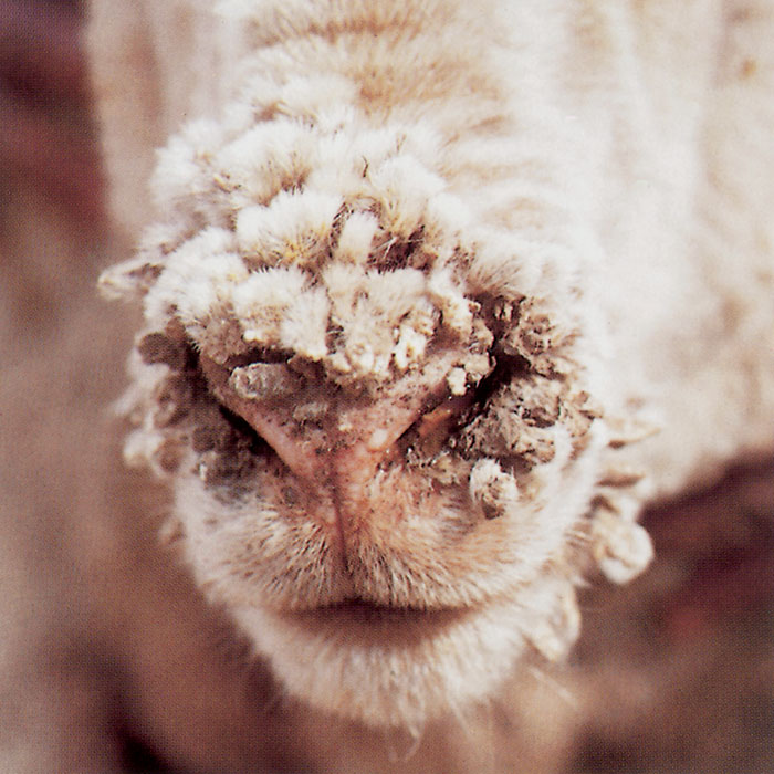
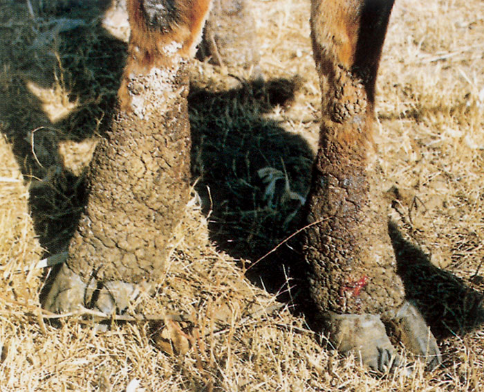
Lesions of the haired areas of the skin in woolled sheep may occur on the face, lips, chin, nose, ears, axillae, inguinal region, udder, scrotum, and legs, from the carpus and tarsus to the hooves but particularly around the coronets.6, 33, 41, 222, 256 When the lower legs are affected, the condition is generally referred to as strawberry footrot.13, 19, 26, 263 On the lower legs the lesions may become confluent; the affected areas are covered by thick crusts, while dense wart-like growths are sometimes evident around the coronets. On removal of the crusts, granular and bleeding surfaces that somewhat resemble strawberries are exposed. Secondary infection of the lesions is common. In long-standing cases the lesions on the lips, chin, nose, forehead, and ears tend to be more confined and wart-like.6, 33, 160, 256 Mild chronic lesions are characterized only by a scabiness or slight scab formation on the ears and nose; this occurs particularly in lambs.29
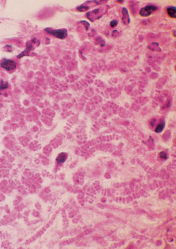
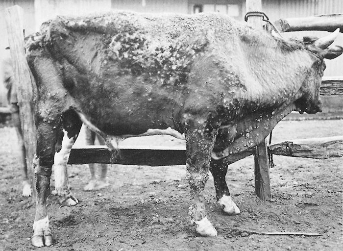
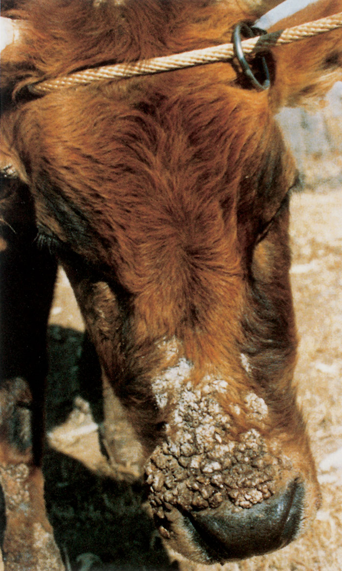
In cattle, the distribution of the lesions varies and is associated with the predisposing cause or causes. In some, the lesions cover almost the entire body, especially the back and sides, perineal region, lower limbs, tail, muzzle and ears.177 Lesions associated with a heavy tick infestation develop mainly on the ventral areas of the body, such as the axillae, brisket, inguinal area, scrotum or udder.270 Lesions of the face, ears and body (Figure 200.6) may suggest that infection has been transferred by thorny bushes. In calves, lesions often occur on the muzzle, neck and head.270
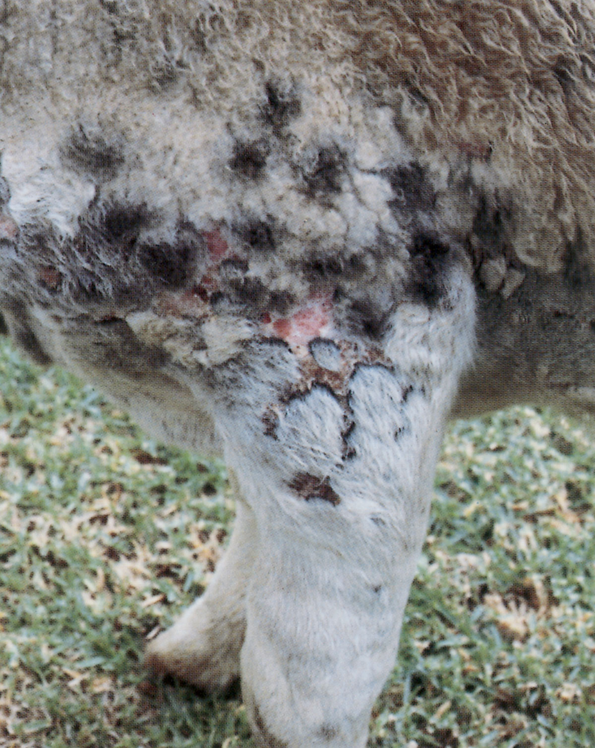
Lesions in cattle may be focal, about 50 mm in diameter or become confluent to form a large, mosaic-like area of the skin (Figures 200.7 and 200.8). There is initially formation of a few moist papules scattered over the affected area which subsequently become covered with hard crusts. Affected areas are thickened, raised, hard, coarse and uneven on palpation. 44 The whitish to yellowish-brown crusts are difficult to remove, but on removal an area of granulation tissue is exposed. The hair coat may contain loose yellowish crusts or flakes resembling severe dandruff.44
In regions where dermatophilosis in cattle is associated with rainfall and moderate humidity but where the rainy season is fairly short, lesions seldom progress further than the crusty or scabby stage during the dry season. However, in those regions where there are extended rainfall periods associated with a prolonged high humidity, the lesions progress to form thick layers of dry, spongy scabs or hard, button-like crusts which protrude above the surface of the hair coat, especially where they are complicated by secondary bacterial infections.177 The spongy type of lesion is usually diffuse and covers large areas, particularly affecting areas on the body where the hair coat is long. The hard, button- like lesions occur more frequently in the regions where the hair coat is shorter, especially along the back, rump, face and perineal regions. Detachment of these dry crusts exposes the moist raw surfaces of the skin; usually no pus is present. These lesions are referred to as the ‘nodular and papillomatous’ stages of the disease.177 The appearance of the lesions may be modified by secondary bacterial invaders. 99 In some cattle a necrotic dermatitis characterized by the presence of a thick, purulent, offensive-smelling exudate may develop, which usually spreads rapidly and covers large areas of the body. This grave complication, caused by a mixed bacterial infection, invariably terminates in the death of the animal.33, 40
Severely affected animals become emaciated and the disease may terminate in death. On the other hand, mild cases may heal spontaneously under conditions such as the advent of the dry season, the feeding of high levels of concentrates, or the improvement of the general body condition by other means.
In equine animals, such as horses, donkeys and mules, the disease is known as ‘rain scald’ or ‘rain rot’, and lesions consist of irregular patches of matted hair or small, localized raised areas which are accumulations of sebum, or areas of alopecia. They occur particularly on the muzzle, especially around the nostrils, and on the ears, withers, back, croup, rump and tail. There is also a considerable amount of scurf on the affected part of the body.62, 99, 235 Horses maintained under muddy conditions or on very wet pastures may develop chronic lesions on the heels. This disease is known as ‘mud fever’.
Other lesions that have occasionally been associated with D. congolensis infections include subepidermal granulomas and oral lesions in cattle,22, 59, 100, 129, 245 and chronic lymphadenitis in goats.178
The lesions in wild animals are similar to those described in livestock.228, 229, 257
Dermatophilosis in humans usually manifests as pustules on the hands and occasionally feet, which may itch.10, 46, 99, 108, 130, 223, 255 It has been suggested that pitted keratolysis or keratoma plantare sulcatum may be associated with D. congolensis.12, 277, 282
Histopathologically, the most striking feature of the lesions of the uncomplicated disease is its superficial nature. Lesions are restricted mainly to the epidermis and upper layer of the dermis. Acute lesions are characterized by congestion and oedema of the dermis;13, 214, 217, 219 degeneration, necrosis, parakeratosis and hyperkeratosis of the cells of the epidermis; accumulation of exudate on the surface of the skin; and infiltration of neutrophils and other inflammatory cells into the dermis and epidermis. Branching, septated, bacterial filaments or packets of parallel rows of coccoid zoospores up to eight rows thick are found in the exudate as well as the epidermis as far down as the stratum basale, but they do not usually invade the dermis. Organisms extend into the follicular sheaths for some distance, causing proliferation of the sheath cells. Wool and hair fibres are not invaded.
Diagnosis
A provisional diagnosis of dermatophilosis can be made on the clinical signs but it should be confirmed by the demonstration of the organism by microscopical examination of Giemsa- or Gram-stained smears prepared from the lesions (Figures 200.1 and 200.2), by histopathological examination of biopsies of lesions, or by cultivation and identification of the organism.76, 83, 98, 114, 274 Smears may be made from the affected skin, the concave base of freshly removed scabs, or aqueous suspensions of some of the most recently formed scabs, the suspensions being prepared by grinding the latter in a mortar with a pestle and adding water. Another method is by animal inoculation of material from clinical specimens onto scarified areas of the skin of laboratory animals such as rabbits, guinea pigs or rats.249, 255
The isolation of the organism by culture is usually facilitated by soaking newly formed scabs or crusts in distilled water for three hours. The material is then transferred to a candle jar for 20 minutes, after which the fluid is inoculated onto culture media such as blood agar slopes or plates. These are incubated in an atmosphere containing 10 per cent carbon dioxide for 48 hours.83 The addition of compounds that inhibit the growth of contaminants to the culture media such as 1 000 units of polymyxin B sulphate (Pfizer) per millilitre of medium, is recommended.
Serological tests which have been applied include the direct agglutination, indirect haemagglutination, enzymelinked immunosorbent assay,74, 125, 143, 155, 159 complement fixation and fluorescent antibody.29, 92, 119, 142, 143, 145–147, 149, 150, 152, 153, 155, 202, 205 Serology is, however, less reliable in the diagnosis of dermatophilosis than the tests mentioned above since most animals have circulating antibodies, but elevated antibody concentration is indicative of a recent clinical infection.
Differential diagnosis
In cattle, ringworm lesions (which are usually characterized by circular skin leisons on the face or neck), mange (which is usually pruritic), subacute to chronic photodermatitis (which only affects the non-pigmented areas of the body), skin lesions associated with lumpy skin disease, pseudolumpy skin disease, and subacute to chronic sweating sickness may be confused with dermatophilosis.
In sheep, skin and fleece disorders, such as contagious pustular dermatitis, sheep scab, sarcoptic and chorioptic mange, sheep-itch mite infestation, ringworm, dermatitis as a result of photosensitization, fleece rot, fleece discoloration and Bolo disease should be differentiated from dermatophilosis. Differentiation from dermatophilosis often depends on the microscopic and cultural demonstration of D. congolensis in lesions.270
Control
Preventive measures, if at all attainable, should aim at interrupting the chain of epidermiological events required for the occurrence of outbreaks of dermatophilosis.208, 209–212, 219, 220 These measures will therefore mainly involve methods of husbandry.22 Tick control, when tick infestations are the predisposing cause, and early treatment are the most important managemental factors influencing the prevalence of dermatophilosis in cattle.61, 169 The culling of animals with advanced chronic lesions should also be considered.
At present there is no commercial vaccine available.259 In cattle and sheep, because of the thick and firmly adherent scabs which cover the lesions, and the depth to which the filaments of D. congolensis penetrate into the affected skin and follicle sheaths, topical treatment is usually unrewarding. Nevertheless, several preparations have been used topically to ‘spot treat’ individual lesions with varying degrees of success. These include preparations in the form of an ointment, lotion or solution containing sulphur, iodine, arsenic, copper sulphate, mercuric chloride, zinc sulphate, methyl violet, penicillin or terramycin.18, 172 Where ticks are involved in the pathogenesis of the disease in individual animals, a mixture comprising an acaricide and aluminium sulphate has been found to have beneficial effects.90, 91
A new formulation called ‘Lamstreptocide’ and which is used topically has been prepared by the National Veterinary Research Institute in Vom, Nigeria. This preparation consists of two parts: Lamstreptocide A, which contains palmitic, stearic, oleic and linoleic acids, while the second compound, Lamstreptocide B, contains salts of zinc, iron, copper, manganese, cobalt, calcium, magnesium, sodium, potassium and phosphorus. In in vitro studies Lamstreptocide was found to have inhibitory properties on D. congolensis at very low concentrations, although it did not inhibit such organisms as Staphylococcus aureus, Bacillus cereus, Bacillus subtilis and a Corynebacterium sp. Some of these organisms are found on the skin and often cause secondary infections, hence the need to support topical Lamstreptocide treatment by the parenteral administration of broadspectrum antibiotics, especially in chronically infected animals.59
In clinical trials in cattle it was demonstrated that Lamstreptocide penetrated deeply into skin scabs and softens them to such an extent that they fall off without them having to be removed mechanically; this occurred seven days after its application.
This facilitated contact between the agent and D. congolensis. Lamstreptocide has, in addition, a repellant effect on flies which impedes healing of the lesions. 59 The preparation was proved to be non-toxic and non-corrosive; fewer applications of Lamstreptocide are required to cure mild and acute cases of the disease than chronic cases.
In horses, mild cases are treated topically with 10 per cent povidone iodine and chlorhexidine soap. Severe cases should, in addition, be treated with parenterally administered antimicrobials.59
The failure of many topically applied medications to provide effective treatment for the disease is probably because they cannot be brought into direct contact with the causative agent because of the nature of the lesion. In the field, several methods have, however, been attempted to achieve this. These include the mechanical removal of the crusts by pulling them off the skin; this is inhumane because of the pain and may induce severe shock and even the death of the animal. Another method is the softening of the lesion by the repeated application of mineral oil, but this is time-consuming and has been found not to be very effective and not much success has been achieved with it.
In contrast to topical treatment, parenteral therapy has proved to be more rewarding.181 The organism is highly sensitive in vitro to a wide range of antibiotics and sulphonamide agents.4, 181, 220, 271 A single treatment of penicillin and streptomycin or dihydrostreptomycin given intramuscularly at dosage rates of 70 000 IU/kg and 70 mg/kg respectively is usually effective.61, 173, 224 Good results have been obtained in the treatment of affected cattle 95, 101, 181, 187 and sheep 80 with a single dose of long-acting oxytetracycline at 20 mg/kg. Shearing of severely affected sheep, provided that the lesions are inactive and separated from the skin, is often an important additional measure to further reduce the development of lesions and to lessen the economic impact of the disease on the quality of the shorn wool. If animals are continually reinfected, treatment is unlikely to influence the course of the disease. Recurrence of the disease after treatment in those parts where rainfall is a major predisposing factor, however, is common. Acute infections sometimes do heal spontaneously and about 10 per cent of cattle that are not severely affected may recover without treatment.
Although it has been proved that protection of livestock against excessive wetting is highly successful in the prevention of dermatophilosis, this prophylactic measure is generally impractical when livestock are maintained under extensive systems of husbandry.
The most effective method of controlling dermatophilosis in cattle is probably tick control and the reduction in the number of other ectoparasites, especially flies. By these means the prevalence of the disease can be considerably decreased. 84, 156
Other control measures include draining dipping or spray tanks of contaminated solutions and replacing it with fresh fluids. Although it is often recommended that zinc sulphate be added to acaricidal dips at a concentration of 1:1 000, its efficacy is in some doubt.61 The quarternary ammonium compounds have been found to greatly reduce the contamination of dipping fluids by killing D. congolensis.90, 91, 99 It has been recommended that sheep should be sprayed with a 0,5 per cent zinc sulphate solution to prevent infection of shearing cuts.61
To control the infection in working horses, potentially contaminated implements, harness and bedding should be disinfected or destroyed.
Modification of management practices such as changing the timing and dates of shearing and lambing in relation to weather, as well as dipping techniques, may produce significant effects on transmission of the disease and thereby reduce the prevalence of the disease.
References
- abu-samra, m.t., 1978. Dermatophilus infection: The clinical disease and diagnosis. Zentralblatt für Veterinärmedizin, 25B, 641–651.
- abu-samra, m.t., 1980. The epizootiology of Dermatophilus congolensis infection (a discussion article). Revue d’Elevage et de Medecine Veterinair des Pays Tropicaux, 33, 23–32.
- abu-samra, m.t. & ibrahim, k.e.e., 1989. Bovine cutaneous streptothicosis and demodicosis: Studies on the defects and economic losses on leather resulting from damage caused by these diseases. J. Soc. Leath. Tech. Chem., 73, 74–78.
- abu-samra, m.t., imbabi, s.e. & mahgoub, e.s., 1976. A bacteriological in vitro antibiotic sensitivity and histopathological study of natural Dermatophilus infection in Sudanese cattle. British Veterinary Journal, 132, 627–631.
- abu-samra, m.t. & walton, g.s., 1981. The inoculation of rabbits with Dermatophilus congolensis and the simultaneous infection of sheep with Dermatophilus congolensis and orf virus. Journal of Comparative Pathology, 91, 317–329.
- aghomo, h.o. & oduye, o.o., 1986. Epidemiological survey of Dermatophilus congolensis antibodies in man and dogs in Ibadan area. Paper presented at the National Workshop on Streptothricosis Control, N.V.R.I., Vom, Nigeria. pp. 1–11.
- ainsworth, g.c. & austwick, p.k.c., 1959. Fungal Diseases of Animals. Commonwealth Bureau of Animal Health, Farnham Royal, England. p. 73.
- aisha, b.d., 1997. Prevalence of bovine dermatophilosis in Borno State, Nigeria. DVM thesis, University of Maiduguri. p. 25.
- albisto, h.e., 1933. Mycotic dermatitis in the calf. Australian Veterinary Journal, 9, 107–109.
- albrecht, r., horowitz, s. gilbert, e. hong, r. richard, j. & connor, d.h., 1974. Dermatophilus congolensis: Chronic nodular disease in man. Paediatrics, 53, 907–912.
- alcock, e.r., woods, d.r., webster, j.r. & harrison, j., 1980. Aerobic and facultative bacterial population from cattle dip tanks. South African Journal of Science, 76, 561.
- allen, a.m. & taplin, d., 1973. Tropical immersion foot. Lancet, 11, 1185–1189.
- allworth, m.b., west, d.m. & bruere, a.n., 1985. Ovine dermatophilosis in young sheep associated with the grazing of Brassica spp. crops. New Zealand Veterinary Journal, 33, 210–212.
- amakiri, s.f., 1974. Extent of skin penetration by Dermatophilus congolensis in bovine streptothricosis. Tropical Animal Health and Production, 6, 99–105.
- amakiri, s.f., 1976. Anatomical location of Dermatophilus congolensis in bovine cutaneous streptothricosis. In: lloyd, d.h. & sellers, k.c., (eds). Dermatophilosis infection in Animals and Man. London: Academic Press. pp. 163–171.
- amakiri, s.f., 1977. Electrophoretic studies of serum proteins in healthy and streptothricosis infected cattle. British Veterinary Journal, 133, 106–107.
- arantes, i.g., fischman, o., portugal, m.a.s.c., calik, e.b. & oliveira, m., 1977. Dermatophilosis in sheep from Sao Paulo (Brazil). Mykosen, 20, 83–88.
- asagba, m.o., lamorde, a.g., ajayi, s.a. & gagut, s.g., 1991. Application of sulfur oil for the treatment of some dermatophilosis related diseases. Paper presented at the Second International Symposium on Dermatophilosis, N.V.R.I., Vom, Nigeria. pp. 1–8.
- austwick, g.c. & austwick, p.k.c., 1959. Fungal Diseases of Animals. Commonwealth Bureaux of Animal Health, Farnham Royal, England.
- austwick, p.k.c., 1976. The probable relationship of rainfall to Dermatophilus congolensis infection in sheep. In: lloyd, d.h. & sellers, k.c., (eds). Dermatophilus Infection in Animals and Man. Proceedings of a symposium held at the University of Ibadan, Nigeria, 1973. London: Academic Press. p. 89.
- austwick, p.k.c. & davies, e.t., 1958. Mycotic dermatitis in Great Britain 1954–58. The Veterinary Record, 70, 1081–1086.
- baker, g.j., breeze, r.g. & dawson, c.d., 1972. Oral dermatophilosis in a calf: A case report. Journal of Small Animal Practice, 13, 11–15.
- barre, n., matheron, g., lefedre, p.c., leggott, c., rogez, b., roger, f., martine, d. & sheikboudon, c., 1988. La dermatophilose des bovins a Dermatophilus congolensis dams les Antilles trancaises. 1. Characteristiques des lesions et de la repose serologique. Revue d’Élevage et de Médécine Veterinaire des Pays tropicaux, 41, 129–138.
- beaton, w.g., 1933. Annual Report of Veterinary Department of Nigeria. pp. 2–34.
- bentick-smith, j., fox, f.h. & baker, d.w., 1961. Equine dermatitis (cutaneous streptothricosis): Infection with Dermatophilus in the United States. Cornell Veterinarian, 51, 334–339.
- bekker, j.g., 1927. Undescribed skin diseases of sheep in South Africa. Journal of the South African Veterinary Medical Association, 1, 51–57.
- bida, s.a., 1973. Epidemiology and pathological study of bovine dermatophilosis in Northern Nigeria. PhD thesis, Ahmadu Bello University. pp. 85–109.
- bida, s.a. & dennis, s.m. Dermatophilosis in Northern Nigeria. Veterinary Bulletin, 46, 471–478.
- bida, s.a. & kelley, d.c., 1976. Immunological studies of antigenic components of Dermatophilus congolensis. In: lloyd, d.h. & sellers, k.c., (eds). Dermatophilus infection in Animals and Man. Proceedings of a Symposium held at the University of Ibadan, Nigeria, 1973. London: Academic Press. pp. 229–242.
- blancou, j.m., 1973. Infection of a dog with Dermatophilus congolensis (Van Saceghem 1915). Revue d’Élevage et de Médécine Veterinair Pays Tropicaux, 26, 289–291.
- bridges, c.h. & roman, w.m., 1961. Streptothricosis in cattle. Journal of the American Veterinary Medical Association, 138, 153.
- buck, g., 1984. Actinomycose on streptothricose cutanee de bovine a Madagascar (Drodo-Boka). Bulletin of International Epizootics, 29, 117–122.
- bull, l.b., 1929. Dermatomycosis of sheep (lumpy or matted wool) due to Actinomyces dermatonomus (n. sp.). Australian Journal of Experimental Biology and Medical Sciences, 6, 301–314.
- butler, m.c., 1975. Dermatophilosis of cattle in St. Lucia. State Veterinary Journal, 30, 279–283.
- burridge, m.j., barre, n., birnie, e.f., canys e. & uilenberg g., 1984. Epidemiological studies on heartwater in the Caribbean with observation on tick associated bovine dermatophilosis. Proceedings of the Thirteenth World Congress on Diseases of Cattle. South African Veterinary Association. Durban. p 24.
- bussieras, j., chermette, r. & marchand a., 1978. Un eas de dermatophilose equine en France. Red. Med. Vet. Ec, Afort, 154, 27–30.
- bwangomoi, o., 1976. Dermatophilus infection in cattle, goats and sheep in East Africa. In: lloyd, d.h. & sellers, k.c., (eds). Dermatophilus Infection in Animals and Man. London: Academic Press. pp. 266–269.
- chamoiseau, g., provost, a. & touade, m., 1973. Recherches immunogiques sur la dermatophilose cutanee bovine. II: essais dimmunization due Zebu. Revue d’Elevage et de Medecine Veterinaire des Pays Tropicaux, 26, 7–11.
- chastin, c.b., carithera, r.w. & hogle, r.m., 1976. Dermatophilosis in two dogs. Journal of the American Veterinary Medical Association, 169, 1079–1080.
- chodnik, k.s., 1956. Mycotic dermatitis of cattle in British West Africa. Journal of Comparative Pathology and Theriogenology, 66, 179.
- clements, l.o. & weavers, e.d., 1980. Dermatophilus congolensis infection in lambs. Irish Veterinary Journal, 34, 65–67.
- coleman, c.h., 1961. Cutaneous streptothricosis in West Africa. The Veterinary Record, 79, 251.
- curson, h.h., 1920. Cited by beaton, w.g. Bovine cutaneous streptothicosis. A review of the bibliography. Bulletin of Epizootic Diseases of Africa, 2, 127–129.
- da curz, l.c.h., 1975. Dermatophilus congolensis isolated from cattle. Pesquisa Agropecuaria Brasiliera Serie Veterinária, 9, 13–19.
- daubey, r., 1935. Annual Report of the Agriculture Department of Kenya. p. 148.
- dean, d.j., gordon, m.a., severinghaus, c.w., kroll, e.t. & reilly, j.r., 1961. Streptothricosis: a new zoonotic disease. New York State Journal of Medicine, 61, 1283.
- dennis, s.m., 1966. Perinatal lamb mortality in Western Australia. Thesis, Royal College of Veterinary Surgeons, London. pp. 100–200.
- deryke, j., maccoaster, p. & welte, v., 1991. Global picture of dermatophilosis: FAO point of view. Paper presented at Second International Symposium on Dermatophilosis, N.V.R.I. Vom, Nigeria. pp. 1–14.
- disalvo, a.f. kaplan, w. mccrory, h.f. & bryan, w.m., 1960 Dermatophilosis (cutaneous streptothricosis) in cattle and horses in Mississippi. Veterinary Medicine and Small Animal Clinician, 64, 502–506.
- dumas, r., lhoste, p., chabeut, s. & blancou, j., 1971. Note sur la sensibilite hereditaire des bovins a la streptothricose. Revue d’Élevage et de Médécine Veterinaire des Pays Tropicaux, 24, 349–353.
- edgar, g. & keast, j.g., 1940. Note on susceptibility of horses and cattle to infection with mycotic dermatitis caused by Actinomyces dermatonomus. Australian Veterinary Journal, 9, 107–109.
- edwards, j.r., 1984. Dispelling some myths about ‘Dermo’ (Dermatophilus congolensis). Journal of Agriculture, Western Australia, 25, 62–64.
- edwards, g.r., gardner, j.j., norris, r.t., love, r.a., spicer, p., bryant, r., gwynn, r.v.r., hawkins, c.d. & swan, r.a., 1985. A survey of ovine dermatophilosis in Western Australia. Australian Veterinary Journal, 62, 361–365.
- egerton, j.r., 1964. Mycotic dermatitis of cattle. Australian Veterinary Journal, 40, 144–147.
- ellis, t.m., masters, s.s., sutherland, j.m., carson, j.m. & gregory, a.r., 1993. Veterinary Microbiology, 38, 1–2, 81, 102.
- ellis, t.m., sutherland, s.s. & gregory, a.r., 1989. Inflammatory cell and immune function in Merino sheep and chronic dermatophilosis. Veterinary Microbiology, 21, 79–93.
- erickson, e.l. 1975. Dermatophilus congolensis infection in man. Cutis, 16, 83–84.
- eze, s.i., amakiri, s.f. & onwaka, s.k., 1986. Streptothricosis infection in Nigeria: Its effect on blood chemistry, ascorbic acid and cholesterol levels. In: uzoukwu, m., (ed.). Streptothricosis in Nigeria. p. 200.
- fennel, c. & vas, r.i., 1976. The treatment of equine skin infections. Equine Veterinary Journal, 8, 42–45.
- fischman, o., portugal, m.a.s.c. & arantes, i.g., 1971. Cutaneous streptothricosis in Brazilian cattle. Review on Microbiology, 2, 161–185.
- food and agricultural organisation, 1982. Production Year Book. Rome, Italy. p. 35.
- ford, r.b., cains, r.a. & short, c.d., 1974. Equine dermatophilosis: A 2 year clinico-pathological study. Veterinary Medicine for the Small Animal Clinician, 69, 1557–1561.
- fox, j.g., campbell, l.h., reed, c., synder, s.b. & soove, o.a.d., 1973. Dermatophilosus (cutaneous streptothricosis) in owl monkeys. Journal of the American Veterinary Medical Association, 163, 642–644.
- fraser & horne, 1967 cited by lloyd, d.h., 1980. The effect of climate on the microbial ecology of the skin of cattle and sheep with particular reference to infection by Dermatoplilus congolensis. PhD thesis, University of Glasgow. pp. 1–100.
- frese, k. & weber, a., 1971. Eine Dermatitis bei Mahnerobben lotario bryomia Blainville, hervorgeruten durch Dermatophilus congolensis. Berliner und Münchener Tierärztliche Wochenschrift, 84, 50.
- friot, d. & calvert, h., 1971. Further study on mineral deficiency disease found in herds in Northern Nigeria. Revue d’Élevage et de Médécine Veterinaire des Pays Tropicaux, 24, 393.
- gao, c.g., evans, i.o. & atkins, s.d.j., 1990. Natural Dermatophilus congolensis infection in camels (Camelus dromedarius) from Kenya. Journal of Comparative Pathology, 103, 307.
- gbodi, t.a., 1980. Serum mineral status of normal and Dermatophilus congolensis infected Friesian calves. Bulletin of Animal Health and Production in Africa, 28, 348.
- gbodi, t.a., 1991. Comparative serum mineral status of cattle breeds that are resistant and those susceptible. Paper presented at the Second International Symposium on Dermatophilosis, N.V.R.I., Vom, Nigeria. pp. 1–10.
- gbolagunte, c.d., isitor, g.n., owoeye, l.d., ejiiila, a., salako, m.o. & mshelbwala, a.s., 1987. Comparative study of histopathology and physical quality of chrome-tanned shoe upper leather from hides of treated and untreated dermatophilosis infected cattle. J. Leath. Res., 5, 33–40.
- gbolagunte, g.d. & mshelbwala, a.s., 1991. Concurrent tendency of dermatophilosis with other skin diseases in Nigerian small ruminants and their consequent effect on the physical quality of the leather. Paper presented at the Second International Symposium on Dermatophilosis, N.V.R.I., Vom, Nigeria. pp. 1–23.
- gherardi, s.g., monzu, n., sutherland, s.s., johnson, k.g. & robertson, g.m., 1981. The association between body strike and dermatophilosis of sheep under controlled conditions. Australian Veterinary Journal, 57, 268–271.
- gitao, g., 1993. The epidemiology and control of camel dermatophilosis. Revue d’Élevage et de Médécine Veterinaire des Pays Tropicaux, 46, 1–2, 305–311.
- gitao, c.g., 1993. An enzyme-linked immunosorbent assay for the epidemiological survey of Dermatophilus congolensis infection in camels (Camelus dromedarius). Revue Scientifique et Technique Office International des Epizooties, 12, 639–645.
- gordon, m.a., 1964. The genus Dermatophilus. Journal of Bacteriology, 88, 509–522.
- gordon, m.a., 1976. Characterisation of Dermatophilus congolensis; its affinities with the Actinomycetales and differentiation from Geodermatophilus. In: lloyd, d.h. & sellers, k.c., (eds). Dermatophilosis Infection in Animals and Man. London: Academic Press. pp. 187–201.
- gordon, m.a., 1989. Genus Dermatophilus. In: hensyl, w.r., (ed.). Bergey’s Manual of Systematic Bacteriology. Vol. 4. Baltimore: Williams and Wilkins. pp. 2409–2411.
- gordon, m.a. & edwards, m.r., 1963. Microbioloy of Dermatophilus congolensis. Journal of Bacteriology, 86, 1101–1115.
- gordon, m.a., salkin, i.f. & stone, w.b., 1976. Dermatophilus dermatitis enzootic in deer in New York State and vicinity. Journal of Wildlife Diseases, 13, 185–190.
- graber, m., 1969. Existence au Tchad de taurin et de zebus posteurs sains de D. congolensis. Revue d’Élevage et de Médécine Veterinaire des Pays Tropicaux, 22, 41–45.
- green, h.f.a., 1956. A survey of skin diseases in goats in Kenya and their effects on the finished leather. J. Boc. Leath. Tech. Chem., 40, 259.
- gyang, e.o., ilemobade, a.a. & shannon, d., 1980. Treatment of ovine dermatophilosis with long-acting oxytetracycline. The Veterinary Record, 106, 106.
- haalstra, r.t., 1965. Isolation of Dermatophilus congolensis from skin lesions in the diagnosis of streptothricosis. The Veterinary Record, 77, 824–825.
- hadrill, d.j. & walker, a.r., 1994. Effect of acaricide control of Amblyomma variegatum ticks on bovine dermatophilosis. Tropical Animal Health and Production, 26, 28–34.
- haines, b.m., 1983. Microscopy of grains defects of bovine leather. J. Soc. Leath. Tech. Chem., 67, 25–30.
- hall, g.n.a., 1932. Annual Report of the Veterinary Department, Nigeria. pp. 1–50.
- harris, s.t., 1948. Proliferative dermatitis of the legs (strawberry foot rot) in sheep. Journal of Comparative Pathology and Therapeutics, 58, 315–328.
- hart, c.b., 1976. Dermatophilus infection in the United Kingdom. In: lloyd, d.h. & sellers, k.c., (eds). Dermatophilus Infection of Animals and Man. London: Academic Press. p. 77.
- hart, c.b., 1967. Mycotic dermatitis in sheep. 1. Clinical observations in Great Britain. The Veterinary Record, 79, 36–37.
- hart, c.b. & tyszkiewicz, k., 1968. Mycotic dermatitis in sheep III: Chemotherapy with potassium aluminium sulphate. The Veterinary Record, 82, 272.
- hart, c.b., tyszkiewicz, k. & kane, g.j., 1967. Mycotic dermatitis in sheep. II. Dermatophilus congolensis and its reaction to compounds in vitro. The Veterinary Record, 81, 623.
- hermoso-de-mendoza, m., niet, c.g. arenas, a., alonso, j.m., rey, j. & gil, m.c., 1994. An indirect fluorescent antibody technique for detection of anti Dermatophilus congolensis antibodies in sheep. Tropical Animal Health and Production, 26, 2, 74–78.
- herron, i.d. & morrow, a.n., 1989. The bacterial activity of acaricides in relation to streptothricosis. Journal of Veterinary Medicine, B, 36, 69.
- hobday, 1956. Streptothricosis (Senkobo skin disease). Annual Report of the Department of Veterinary Services, Lusaka: Northern Rhodesia. p. 12.
- horner, r.f., 1986. Dermatophilus congolensis infection in cattle. Journal of the South African Veterinary Association, 57, 121–123.
- how, s.j. & lloyd, d.h., 1988. Use of a monoclonal antibody in the diagnosis of infection by Dermatophilus congolensis. Research in Veterinary Science, 45, 416–417.
- how, s.j. & lloyd, d.h., 1990. The effect of recent vaccination on the dose response to experimental of Dermatophilus congolensis infection in rabbits. Journal of Comparative Pathology, 102, 157–163.
- hudson, j.r., 1937. Cutaneous streptothricosis. Proceedings of the Royal Society of Medicine, 30, 1457–1460.
- hyslop, n. st. g., 1980. Dermatophilosis (Streptothricosis) in animals and man. Comparative Immunology Microbiology and Infectious Diseases, 2, 389–404.
- ibu, j., makinde, a.a. & nawathe, d.r., 1987. Oral dermatophilosis in imported cattle in Nigeria. The Veterinary Record, 120, 42.
- ilemobade, a.a. gyang, e.o. bida, s.a. & addo, p.b., 1979. Cure of Dermatophilus congolensis infection in cattle by long-acting oxytetracycline. Research in Veterinary Science, 27, 302–305.
- isitor, g.n. & gbolagunte, g.d., 1987. Fine structural features of dermatophilosis infected bovine hides and their implication in the quality of leather. Journal of the American Leather Chemical Association, 82, 351–359.
- isitor, g.n. kazeem, h.m. njoku, c.o., adegboye, d.s. & dillman, h.d., 1988. Frequency of involvement of pox virions in lesions of bovine dermatophilosis. Tropical Animal Health and Production, 20, 2–10.
- jenkinson, d.m., 1976. The skin surface. An environment of Dermatophilus congolensis. In: lloyd, d.h. & sellers, k.c., (eds). Dermatophilus Infection in Animals and Man. London: Academic Press. pp. 146–158.
- jones, r.t., 1976. Subcutaneous infection with Dermatophilus congolensis in a cat. Journal of Comparative Pathology and Therapeutics, 86, 415–421.
- jubb, k.v.f., kennedy, p.c. & palmer, n., 1985. Pathology of Domestic animals. 3rd edn. Vol. 1. Orlando, Florida: Academic Press, Inc.
- kamijalo, k.s. & karua, s.l., 1981. Dermatophilosis in Malawi and Kenya. Veterinarian, 5.
- kaminski, g.w., 1971. Dermatophilus congolensis: human infection in Australia. Comptes Rendus. V. Congress of the International Society of Human and Animal Mycology, pp. 67–77.
- kaplan, w., 1976. Dermatophilosis in primates. In: lloyd, d.h. & sellers, k.c., (eds). Dermatophilus Infection in Animals and Man. London: Academic Press. pp. 128–137.
- kaplan, w. & johnson, w.s., 1966. Equine dermatophilosis (cutaneous streptothricosis) in Georgia. Journal of the American Veterinary Medical Association, 149, 1162–1172.
- kane, g.j., downing, w. & witsdon, a.j., 1955. Mycotic dermatitis in Great Britain. The Veterinary Record, 67, 779.
- kapu, m., 1975. Mineral composition of serum from dermatophilous Zebu cattle under grazing conditions in Nigeria. Nigerian Journal of Animal Production, 2, 247.
- kelley, d.c., 1977. Epidemiology of streptothricosis (dermatophilosis) in the United States and Nigeria. In: Recent Advances in Medical and Veterinary Mycology. Baltimore: University Park Press. pp. 25–50.
- kelly, d.c. & bida, s.a., 1970. Epidemiological survey of streptothricosis (kirchi) in Northern Nigeria. Bulletin of Epizootic Diseases of Africa, 18, 325.
- kharole, m.u., chemchan, h.u.s., dixit, s.n. & lai, r.l., 1975. Oral streptothricosis in a cow, calves and a buffalo calf. Indian Journal of Animal Science, 85, 119–122.
- koney, e.b.m. & morrow, a.n., 1990. Streptothricosis in cattle on the coastal plains of Ghana: A comparison of disease in animals reared under two different management systems. Tropical Animal Health and Production, 22, 89.
- kruse, j., zomora, j. & norambuena, l., 1975. Dermatophilosis bovina en Chile. Zebtvk. Vet. Med., B22, 230–238.
- kwapinski, j.b.g., 1969. Serological characterisation of particulate antigens of Dermatophilus. Canadian Journal of Microbiology, 15, 1141–1144.
- kwapinski, j.b.g. & simmons, g.c., 1979. Serological and chemical properties of Dermatophilus endoplasm antigen. V. Leeuwenhock Journal of Microbiology and Serology, 33, 100–106.
- laurent, c.k., 1968. The use of bullocks for power on farms in Northern Nigeria. Bulletin of Rural Economic and Sociology, 3, 2.
- le riche, p.d., 1967. The activity of dipping fluids in the treatment and prevention of mycotic dermatitis in sheep. Australian Veterinary Journal, 43, 265–269.
- le riche, p.d., 1968. The transmission of dermatophilosis (mycotic dermatitis) in sheep. Australian Veterinary Journal, 44, 64–67.
- leroy, p. & marchot, p., 1987. The resistance to dermatophilosis of Dinka cattle, Dinka crossbreeds and Boran, Friesian, Jersey and Sahiwal crossbreeds. Annals of Research in Veterinary Science, 18, 107–109.
- lloyd, d.h., 1976. The economic effect of bovine streptothricosis. In: lloyd, d.h. & sellers, k.c., (eds). Dermatophilus infection in Animals and Man. Proceedings of a Symposium held at the University of Ibadan, Nigeria, 1973. London: Academic Press. pp. 274–291.
- lloyd, d.h., 1981. Measurement of antibody to Dermatophilus congolensis using enzyme-linked immunosorbent assay. The Veterinary Record, 109, 426–427.
- lloyd, d.h., 1984. Immunology of dermatophilosis: Recent development and prospect for control. Preventive Veterinary Medicine, 2, 93–102.
- lloyd, d.h. & ojo, m.o., 1975. Streptothricosis in the domestic donkey (Equus asinus asinus). II. Bacteriology and immunological relationships of the strains of Dermatophilus congolensis isolated. British Veterinary Journal, 131, 108–114.
- lomax, l.g., & cole, j.r., 1983. Porcine epidermitis and dermatitis associated with Staphylococcus hyicus and Dermatophilus congolensis. Journal of the American Veterinary Medical Association, 183, 1091–1092.
- londero, a.t., 1976. Dermatophilus infection in the subtropical zone of South America: In: lloyd, d.h & sellers, k.c., (eds). Dermatophilus Infection of Animals and Man. London: Academic Press. pp. 110.
- londero, a.t., ramos, c.d. & souza, l.p., 1974. Human dermatophilosis: Its occurrence in Brazil. Mykosen, 17, 111.
- louw, d.f.j., Voorkoms van klontwol by skape. Department of Wool Production, South African Wool Board, Information Series No. 4.3.1.
- macadam, i., 1961. The effect of humidity on the lesions of streptothricosis. The Veterinary Record, 73, 1039–1041.
- macadam, i., 1962. Bovine streptothricosis: Production of lesions by bites of the tick Amblyomma variegatum. The Veterinary Record, 74, 643–646.
- macadam, i., 1964. Observation on the effects of flies and humidity on the natural lesions of streptothricosis. The Veterinary Record, 76, 194–198.
- macadam, i., 1976. Some observations on dermatophilus infection in the Gambia with particular reference to the disease in sheep. In: lloyd, d.h. & sellers, k.c., (eds). Dermatophilus infection in Animals and Man. Proceedings of a Symposium held at the University of Ibadan, Nigeria, 1973. London: Academic Press. p. 33.
- macadam, i., 1977. Control of Dermatophilus congolensis infection. The Veterinary Record, 100, 411.
- macadam, i. & haalstra, r.t., 1971. Bacteriology of Nigerian strains of Dermatophilus congolensis. Tropical Animal Health and Production, 3, 225–231.
- magallanes, n. bellagamba, c. marino, h. & mackinnon, s.e., 1959 Comprobacion de la dermatitis micotica en ovinos del Uruguay. Publication del Ministero de Ganaderia, Y. Agriculture, Montevideo, p. 13.
- maillard, j.c., kemp., s.j., naves, m., palin, c., demangel, c., accipe, a., maillard, n. & bensaid, a., 1993. An attempt to correlate cattle breeds and diseases associated with or transmitted by the tick Amblyomma variegatum in French West Indies. Revue d’Élevage et de Médécine Veterinaire des Pays Tropicaux, 46, 1–2, 283–290.
- maillard, j.c., palin, g., tral, i. & bensaid, a., 1993 An attempt to identify genetic markers of resistance or susceptibility to dermatophilosis in the Zebu Brahman population of Martinique. Revue d’Élevage et de Médécine Veterinaire des Pays Tropicaux, 46, 1–2, 291–295.
- majiyagbe, k.a., makinde, a.a. & akoma, m., 1986. Simultaneous presence of Dermatophilus congolensis and lumpy skin disease virus antibodies. In: uzoukwu, m., (ed.). Streptothricosis in Nigeria. pp. 244–251.
- makinde, a.a., 1979. Detection of Dermatophilus congolensis antibody in the milk of streptothricosis infected cows. Research in Veterinary Science, 30, 374–375.
- makinde, a.a., 1986. Development of an enzyme-linked immunosorbent assay (ELIZA) for detecting Dermatophilus congolensis specific IgG antibody in bovine dermatophilosis. Paper presented at the Fourth International Symposium of Veterinary Laboratory Diagnosticians. pp. 320–323.
- makinde, a.a., 1981. Detection of antibody in the milk of streptothricosis infected cows. Research in Veterinary Science, 30, 374–375.
- makinde, a.a. & ezeh, a.o., 1981. Primary and secondary immune response in cattle experimentally infected with Dermatophilus congolensis. Bulletin of Animal Health and Production in Africa, 29, 19–23.
- makinde, a.a., ezeh, a.o., majiyagbe, k.a., eze, a.n., orhue, p.n. & garba, a., 1986. Serological surveillance of pigs, camels and man for antibodies to Dermatophilus congolensis. In: uzoukwu, m., (ed.). Streptothricosis in Nigeria. p. 254.
- makinde, a.a. & majiyagbe, k.a., 1982. Serodiagnosis of Dermatophilus congolensis by counter-electrophoresis. Research in Veterinary Science, 33, 265–269.
- makinde, a.a., majiyagbe, k.a., garba, a. & orhue, p.n., 1986. Antigenic selection for the detection of antibodies to Dermatophilus congolensis using counterimmunoelectrophoresis. In: uzoukwu, m., (ed.). Streptothricosis in Nigeria. p. 253.
- makinde, a.a., majiyagbe, k.a., ibu, o.j. & garba, a., 1991. Serodiagnosis of dermatophilosis V: A comparative study of passive haemagglutination, double immunodiffusion and counterimmunoelectrophoresis of an outbreak of dermatophilosis in Santa Gertrudis cattle in Nigeria. Nigerian Journal of Immunology.
- makinde , a.a., majiyagbe, k.a., orhue, p. & garba, a., 1991. Serodiagnosis of dermatophilosis IV: Antigenic selection for optimal antibody response determined by counterimmunoelectrophoresis. Nigerian Journal of Immunology.
- makinde, a.a., molokwu, j.u. & ezeh, a.o., 1986. Humoral antibody response to glutaraldehyde-treated antigens of Dermatophilus congolensis. Veterinary Quarterly, 8, 145–149.
- makinde, a.a. & ojo, m.o., 1987. Serodiagnosis of dermatophilosis II: Immunodiffusion analysis of antigenic extracts of Dermatophilus congolensis. Tropical Veterinarian, 5, 89–92.
- makinde, a.a. & wilkie, b.n., 1979. Humoral and cell-mediated immune response to crude antigens of Dermatophilus congolensis during experimental infection of rabbits. Canadian Journal of Comparative Medicine, 43, 68–77.
- mammerickx, m., 1961. Observations sur la dermatose contagieuse des ruminant au Congo. Annales de la Sociéte Belde de Médecine Tropicale, 41, 133.
- martine, d., mari, b., aumont, g. & vidalenc, t., 1993. Development of a single dilution ELISA test to detect antibody to Dermatophilus congolensis in goat and cattle sera. Veterinary Microbiology, 34, 47–62.
- martinez, d., 1989. Geographical correlation between occurrence of Amblyomma variegatum ticks and severe dermatophilosis. Contribution to Discussion Workshop on Dermatophilosis. International Veterinary Dermatophilosis Conference, Dijou, France. pp. 1–10.
- martinez, d., 1991. Extent of skin penetration by Dermatophilus in the Caribbean and prospect for control. Paper presented at the Second International Symposium on Dermatophilosis, N.V.R.I. Vom, Nigeria. pp. 1–10.
- martinez, d., aumont, g., moutoussamy, m., gabriel, d., tatareau, a.h., barre, n., vallea, f. & mari, b., 1993. Epidemological studies on dermatophilosis in the Caribbean. Revue d’Élevage et de Médécine Veterinaire des Pays Tropicaux, 46, 1–2, 323–327.
- martinez, d., coinsne, s., sheikboudou, c., moutoussamy, m.b., vidalene, t., uilenberg, g. & hamers, r., 1993. The development of an ELISA test for the serodiagnosis of dermatophilosis and cowdriosis and their use in epidimiological studies. Resistance or Tolerance of Animals to Disease and Veterinary Epidemiology and Diagnostic Methods, 123–131.
- mason, j.h. & bekker, j.g., 1934. Further notes on lumpy wool in South Africa. Onderstepoort Journal of Veterinary Science and Animal Industry, 3, 211–216.
- matheron, g., barre, n., roger, f., roget, b., martinez, d. & sheikboundou, c., 1989. La dermatophilose des bovins a Dermatophilus congolensis dan les. Antilles francaries. III. Comparison entere elevages infectes et indemners. Revue d’Élevage et de Médécine Veterinaire des Pays Tropicaux, 42, 331.
- memery, g., 1961. La streptothricose cutane III. Bacteriologie. Revue d’Elevage et de Medecine Veterinaire des Pays Topicaux, 14, 141.
- memery, g. & thiery, g., 1960. La strepthincosis cutaaenee. 1 Etude de la maladie naturelle et experimentale des bovins. Revue d’Élevage et de Médécine Veterinaire des Pays Tropicaux, 13. 2.
- merkal, r.s., richard, s.l., thurston, j.r. & ness, r.d., 1972. Effects of methotrexate on rabbits infected with Mycobacterium paratuberculosis or Dermatophilus congolensis. American Journal of Veterinary Research, 33, 401–407.
- momotani, e. yoshino t., ishikawo y. & azuwa r., 1983. Morphological appearance of experimental actinomycotic abscesses in mice infected with a Dermatophilosis-like microorganism from porcine tonsils. Mycopathologia, 81, 99–105.
- montali, r.j., smith, e.e. davenport, m. & bush, m., 1975. Dermatophilus in Australian bearded lizards. Journal of the American Veterinary Medical Association, 167, 553–555.
- moreira, e.c. & barbosa, m., 1976. Dermatophilosis in tropical South America. In: lloyd, d.h. & sellers, k.c., (eds). Dermatophilus Infection in Animals and Man. London: Academic Press. p. 102.
- moreira, e.c., barbosa, m. & moreira, y.k., 1974. Dermatophilosis in South America. Archives Esc. Sup. Vet. Est. Minas Gerais, 26, 77–74.
- morrow, a.n., arnoh, j.l., heron, i.d., koney, e.b.m. & walker, a.r., 1993. The effect of tick control on the prevalence of dermatophilosis on indigenous cattle in Ghana. Revue d’Élevage et de Médécine Veterinaire des Pays Tropicaux, 46, 12.
- morrow, a.n. & compton, e.a.e., 1989. The occurrence of streptothricosis and its association with Amblyomma variegatum ticks in St. Lucia. Cited by oppong, E.N.W., 1991.
- morrow, a.n., heron, i.d., walker, a.r. & robinson, s.l., 1989. Amblyomma variegatum ticks and the occurrence of bovine streptothricosis in Antigua. Journal of Veterinary Medicine Series B, 36, 241.
- moule, g.r. & sutherland, a.k., 1947. Mycotic dermatitis in cattle. Australian Veterinary Journal, 13, 44.
- muhammad, l.u., 1991. The economic significance of dermatophilosis on arable farming. The Gombe Model. Paper Presented at the Second International Symposium on Dermatophilosis, N.V.R.I. Vom, Nigeria. pp. 1–7.
- narayani, k., gopinathan, t. & ipe, p.t., 1981. Pitted keratolysis. Indian Journal of Dermatology, Venereology and Leptrology, 47, 151–154.
- newman, m.s., cook, r.w., appelhof, w.k. & kitchen, h., 1975 Dermatophilosis in two polar bears. Journal of the American Veterinary Medical Association, 167, 561–564.
- nigerian tanners council, 1989. Domestic utilization of hides and skin in Nigeria. Paper presented at the First National Seminar/Workshop on Economic Impacts of Hides and Skins Production, Improvement and Utilization, Kano, Nigeria. pp. 1–14.
- njoku, c.o. & alafiatayo, r.a., 1986. Comparative pathology of the main bovine skin disease in Nigeria. In: uzoukwu, m., (ed.). Streptothricosis in Nigeria. pp. 130–143.
- nobel, t.a., klopter, u. & neuman, f., 1976. Cutaneous streptothricosis (dermatophilosis) of cattle in Israel. In: lloyd, d.h. & sellers, k.c., (eds). Dermatophilus Infection in Animals and Man. London: Academic Press. p. 70.
- noorudin, m. & khaleque, m.a., 1986. Prevalence of bovine dermatophilosis in Bangladesh. Indian Journal of Animal Science, 56, 337.
- norval, r.a.i., 1986. Amblyomma variegatum and acute bovine dermatophilosis. Parasitology Today, 2, 23.
- nwufor, k.j., amakiri, s.f. & ojo, m.o., 1981. The pattern of sensitivity of a Dermatophilus congolensis stain to various antibiotics in vitro. Revue d’Élevage et de Médécine Veterinaire des Pays Tropicaux, 24, 19–22.
- obeid, h.m.a., 1976. Streptothricosis of cattle in Sudan. In: lloyd, d.h. & sellers, k.c., (eds). Dermatophilus Infection in Animals and Man. London: Academic Press. p. 44.
- oduye, o.o., 1974. A comparative serologic study of Dermatophilus congolensis antigens prepared by different methods. British Veterinary Journal, 130, 59–63.
- oduye, o.o., 1976. Bovine strepthricosis in Nigeria. In: lloyd, d.h. & sellers, k.c., (eds). Dermatophilus Infection in Animals and Man. Proceedings of a Symposium held at the University of Ibadan, Nigeria, 1973. London: Academic Press. pp. 2–16.
- oduye, o.o., 1978. Clinical observation in natural outbreaks of bovine cutaneous streptothricosis in Nigeria. Paper presented at the Workshop for Professional Staff, N.V.R.I., Vom, Nigeria. pp. 1–5.
- oduye, o.o. & lloyd, d.h., 1971. Incidence of bovine cutaneous streptothricosis in Nigeria. British Veterinary Journal, 127, 505–510.
- ogwu, d., alhavji, i. & osori, i.k., 1981. Effectiveness of long-acting terramycin injectable solution in the treatment of streptothricosis in cattle. British Veterinary Journal, 137, 585–589.
- ogwu, d., osori, d.j.k. & kumi-diaka, j., 1981. Bovine streptothricosis and reproduction in Northern Nigeria: A case study. Theriogenology, 15, 469–475.
- oppong, e.n.w., 1973. Bovine streptothricosis in the Accra plain, Ghana. Journal of Science, 13, 44.
- oppong, e.n.w., 1976. Epizootiology of Dermatophilus infection in cattle in the Accra plains of Ghana. In: lloyd, d.h. & sellers, k.c., (eds). Dermatophilus Infection in Animals and Man. London: Academic Press. p. 17.
- o’sullivan, b.m., 1968. Lumpy wool in sheep. Queensland Agricultural Journal. January 1968.
- oxer, d.t., 1930. Mycotic dermatitis in sheep and green staining of wool. Tasmanian Journal of Agriculture, 1, 120–121.
- oyijide, a., makinde, a.a. & ezeh, a.o., 1984. Prevalence of antibodies to Dermatophilus congolensis in sheep and goats in Nigeria. Veterinary Quarterly, 601, 44.
- pascoe, r.r., 1972. Further observation on Dermatophilus infection in horses. Australian Veterinary Journal, 48, 32–34.
- perez-catan, e. & di-rocco, m., 1963. Primer aislamiento en la Republica Argentina del Dermaphilus congolensis comp. Agente etiologico de la dermitis contagiosa de los congress (tipo lama de plalo). Revista de Investigaciones Ganaderas, 16, 25–32.
- perreau, p. & chambron, j., 1966. Immunologie de la streptothricose cutanee des bovines essai de vaccination. Revue d’Élevage et de Médécine Veterinaire des Pays Tropicaux, 19, 263–274.
- pier a.c.w., neal, f.c. & cysewski, s.j., 1963. Cutaneous streptothricosis in lowa cattle. Journal of American Veterinary Medical Association, 142, 995–1000.
- plowright, w., 1956. Cutaneous streptothricosis of cattle. I. Introduction and epizootiological features in Nigeria. The Veterinary Record, 66, 350–355.
- plowright, w. & ferris, r.d., 1959. Papular stomatitis of cattle in Kenya and Nigeria. The Veterinary Record, 71, 718–724.
- pridham, t.g., anderson, p., foley, c., linderfelser, l.a., hesseltine, c.w. & benedict, r.g.a., 1957. A selection of media for maintenance and taxonomic study of streptomyces. Antibiotics Annual, 947–953.
- provost, a., touade, m.m., cuilame, m., peleton, h. & damson, f., 1974. Vaccination test against bovine dermatophilosis in Southern Chad. Bulletin of Epizootic Diseases of Africa, 22, 223–229.
- pulliam, j.d., kelley, d.c. & coles, e.h., 1967. Immunological studies of natural and experimental cutaneous streptothricosis infection in cattle. American Journal of Veterinary Research, 28, 447–455.
- ranaivonson, a., ranaivonson, r. & rambelomanana, d., 1986. Epizootiologie et incidence de la dermatophilose bovine. A Madagascar. Revue d’Élevage et de Médécine Veterinaire des Pays Tropicaux, 39, 279–287.
- richard, j.r. & pier, a.c., 1966. Transmission of Dermatophilus congolensis by Stomoxys calcitrans and Musca domestica. American Journal of Veterinary Research, 27, 419–213.
- richard, s.l., thurston, i.r. & pier, a.c., 1976. Comparison of antigen of Dermatophilus congolensis isolates and their use in serologic tests in experimental and natural infections. In: lloyd, d.h. & sellers, k.c., (eds). Dermatophilus Infection in Animals and Man. London: Academic Press. p. 216.
- roberts, d.s., 1957. Some features of the mycotic dermatitis organism. Australian Veterinary Journal , 33, 141–143.
- roberts, d.s., 1961. The life cycle of Dermatophilus dermatonomus, the causal agent of ovine mycotic dermatitis. Australian Journal of Experimental Biology, 36, 463–476.
- roberts, d.s., 1962. An approach to the control of mycotic dermatitis. Wool Technology and Sheep Breeding, 9, 101–103.
- roberts, d.s., 1963. Properties of Dermatophilus dermatonomus zoospores in relation to the transmission of mycotic dermatitis. Australian Journal of Agricultural Research, 14, 373–385.
- roberts, d.s., 1963. Chemotactic behaviour of the infective zoospores of Dermatophilus dermatonomus. Australian Journal of Agricultural Research, 14, 400–411.
- roberts, d.s., 1963. The influence of carbon dioxide on the growth and sporulation of Dermatophilus dermatonomus. Australian Journal of Agricultural Research, 14, 412–423.
- roberts, d.s., 1963. The release and survival of Dermatophilus dermatonomus zoospores. Australian Journal of Agricultural Science, 14, 386–399.
- roberts, d.s., 1963. Barrier to Dermatophilus dermatonomus infection of the skin of sheep. Australian Journal of Agricultural Research, 14, 492–508.
- roberts, d.s., 1965. The histopathology of epidermal infection with the actinomycete, Dermatophilus congolensis. Journal of Pathology and Bacteriology, 90, 213–216.
- roberts, d.s., 1965. Penetration and irritation of the skin by Dermatophilus congolensis. British Journal of Experimental Pathology, 46, 635–642.
- roberts, d.s., 1965. The role of graulocytes in resistance of Dermatophilus congolensis. British Journal of Experimental Pathology, 46, 643–648.
- roberts, d.s., 1966. The influence of delayed hypersensitivity on the course of infection with Dermatophilus congolensis. British Journal of Experimental Pathology, 47, 9–16.
- roberts, d.s., 1967. Chemotherapy of epidermal infection with Dermatophilus congolensis. Journal of Comparative Pathology and Therapeutics, 77, 129–136.
- roberts, d.s., 1967. Dermatophilus infection. Veterinary Bulletin, 37, 513–521.
- roberts, d.s. & graham, n.p.h., 1966. Control of ovine cutaneous actinomycosis. Australian Veterinary Journal, 42, 74–78.
- roscoe, d.e., lund, r.c., gordon, m.a. & salkin, i.f., 1975. Spontaneous dermatophilosis in twin white-tailed deer fawns. Journal of Wildlife Disease, 11, 398–401.
- rossiter, l.w., 1956. Experiments in the treatment of lumpy wool. Journal of the South African Veterinary Medical Association, 27, 179–182.
- rubel, l.r., 1972. Pitted keratolysis and Dermatophilus congolensis. Archives of Dermatology, 105, 584.
- saidu, s.n.a., 1987. Role of ticks and acaricides in the epidermiology of dermatophilosis in Nigeria. PhD thesis, Ahmadu Bello University, Zaria, Nigeria. pp. 50–100.
- saidu, s.n.a., abdullahi, s.u. & abdulkadir, i.a., 1986. The effects of selected acaricides on dermatophilosis. In: uzoukwu, m., (ed.)., Streptothricosis in Nigeria. pp. 105–114.
- saidu, s.n.a., abdulkadir, i.a. & addo, p.b., 1986. Effects of chemicals on Dermatophilus congolensis and Amblyomma variegatum. In: uzoukwu, m. (ed.). Streptothricosis in Nigeria. pp. 270–274.
- salkin, i.f., gordon, m.a. & stone, w.b., 1975. Dual infection of whitetailed deer by Dermatophilus congolensis and Alternaria alternata. Journal of the American Veterinary Medical Association, 167, 571–573.
- salkin, i.f., gordon, m.a. & stone, w.b., 1976. Dermatophilosis among wild racoons in New York State. Journal of the American Veterinary Medical Association, 169, 949–951.
- salkin, i.f., stone, w.b. & gordon, m.a., 1981. Dermatophilus congolensis infection in wildlife in New York State. Journal of Clinical Microbiology, 14, 604–606.
- samui, k.l., 1991. Bovine dermatophilosis in Zambia: Epidemiology, financial and production impacts and future perspective. Paper Presented at the Second International Symposium on Dermatophilosis. N.V.R.I. Vom, Nigeria. pp. 1–5.
- samui, k.l. & hugh jones, m.e., 1990. The epidemiology of bovine dermatophilosis in Zambia. Veterinary Research Communication, 14, 267–278.
- sansi, k.o., 1972. Experimental cutaneous streptothricosis in chickens and rabbits influenced by corticosteroids. Bulletin of Epizootic Diseases of Africa, 20, 161–166.
- saunders, p. t., 1915. Skin diseases of cattle in Antigua. West Indies Bulletin, 15, 36.
- scanlan, c.m., garrett, p.d. & geiger, d.b., 1984. Dermatophilus congolensis infections of cattle and sheep. Compendium of Continuing Education for the Practising Veterinarian, 6, 54–59.
- scarnell, j., 1961. Clinical observations on dermatitis of the horse caused by Dermatophilus species. The Veterinary Record, 73, 795–797.
- schandevyl, p., 1982. Occurrence of lumpy wool disease in Nepal. Indian Veterinary Journal, 59, 325.
- schultz, k.c.a., 1955. Mycotic dermatitis (Senkobo skin disease) of cattle in the Union of South Africa. Bulletin of Epizootic Diseases of Africa, 3, 244–261.
- searcy, d.p. & hulland, j., 1968. Dermatophilus dermatitis (streptothricosis) in Ontario. 1. Clinical observation. Canadian Veterinary Journal, 9, 7–15.
- searcy, d.p. & hulland, t.j., 1968. Dermatophilus dermatitis (streptothricosis) in Ontario. II. Laboratory findings. Canadian Veterinary Journal, 9, 16–21.
- seddon, h.r., 1929. Mycotic dermatitis of sheep. Veterinary Research Report in Reproduction, New South Wales, 1927–1928, 5, 10–18.
- seddon, h.r., 1965. Mycotic dermatitis. In: albiston, h.e., (ed.). Diseases of Domestic Animals in Australia. Part 5. Bacterial Diseases. Vol. II. 2nd edn. Department of Health, Commonwealth of Australia, Service Publications (Veterinary Hygiene) No. 10.
- skalka, b. & pospisil, i., 1993. Antigenicity of Dermatophilus congolensis hemolysin. Journal of Veterinary Medicine, Series B, 40, 215–221.
- shoyinka, s.v.o. & uzoukwu, m., 1986. The relationship between oral mineral supplementation (zinc and copper) and experimental cutaneous dermatophilosis in rabbits. In: uzoukwu, m., (ed.). Streptothricosis in Nigeria. p. 188.
- simmons, g.c., sullivan, c.n.d. & green, p.e., 1972. Dermatophilosis in a lizard (Amphibolus barbatus). Australian Veterinary Journal, 48, 465–466.
- simpson, b.h. & cuming, g., 1976. An case of dermatophilosis in a calf. New Zealand Veterinary Jorunal, 24, 21–22.
- singh, v.p. & murty, d.k., An outbreak of Dermatophilus congolensis infection in goats. Indian Veterinary Journal, 55, 674–676.
- singh, g. & sikdar, j., 1971. A note on cutaneous streptothricosis among cattle in India. Indian Journal of Animal Science, 41, 825–826.
- singh, m.p. & singh, c.m., 1969. Mycotic dermatitis in camels. Indian Veterinary Journal, 46, 854–856.
- sloet, m., 1989. Discussion at a workshop on dermatophilosis. International Veterinary Dermatophilosis Conference, Dijourn, France. pp. 1–20.
- smith, c.f., 1973. Dermatitis caused by Dermatophilus congolensis in polar bears. The Veterinary Record, 92, 533–534.
- smith, c.f. & codes, d.o., 1972. Dermatitis caused by Dermatophilus congolensis in polar bears. British Veterinary Journal, 128, 366–371.
- smith, j.m.b., daniel, r.c.w. & bruere, a.n., 1967. Dermatophilosis an emerging disease in New Zealand. New Zealand Veterinary Journal, 15, 88–89.
- solstys, m.a., 1964. Cutaneous streptothricosis of cattle in Sudan. Sudan Journal of Veterinary and Animal Husbandry, 5, 20.
- stankusher, h.s., 1968. Zmicoticinite dermatii posvinette vet. Shirka Sofia, 1, 3.
- stewart, g.h., 1972. Dermatophilosis: A skin disease of animals and man. Part II. The Veterinary Record. 91, 555–561.
- steyn, d.g., 1931. Investigations into the cause and transmission of lumpy wool affecting Merino sheep and its treatment. Seventeenth Report of the Director of Veterinary Services and Animal Industry, Union of South Africa, 205–212.
- stone, w.b., 1972. Dermatophilosis (Cutaneous streptothricosis) in a white-tailed deer fawn. New York Fish and Game Journal, 19, 92–95.
- sutherland, s.s., ellis, t.m. & edwards, j.r., 1991. Evaluation of vaccines against ovine dermatophilosis. Veterinary Microbiology, 27, 91–99.
- sutherland, s.s. & robertson, g.m., 1988. Vaccination against dermatophilosis. Veterinary Microbiology, 18, 285–295.
- tancous, j.j., roddy, w.t. & oflaherty, (eds). 1959. In: Skin, hide and leather defects. p. 242.
- tap, d.f.d., lloyd, d.h., sasiak, a.b., tarilengar, n., fiabra-djok, tap, d., uilenbgerg, g. & hamers, r., 1993. Vaccination trial against bovine dermatophilosis in Chad. Evaluation by a disc-response method and effects of malnutrition on the immune response in Zebus. Resistance or Tolerance of Animals to Disease and Veterinary Epidemiology and Diagnostic Methods, pp. 53–62.
- thoen, c.o., jarnagen, j.l., saari, d.a., ortiz, b. & harrington, r., 1980. Pathogenicity and transmission of Dermatophilus congolensis isolated from cattle in Puerto Rico. Proceedings of the U.S. Animal Health Association, 84, 232, 45, 416–417.
- thompson, r.e., 1954. A species of Rhizobium isolated from strawberry footrot in the sheep. Journal of Pathology and Bacteriology, 68, 445–452.
- turner, a.w., 1954. Bacterial oxidation of arsenite. Australian Journal of Biological Sciences, 7, 452.
- ulvund, m.j., 1975. Dermatophilosis in sheep. Norsk Veterinaertidsskrift, 87, 537–543.
- vagus, l. & mendoza, i., 1982. Resena epizootiologica de la dermatophilosis en Costa Rica. Gencias Veterinarias, 4, 117.
- vandemaele, f.p., 1961. Enquête sur la streptothicose cutanée en Afrique. Bulletin of Epizootic Diseases of Africa, 9, 251–258.
- van dorsen, c.a. & tydschrift, d., 1948. Cited by macadam, i. & haalstra, r.t.,1971. Bacteriology of Nigerian strains of Dermalophilus congolensis. Tropical Animal Health and Production, 3, 255.
- van saceghem, r., 1915. Dermatose contagieuse (Impetio contagieux) Bulletin de la Sociéte de Pathologie Exotique, 8, 354–359.
- van tonder, e.m. & horner, r.f., 1994. Dermatophilosis. In: coetzer, j.a.w., thomson, g.r. & tustin, r.c., (eds). Infectious Diseases of Livestock with Special Reference to Southern Africa. Cape Town: Oxford University Press Southern Africa.
- verdes, n., pop, t. & verden, e., 1990. Infection with Dermatophilus congolensis in pigs. Bred Zaleanu I. Archs Vet. Bucuresii, 19, 83–87.
- walker, a.r. & lloyd, c.m., 1993. Experiments on the relationship between feeding of the tick Amblyomma variegatum (Acari Ixodidae) and dermatophilosis skin disease in sheep. Journal of Medical Entomology, 30, 136–143.
- walker, a.r., lloyd, c.m. & ambrose, n., 1991. Experiments on the role of Amblyomma variegatum in the pathogenesis of dermatophilosis. Paper Presented at the Second International Symposium on Dermatophilosis, N.V.R.I., Vom, Nigeria. pp. 1–9.
- weber, a., hoffman, w. & frose, k., 1977. Dermatophilosis in cattle— diagnosis and differential diagnosis. Mykosen, 20, 75.
- wilkinson, f.c., 1979. Dermatophilosis of sheep: Association with dipping and effect on reproduction. Australian Veterinary Journal, 55, 74–76.
- wilson, o.b., ette, s.i., onwuka, s.k., amakiri, s.f., 1990 Plasma levels of cholesterol and inorganic phosphate in normal and streptothricosis infected Fulani cattle. Tropical Veterinarian, 8, 3–4, 128–132.
- woodgyer, a.j., baxter, m., rush-munro, f.m., brown, j. & kaplan, w., 1985. Isolation of Dermatophilus congolensis from two New Zealand cases of pitted keratolysis. Australian Journal of Dermatology,. 26, 29–35.
- wright, m.w. & pelham, p.e., 1968. Prevention of post-dipping lameness in sheep: A laboratory method for the evaluation of dip bacteriostat. British Veterinary Journal, 124, 393.
- yip, t.k.s.,cheng, n., mak, k. s. wong, y.c. & tom. y.y., 1973. Progress Report of the Livestock Husbandry and Health Division, Hong Kong. p. 15.
- zamri-saad, m. sharif, h. & basri, k., 1986. Histological evidence for the presence of Dermatophilus congolensis in Boophilus microplus ticks of Malaysian cattle. Tropical Veterinarian, 6, 11–112.
- zaria, l.t., 1993. Dermatophilus congolensis infection in animals and man. Comparative Immunology and Microbiology of Infectious Diseases, 3, 179–222.
- zias, n.t. & rebell, g., 1965. Pitted keratolysis. Archives of Dermatology, 92, 151.


