- Infectious Diseases of Livestock
- Part 3
- Bolo disease
- GENERAL INTRODUCTION: SPIROCHAETES
- Swine dysentery
- Borrelia theileri infection
- Borrelia suilla infection
- Lyme disease in livestock
- Leptospirosis
- GENERAL INTRODUCTION: AEROBIC ⁄ MICRO-AEROPHILIC, MOTILE, HELICAL ⁄ VIBROID GRAM-NEGATIVE BACTERIA
- Genital campylobacteriosis in cattle
- Proliferative enteropathies of pigs
- Campylobacter jejuni infection
- GENERAL INTRODUCTION: GRAM-NEGATIVE AEROBIC OR CAPNOPHILIC RODS AND COCCI
- Moraxella spp. infections
- Bordetella bronchiseptica infections
- Pseudomonas spp. infections
- Glanders
- Melioidosis
- Brucella spp. infections
- Bovine brucellosis
- Brucella ovis infection
- Brucella melitensis infection
- Brucella suis infection
- Brucella infections in terrestrial wildlife
- GENERAL INTRODUCTION: FACULTATIVELY ANAEROBIC GRAM NEGATIVE RODS
- Klebsiella spp. infections
- Escherichia coli infections
- Salmonella spp. infections
- Bovine salmonellosis
- Ovine and caprine salmonellosis
- Porcine salmonellosis
- Equine salmonellosis
- Yersinia spp. infections
- Haemophilus and Histophilus spp. infections
- Haemophilus parasuis infection
- Histophilus somni disease complex in cattle
- Actinobacillus spp. infections
- Actinobacillus equuli infections
- Gram-negative pleomorphic infections: Actinobacillus seminis, Histophilus ovis and Histophilus somni
- Porcine pleuropneumonia
- Actinobacillus suis infections
- Pasteurella and Mannheimia spp. infections
- Pneumonic mannheimiosis and pasteurellosis of cattle
- Haemorrhagic septicaemia
- Pasteurellosis in sheep and goats
- Porcine pasteurellosis
- Progressive atrophic rhinitis
- GENERAL INTRODUCTION: ANAEROBIC GRAM-NEGATIVE, IRREGULAR RODS
- Fusobacterium necrophorum, Dichelobacter (Bacteroides) nodosus and Bacteroides spp. infections
- GENERAL INTRODUCTION: GRAM-POSITIVE COCCI
- Staphylococcus spp. infections
- Staphylococcus aureus infections
- Exudative epidermitis
- Other Staphylococcus spp. infections
- Streptococcus spp. infections
- Strangles
- Streptococcus suis infections
- Streptococcus porcinus infections
- Other Streptococcus spp. infections
- GENERAL INTRODUCTION: ENDOSPORE-FORMING GRAM-POSITIVE RODS AND COCCI
- Anthrax
- Clostridium perfringens group infections
- Clostridium perfringens type A infections
- Clostridium perfringens type B infections
- Clostridium perfringens type C infections
- Clostridium perfringens type D infections
- Malignant oedema⁄gas gangrene group of Clostridium spp.
- Clostridium chauvoei infections
- Clostridium novyi infections
- Clostridium septicum infections
- Other clostridial infections
- Tetanus
- Botulism
- GENERAL INTRODUCTION: REGULAR, NON-SPORING, GRAM-POSITIVE RODS
- Listeriosis
- Erysipelothrix rhusiopathiae infections
- GENERAL INTRODUCTION: IRREGULAR, NON-SPORING, GRAM-POSITIVE RODS
- Corynebacterium pseudotuberculosis infections
- Corynebacterium renale group infections
- Bolo disease
- Actinomyces bovis infections
- Trueperella pyogenes infections
- Actinobaculum suis infections
- Actinomyces hyovaginalis infections
- GENERAL INTRODUCTION: MYCOBACTERIA
- Tuberculosis
- Paratuberculosis
- GENERAL INTRODUCTION: ACTINOMYCETES
- Nocardiosis
- Rhodococcus equi infections
- Dermatophilosis
- GENERAL INTRODUCTION: MOLLICUTES
- Contagious bovine pleuropneumonia
- Contagious caprine pleuropneumonia
- Mycoplasmal pneumonia of pigs
- Mycoplasmal polyserositis and arthritis of pigs
- Mycoplasmal arthritis of pigs
- Bovine genital mycoplasmosis
- Neurotoxin-producing group of Clostridium spp.
- Contagious equine metritis
- Tyzzer's disease
- MYCOTIC AND ALGAL DISEASES: Mycoses
- MYCOTIC AND ALGAL DISEASES: Pneumocystosis
- MYCOTIC AND ALGAL DISEASES: Protothecosis and other algal diseases
- DISEASE COMPLEXES / UNKNOWN AETIOLOGY: Epivag
- DISEASE COMPLEXES / UNKNOWN AETIOLOGY: Ulcerative balanoposthitis and vulvovaginitis of sheep
- DISEASE COMPLEXES / UNKNOWN AETIOLOGY: Ill thrift
- Eperythrozoonosis
- Bovine haemobartonellosis
Bolo disease
This content is distributed under the following licence: Attribution-NonCommercial CC BY-NC  View Creative Commons Licence details here
Bolo disease
P A COLLY AND A L LANGE
Introduction
Bolo disease is a localized, mild, subacute to chronic, superficial dermatitis of woolled sheep and is caused by an unclassified Corynebacterium sp.2 Clinically it is characterized by dark grey to blackish discoloration of the fleece overlying affected areas of the skin.
The condition was first reported in sheep in 1974 on the farm ‘Bolo’ in the Eastern Cape Province of South Africa. It is now widely distributed throughout the Cathcart and Stutterheim districts, where it has been diagnosed on approximately 8 per cent of the farms. Isolated cases have also been reported from the East Griqualand district and elsewhere in KwaZulu-Natal, but the exact distribution of the disease in southern Africa is unknown.3
Bolo disease is of considerable economic importance to wool farmers who own flocks in which it occurs. Economic losses in affected flocks occur because many of the fleeces are down-graded, labour costs increase because of the timeconsuming plucking of affected wool from the shorn fleeces, and chronically affected sheep are ‘poor doers’ and show a loss in production.1
Much investigation and research remains to be done on this disease, and the information contained in this chapter is based largely on personal observations and unpublished data. 2, 3–6
Aetiology
Initial field investigation of the condition yielded a number of different bacterial species from affected animals, but the as yet unclassified Corynebacterium sp. is believed to be the causative organism. In a further survey involving 15 farms and 750 sheep, from which 1 168 samples were taken for bacterial isolation, the bacterium was isolated from 94 per cent of severely affected sheep, 83,7 per cent of mildly affected sheep, and only 1,36 per cent of apparently unaffected sheep.6 No evidence could be found of arthropod, helminth or fungal involvement.2 The application of cultures of this organism in normal saline to the skin of sheep led to a condition which resembled Bolo disease both clinically and histopathologically.2, 5
The unclassified Corynebacterium sp. is a Gram-positive, non-acid fast, pleomorphic, beaded, unbranched rod which does not undergo a rod/coccus cycle. It is a facultative anaerobe, is catalase-positive and oxidase-negative. Unpigmented, pinpoint-sized colonies grow after 48 hours’ incubation at 37 °C on Columbia blood agar.2, 5, 6
Epidemiology
The epidemiology of Bolo disease has not been thoroughly investigated. It has only been diagnosed in Merino and Dohne Merino sheep.1 The prevalence of the disease is not related to seasonal changes or to prevailing weather conditions, although it appears to be more prevalent during years of drought.2
Preliminary surveys have indicated that the disease is most common in ewes and wethers. It affects sheep older than two to three years, and is more severe and of longer duration in older animals; lambs are rarely affected. Sheep with medium-fine to medium-strong wool and whose fleeces have a high yolk content are more frequently affected.
Themodeof transmission has not yet been established, but close contact, dipping and shearing are likely factors. Modern dips rarely contain bacteriostats and are thus ideal reservoirs for bacteria. Shearing has an irritant and disruptive effect on the superficial layers of the skin, thus allowing organisms to penetrate with greater ease and the infection to be established. Floors of shearing sheds and the equipment and clothes of shearers contaminated with scales and other skin debris from affected sheep may act as potential sources of infection.
Clinical signs and pathology
Bolo disease usually starts as a well-circumscribed, localized, mild lesion in two- to three-year-old animals. The condition is chronic and progressive, and lesions increase in size and severity as the animal ages, with the most outspoken lesions seen in animals in a poor condition.3
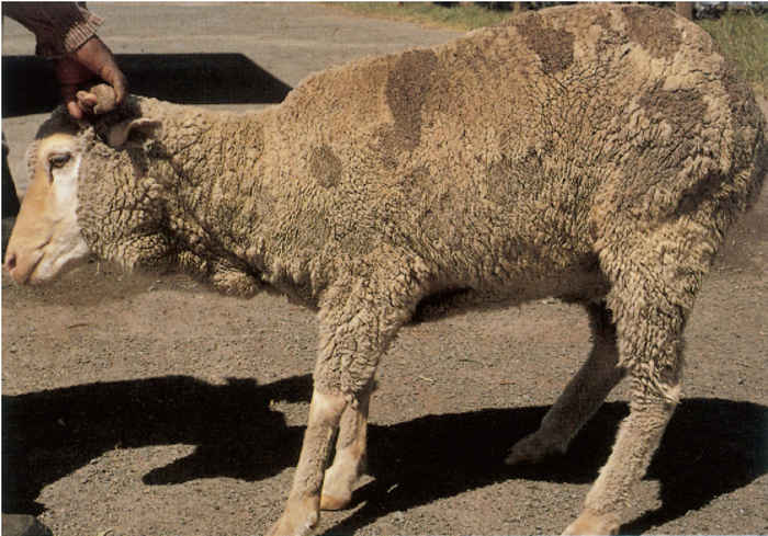
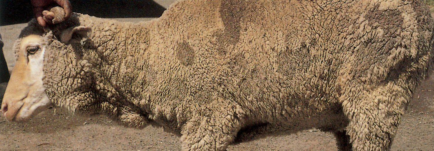
The lesions are most frequently distributed on the neck and shoulders, but are also relatively common on the rump and flanks. In advanced cases with extensive lesions the skin of the back may be involved, but even in such animals the abdominal skin is rarely affected.1–3 The fleece over the affected areas has dark grey to blackish, usually wellcircumscribed spots, patches or bands. These areas vary considerably in size, from 20 mm in diameter to large patches covering a greater part of the body, (Figure 191.1 and Figure 191,2).
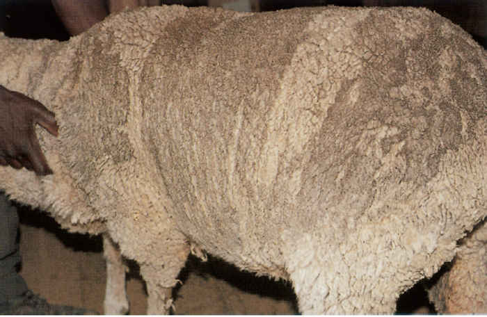
The tips of the wool in the affected areas are spiky and the wool is shorter and less dense, while the skin is often tender.1, 2 On parting the wool in affected areas, yellow to greyish-white, sticky, scaly deposits are found adjacent to the surface of the skin. These deposits cause the wool fibres to adhere to each other, resulting in the spiky staple formation. Dirt and dust adhere to the sticky deposits, thus causing the dark colour of the fleece. Loss of crimp in the staple of the wool fibres also occurs in the affected areas. In recently shorn sheep the lesions are well-circumscribed, pearly-white plaques and patches (Figure 191.3). The fleece of affected sheep has a characteristic, unique odour.
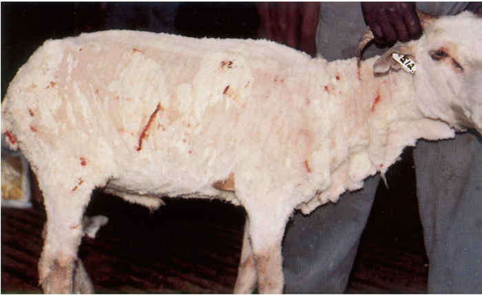
In mild cases, the affected skin is reddish-purple, swollen, very sensitive, and fragile, which causes it to tear when handled, particularly during shearing. In more severe cases, it is often pigmented grey.
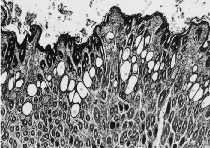
In both the early and more chronic lesions, the most consistent histopathological changes are a mild, mainly lymphocytic, dermatitis; superficial and follicular hyperkeratosis; prominent dilatation of the hair follicles (Figure 191.4); and an increase in the size and/or number of sebaceous glands. There is no exudate present on the surface of the epidermis. The sticky, scaly deposits present macroscopically are probably due to a combination of increased sebum secretion and hyperkeratotic flakes.
Diagnosis and differential diagnosis
The disease is confirmed by isolation of the suspected causative bacterium from sterile cotton-wool swabs used on animals with dry skin and fleece. To obtain the specimen the wool is parted and a swab is rubbed firmly on the affected skin. Skin scrapings and wool samples may also be cultured but they are often severely contaminated by other micro-organisms.6
Lumpy wool and fleece rot are the most important differential diagnoses.
Control
Bolo disease is very difficult to control and no satisfactory cure has yet been found. The causative bacterium shows marked in vitro sensitivity to several antibiotics, yet systemic antibiotic treatment in affected animals is unsuccessful.3
With the exception of sulphur-containing compounds, topical treatments, including dipping agents, have little effect.3 Fairly good results are obtained by dipping sheep twice in 3 per cent flowable sulphur. The first dipping should be carried out two weeks after shearing and the second 10 days after the first. Sheep may be dipped immediately after they have been shorn, provided that the dipping solution is made up daily so as to prevent the occurrence of post-dipping lameness if shearing extends over several days.4 Longstanding cases do not respond well to treatment and should be culled.3
Separation of age groups, dipping and shearing of young animals before the rest of the flock, and regular disinfection of shearing sheds and equipment with 3 per cent formalin, are additional preventive measures that may be instituted.
References
- COLLY, P.A., 1985. Bolosiekte; Geheimsinnige siekte wat skape aantas. Die Landbouweekblad, 26 April 1985. pp. 24–26.
- COLLY, P.A., 1989. State Veterinarian, Lydenburg, South Africa. Unpublished data.
- COLLY, P.A., LANGE, L.A., DE RUITER, A., VAN TONDER, E.M., VERMEULEN, S.0., WHITEHEAD, C.J. & KELLERMAN, G.E., 1989. Bolo disease: A specific, localized skin disease of woolled sheep. Journal of the South African Veterinary Association, 61, 90–95.
- HENTON, M.M., 1989. Onderstepoort Veterinary Institute, Pretoria, South Africa. Unpublished data.
- LANGE, L.A., 1989. Faculty of Veterinary Science, University of Pretoria, South Africa. Unpublished data.
- VAN TONDER, E.M., COLLY, P.A., VERMEULEN, S.0., KELLERMAN, G.E., DE RUITER, A. & WHITEHEAD, C.J., 1989. Bolo disease: A bacteriological survey. Journal of the South African Veterinary Association, 61, 96–101.


