- Infectious Diseases of Livestock
- Part 3
- Genital campylobacteriosis in cattle
- GENERAL INTRODUCTION: SPIROCHAETES
- Swine dysentery
- Borrelia theileri infection
- Borrelia suilla infection
- Lyme disease in livestock
- Leptospirosis
- GENERAL INTRODUCTION: AEROBIC ⁄ MICRO-AEROPHILIC, MOTILE, HELICAL ⁄ VIBROID GRAM-NEGATIVE BACTERIA
- Genital campylobacteriosis in cattle
- Proliferative enteropathies of pigs
- Campylobacter jejuni infection
- GENERAL INTRODUCTION: GRAM-NEGATIVE AEROBIC OR CAPNOPHILIC RODS AND COCCI
- Moraxella spp. infections
- Bordetella bronchiseptica infections
- Pseudomonas spp. infections
- Glanders
- Melioidosis
- Brucella spp. infections
- Bovine brucellosis
- Brucella ovis infection
- Brucella melitensis infection
- Brucella suis infection
- Brucella infections in terrestrial wildlife
- GENERAL INTRODUCTION: FACULTATIVELY ANAEROBIC GRAM NEGATIVE RODS
- Klebsiella spp. infections
- Escherichia coli infections
- Salmonella spp. infections
- Bovine salmonellosis
- Ovine and caprine salmonellosis
- Porcine salmonellosis
- Equine salmonellosis
- Yersinia spp. infections
- Haemophilus and Histophilus spp. infections
- Haemophilus parasuis infection
- Histophilus somni disease complex in cattle
- Actinobacillus spp. infections
- Actinobacillus equuli infections
- Gram-negative pleomorphic infections: Actinobacillus seminis, Histophilus ovis and Histophilus somni
- Porcine pleuropneumonia
- Actinobacillus suis infections
- Pasteurella and Mannheimia spp. infections
- Pneumonic mannheimiosis and pasteurellosis of cattle
- Haemorrhagic septicaemia
- Pasteurellosis in sheep and goats
- Porcine pasteurellosis
- Progressive atrophic rhinitis
- GENERAL INTRODUCTION: ANAEROBIC GRAM-NEGATIVE, IRREGULAR RODS
- Fusobacterium necrophorum, Dichelobacter (Bacteroides) nodosus and Bacteroides spp. infections
- GENERAL INTRODUCTION: GRAM-POSITIVE COCCI
- Staphylococcus spp. infections
- Staphylococcus aureus infections
- Exudative epidermitis
- Other Staphylococcus spp. infections
- Streptococcus spp. infections
- Strangles
- Streptococcus suis infections
- Streptococcus porcinus infections
- Other Streptococcus spp. infections
- GENERAL INTRODUCTION: ENDOSPORE-FORMING GRAM-POSITIVE RODS AND COCCI
- Anthrax
- Clostridium perfringens group infections
- Clostridium perfringens type A infections
- Clostridium perfringens type B infections
- Clostridium perfringens type C infections
- Clostridium perfringens type D infections
- Malignant oedema⁄gas gangrene group of Clostridium spp.
- Clostridium chauvoei infections
- Clostridium novyi infections
- Clostridium septicum infections
- Other clostridial infections
- Tetanus
- Botulism
- GENERAL INTRODUCTION: REGULAR, NON-SPORING, GRAM-POSITIVE RODS
- Listeriosis
- Erysipelothrix rhusiopathiae infections
- GENERAL INTRODUCTION: IRREGULAR, NON-SPORING, GRAM-POSITIVE RODS
- Corynebacterium pseudotuberculosis infections
- Corynebacterium renale group infections
- Bolo disease
- Actinomyces bovis infections
- Trueperella pyogenes infections
- Actinobaculum suis infections
- Actinomyces hyovaginalis infections
- GENERAL INTRODUCTION: MYCOBACTERIA
- Tuberculosis
- Paratuberculosis
- GENERAL INTRODUCTION: ACTINOMYCETES
- Nocardiosis
- Rhodococcus equi infections
- Dermatophilosis
- GENERAL INTRODUCTION: MOLLICUTES
- Contagious bovine pleuropneumonia
- Contagious caprine pleuropneumonia
- Mycoplasmal pneumonia of pigs
- Mycoplasmal polyserositis and arthritis of pigs
- Mycoplasmal arthritis of pigs
- Bovine genital mycoplasmosis
- Neurotoxin-producing group of Clostridium spp.
- Contagious equine metritis
- Tyzzer's disease
- MYCOTIC AND ALGAL DISEASES: Mycoses
- MYCOTIC AND ALGAL DISEASES: Pneumocystosis
- MYCOTIC AND ALGAL DISEASES: Protothecosis and other algal diseases
- DISEASE COMPLEXES / UNKNOWN AETIOLOGY: Epivag
- DISEASE COMPLEXES / UNKNOWN AETIOLOGY: Ulcerative balanoposthitis and vulvovaginitis of sheep
- DISEASE COMPLEXES / UNKNOWN AETIOLOGY: Ill thrift
- Eperythrozoonosis
- Bovine haemobartonellosis
Genital campylobacteriosis in cattle
This content is distributed under the following licence: Attribution-NonCommercial CC BY-NC  View Creative Commons Licence details here
NJ Maclachlan and M-L Penrith (Editors). C H Annandale, D E Holm and P Irons, Genital campylobacteriosis in cattle, 2018.
Genital campylobacteriosis in cattle
Previous authors: P C IRONS, A P SCHUTTE, M L VAN DER WALT AND G C BISHOP*
Current authors:
C H ANNANDALE - Associated Professor, Veterinary Specialist in Reproduction, BCom, BVSc (Tons), MMedVet (Gyn), MBA Dip ACT, Department of Production Animal Studies, Faculty of Veterinary Science, Private Bag X04, Onderstepoort, Gauteng, 0110, South Africa
D E HOLM - Deputy Dean: Teaching and Learning, BVSc, MSc, PhD, Faculty of Veterinary Science, Private Bag X04, University of Pretoria, Pretoria, Gauteng, 0110, South Africa
P C IRONS - Veterinary Program Director, Faculty of Veterinary Science, Murdoch University, Perth, Australia
![]()
Introduction
Genital campylobacteriosis in cattle is caused by Campylobacter fetus venerealis, which is an obligate pathogen of the bovine genital tract.31, 53 A related species, Campylobacter fetus fetus, is an intestinal inhabitant that may also be found in the genital tract following ascending infection or venereal introduction. 39 The primary mode of transmission of C. fetus venerealis is natural service,67 but due to its ability to survive in cryopreserved semen, it can also be transmitted via artificial insemination.39 Genital campylobacteriosis present with clinical signs in cows predominantly, while bulls are mostly asymptomatic. The clinical signs in cows are associated with genital infection and abortion.75
Campylobacteriosis is regarded as one of the most important causes of poor calving rates in cattle, and closely resembles the clinical appearance of infection caused by Tritrichomonas foetus.43, 73 Campylobacter fetus venerealis and T. foetus often occur together.17
Aetiology
Campylobacter fetus was first described in the 1950’s by Akkermans and colleagues, and initially referred to as Vibrio fetus.4, 90 Within the Campylobacter genus two species are distinguished – C. fetus venerealis and C. fetus fetus. The biotype intermedius is associated with C. fetus venerealis.10
Campylobacter bacteria are slender, curved, Gram-negative rods, 0.01 to 0.08 mm in width and 0.5 to 0.8 mm in length. They appear spiral in shape, and in culture, two or more organisms can join at their ends to form a spiral chain. Older cultures also sometimes display coccoid forms of the organisms. The bacteria are motile due to propulsion from a single polar flagellum at one or both ends of the cells. Campylobacter fetus venerealis bacteria are characterized by their distinctive motion and morphology.
Campylobacter fetus is micro-aerophilic and requires an atmosphere with reduced O2 (10 per cent) and increased (4 – 8 per cent) CO2 to grow on solid media. In semi-solid media, growth can occur under aerobic conditions. While growth will occur at 25°C, the optimum temperature for growth is 37°C. Cultures should be examined for growth after two to three days. Colonies on blood agar attain a diameter of 1 -2 mm, are convex and raised. They are colourless to grey in appearance and non-haemolytic. On moist media, growth may occur along the line of inoculation.
The biochemical properties of C. fetus includes reduction of nitrates and inability to ferment carbohydrates. They are catalase- and oxidase-positive. Campylobacter fetus venerealis is tolerant to 1 per cent glycine and produces H2S.10, 32, 81
Campylobacter fetus incorporates a heat-stable somatic antigen (O-antigen) into its lipopolysaccharide wall. While the subspecies C. fetus fetus contains the heat-stable antigens B and A-2, C. fetus venerealis contains antigens A and A-1 and Campylobacter jejuni carries the heat-stable C antigen. There is no cross-reaction between these antigens.81
Several heat-labile antigens have been identified and many may be present in a single strain.7 They are antiphagocytic63 and undergo structural changes in the animal which aids immune evasion. 29, 78 Antigenic variation is accompanied by genomic rearrangements.45 The genus Campylobacter is characterized by a process of genome decay, made possible by the relatively small genome size (~1.5 Mb) and the loss of metabolic genes.46
Epidemiology
Campylobacter fetus venerealis has a worldwide distribution. It has been reported in a number of countries including the USA,11, 74, 82 Canada,91 Australia,26 South Africa,70, 77 Poland,83 Great Britain,65 and Nigeria.6, 61 While sporadic abortion storms have been reported mostly from developing countries in Africa and South America, it has also been reported in the developed world.49
Genital campylobacteriosis in cattle occurs more common in production systems that make use of natural service.67 Lack of regulatory control schemes and inadequate laboratory facilities are often cited as reasons for the poor control and widespread distribution of C. fetus venerealis, and the underestimation of its profound impact on profitability of beef farming through smaller calf crops, prolonged intercalving periods, replacement costs of infected bulls and reduced weaning weights.66
Prevalence of C. fetus varies greatly across different regions in the world. Some surveys have failed to detect the organism in bulls.59 A prevalence rate of 40 and 47 per cent have been recorded in seropositive cows during a rare outbreak in the USA.1, 3 In Australia, abortions due to C. fetus have been reported on 37 farms, and positive bulls were identified in 87 per cent of herds.49, 62 Across most regions, C. fetus venerealis occurs more frequently than C. fetus fetus.
The sources of infection with C. fetus venerealis are bulls, infected heifers and cows. Bulls can be either permanently or transiently infected. While infection is more commonly seen in bulls older than four years of age, most young bulls either do not become infected or rid themselves of the infection within a period of three to four weeks.36 Spontaneous recovery has also been documented for older bulls, but it is rare. High prevalence rates have been reported in young bulls from a heavily infected population.62 Microscopic evaluation of preputial and penile mucosa of bulls infected with C. fetus venerealis failed to demonstrate a difference between young and old bulls. Due to these conflicting reports, it is presently uncertain whether bull age plays a role in persistence of C. fetus venerealis infection.66 While most heifers and cows rid themselves of the infection within three to five months, a carrier state (for a prolonged period, or permanently) has been identified in up to 10 per cent of infected heifers and cows.21, 38
Campylobacter fetus venerealis is transmitted from bulls to heifers and cows by coitus. The rate of transmission is dependent on the number of organisms present in the preputial cavity at the time of coitus, and varies between 38 to 78 per cent.38 Bulls usually become venereally infected, but also via fomites.80 While C. fetus venerealis can survive semen freezing, transmission via artificial insemination is rare, given biosanitary control measures and improved cryoprotectants.35
Campylobacter fetus fetus is a commensal of the gastrointestinal tract of cattle and sheep and is spread by the ingestion of contaminated material.16, 38 It is occasionally associated with bovine abortion with heavy environmental contamination.33
Pathogenesis
After introduction of the organisms into the genital tract of a heifer or cow, C. fetus venerealis localizes in the vagina. In some cows, the organisms are eliminated from the vagina and no infection results while in others the organisms migrate through the cervix after five to seven days and can go up to the Fallopian tubes.38 The organisms in the reproductive tract induce varying degrees of inflammation, including vaginitis, cervicitis, endometritis and salpingitis.79 The inflammation, in conjunction with the lowered oxygen tensions produced by the bacteria, create an unfavourable environment for embryo survival and results in embryonal death.90, 93 There is no direct effect of the bacteria on fertilization or early embryonic development.8
Campylobacter organisms elicits an immune response in cows. The nature of the response is dependent on the site of presence of the organisms and the site and time of sampling. The bacteria cause an antigen-specific transient IgM response, followed by persistent IgA and IgG antibody responses in cervicovaginal secretions, while an IgG1 response can be detected in uterine secretions.29, 85, 86 The duration of the immune response was limited to weeks in the uterus (where IgG predominates), while in the vagina (where IgA predominates) it has been recorded to last up to 24 months.30 The duration of protective immunity following natural infection is two to four years.38, 58
The genital inflammation and embryonal death result in prolonged oestrus cycles of 24 to 40 days or more. The infection is mostly self-limiting, with cure, and return to normal fertility, within three to five months.33, 38 An exception to this may occur in the case of bilateral salpingitis due to C. fetus venerealis infection, where permanent infertility may result. Up to 10 per cent of infected cows can remain persistently infected, acting as sources of infection during subsequent breeding seasons.21
In the bull, C. fetus venerealis strictly colonizes the superficial epithelium of the preputial and penile lumen and crypts, without producing clinical signs.47, 76 The greatest numbers of organisms can be found in the fornix of the prepuce and on the penis. The colonization of the prepuce and penis elicits a poor, almost undetectable, immune response86 and could explain the prolonged survival of organisms in the prepuce of bulls. A local inflammatory response, characterized by lymphocytes and plasma cells in the preputial and penile subepithelium, can be seen in bulls infected with C. fetus venerealis.9, 42
It has been shown in vitro that C. fetus venerealis is associated with the sperm head or tail resulting in decreased sperm motility.20
Campylobacter fetus fetus produce endotoxins, which are thought to result in hypersensitivity reactions in affected cows, leading to abortion and lowered milk production, but not lowered reproductive efficiency.2, 3, 68
Clinical signs
Clinical signs associated with campylobcateriosis are limited to cows, and even then are often mild or inapparent. Bulls remain breeding sound with no deterioration in semen quality.23, 88 In heifers and cows that do display clinical signs, a mild vaginitis with scant vaginal exudate, can sometimes be seen. In cases where there is copious mucopurulent vaginal discharge, secondary bacterial infections should be suspected.
The main clinical manifestations of C. fetus venerealis infection are irregular oestrus cycles, delayed conception and sporadic abortions. This typically presents as low pregnancy rate during routine pregnancy diagnosis, with some reports documenting a pregnancy rate as low as 20 per cent in the first year of infection.88 The pregnancy rate tends to improve after a period of approximately six months, and with prolonged breeding seasons and more matings, eventually a pregnancy rate of 85 – 95 per cent can be attained.5, 38, 88 It is not uncommon for bulls to be overworked in such a scenario. Most heifers and cows eventually recover, conceive and calve normally but especially heifers can succumb to permanent infertility in up to 11 per cent of infected heifers.62
Abortion is uncommon, occurring in about 5 – 10 per cent of infected animals, generally in the second half of gestation.33, 38, 49 Cases of abortion due to C. fetus fetus have been reported, but is rare.60
Economic losses occur due to delayed conception, decreased calving percentage, delayed calving, culling of infertile animals and abortion.
Pathology
In bulls, infection with C. fetus venerealis causes no pathology, although a lymphocytic-plasmacytic infiltrate may be seen in histologic sections of the preputial and penile mucosa. The plasma cells are general located in the apex of the dermal papilla. The infiltrate is not considered specific to C. fetus venerealis infection.9, 76
While pyometra is not a feature of genital campylobacteriosis, as is the case in trichomonosis, there are noticeable changes in the female reproductive tract. This ranges from a mild vaginitis and cervicitis associated with lymphocyte and neutrophil infiltrates, to a diffuse, subacute to chronic superficial endometritis.21, 94 The lesions first appear five days after experimental infection and peak between the 30th and 60th day. Lesions of a milder nature persist for at least 120 days.34
Abortion (after seven months of gestation) due to C. fetus fetus is characterized by a necrotic placentitis. The aborted foetuses display a fibrinous peritonitis, pleuritis and hepatitis.69 Animals that abort due to C. fetus venerealis show similar placental lesions.49
Diagnosis
The diagnosis of genital campylobacteriosis is often suspected on the basis of the herd history and clinical signs, but identification and isolation of the organisms is ultimately needed. Fluorescent antibody techniques complemented with specific culture methods using preputial material sampled by scraping are currently the diagnostic methods of choice for the isolation and identification of C. fetus venerealis.42, 43, 44
Sample collection and handling
The most suitable samples are vaginal secretions and preputial washes or scrapings. In the case of abortion samples should be collected from foetal lung, liver and stomach contents as well as placentomes.
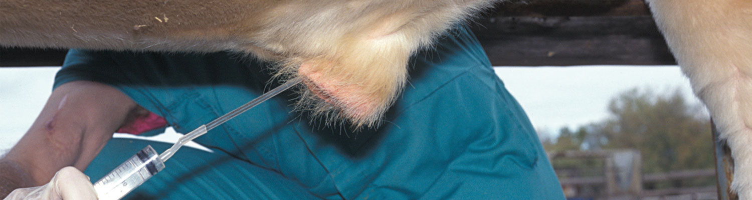
Sheath washes are collected by infusing 50 ml of sterile phosphate-buffered saline into the preputial cavity of a bull through a funnel and silicone tube, vigorously massaging the fluid in the preputial cavity and retrieving it via the tube and funnel into a clear glass bottle. A good sample is opaque in colour and contains visible cellular material. There are some indications that samples collected by sheath washes are more prone to contamination and a higher degree of negative culture results than samples collected via sheath scraping. 37, 38
Sheath scraping is performed with the aid of a plastic artificial insemination pipette, attached to a syringe. The tip of the pipette is inserted into the preputial cavity, advanced to the caudal end of the lamina interna reflection onto the penis, and moderate suction is applied on the syringe. While maintaining the negative pressure on the syringe and pipette, and applying external pressure onto the sheath, the tip of the pipette is directed onto the preputial mucosa and the pipette is moved forwards and backwards, scraping against the mucosa and collecting cellular material into the lumen of the pipette. When the pipette is withdrawn, thick mucoid material should be visible in its lumen; this material is then flushed into a sterile vial by aspirating end ejecting PBS through the pipette to loosen the cellular material. Blood is sometimes seen and is taken to indicate adequate depth of sampling.
Campylobacter fetus organisms remain viable in preputial samples for six to eight hours and should therefore be chilled and reach the laboratory for culture within this period. If the period will be exceeded, a transport medium should be used, and transport should then occur at room temperature.44, 50 The modified Weybridge medium and enrichment medium of Lander is able to maintain viability of samples for a period of up to a week.57
Specimens from cows should be collected from the vagina during oestrus to maximise the number of organisms that could potentially be collected in the sample. Several methods for collection of samples from the vagina has been described.14, 88
Campylobacter fetus can be cultured from placental and foetal tissues following homogenization and inoculation onto non-selective or selective media. Organisms have been shown to survive for up to three days in autolysed tissues.49
Culture
Campylobacter organisms are sensitive organisms, easily contaminated and overgrown with saprophytes and with highly selective growth requirements, making culture difficult.31, 35, 56 While the use of Millipore filtration and selective media has increased sensitivity, isolation of the organism is still problematic and regarded as insensitive. It has been shown that passive filtration onto blood agar yields significantly higher recovery rates of C. fetus venerealis than direct plating onto Skirrow medium. Skirrow medium, even with transport enrichment medium, is also more likely to be affected by Pasteurella overgrowth.19 The presence of polymixin B in many selective media, poses an additional problem since Campylobacter is sensitive to it.50 In the light of these challenges, three negative cultures are generally recommended in order to certify an animal free of C. fetus venerealis.54
Polymerase chain reaction
While many assays have been published claiming successful amplification and identification of C. fetus at subspecies level,18, 46, 52, 64 recent literature have cast doubt on many of these assertions.87 The routine use of PCR assay for diagnosis of C. fetus venerealis is not recommended, especially in conditions of low to moderate risk.92 Due to the economic losses that can accrue when a diagnostic assay produces a false negative C. fetus venerealis, but a false positive C. fetus fetus result, further research of the molecular identification of C. fetus at subspecies level, is indicated. The targeting gene nahE has a 100 per cent specificity and sensitivity for C. fetus, while additionally, accurate primers for ISCfe1 still needs to be developed in order to make the PCR sensitive and specific for C. fetus venerealis identification. Such assays should be evaluated by amplified fragment length polymorphism and multilocus sequence typing.87 The availability of the full genome of C. fetus venerealis should inform development of improved diagnostic tools in future.53
Other diagnostic tools
The vaginal mucus agglutination test is a useful method of diagnosing herd infection,55, 71 despite its low sensitivity and specificity.33 Best results are obtained when mucus is collected during diestrus or anestrus when volumes are smaller and don’t dilute the agglutinins, thereby preventing false negative results. The reliability of the test is influenced by previous vaccination, contamination with blood, inflammatory products or antibodies to C. fetus fetus.22, 38
An ELISA developed for the detection of C. fetus antibodies in vaginal mucus is believed to be more sensitive than the mucus agglutination test, with reported specificity of 98.5 per cent.51 Since the ELISA detects IgA, it isn’t influenced by post-vaccination IgG antibodies.


Differential diagnosis
Since the clinical picture associated with genital campylobacteriosis – irregular return to oestrus, poor pregnancy or calving rate and abortions – so closely resembles trichomonosis, the latter is the most important differential diagnosis to consider. This underlines the importance of identifying the causative organism, either by direct microscopy or by culture. Pyometra is often associated with trichomonosis, whereas that is not a clinical feature of genital campylobacteriosis.
In countries where it is endemic, brucellosis is an important differential diagnosis, particularly in chronically infected herds. Brucella abortus can however, be readily diagnosed by serological or biological tests.
Infectious infertility or “Epivag” (see Epivag) is associated with vaginitis and epididymitis in cattle. It usually causes total infertility of infected bulls, but a number of cows can become fertile again. Abortions are not a clinical feature of “Epivag”.
Control
Since the epidemiology of genital campylobacteriosis is so similar to trichomonosis (see Trichomonosis), many of the control measures for the latter, can also successfully be applied to herd infection with C. fetus venerealis.
The lack of effective control programmes in many countries, contributes to the continuing spread of this important disease and the associated economic losses. Any effective control programme will rely on compulsory testing and reporting, and subsequent monitoring.
Epidemiological models indicate that ensuring bulls are free of the disease prior to introduction into an artificial breeding programme or a breeding herd, is likely to produce the best long-term results. Successful treatment of bulls infected with C. fetus venerealis with dimetridazole has been reported.17 Antimicrobial treatment of cattle infected with C. fetus venerealis is impractical, illegal in many countries and should be discouraged due to the potential effect on human health. However, given the sensitivity of C. fetus venerealis to many antimicrobials (e.g. ampicillin, chloramphenicol, erythromycin, various aminoglycosides and polymixin B), semen is often treated.48, 54, 72 The latter treatments serve to explain the effectiveness of an artificial insemination programme at preventing spread of the disease.
Infected heifers and cows may be treated successfully, within the constraints of the logistics associated with treatment of a herd, by intra-uterine antibiotic infusion. A more pragmatic approach, taking the epidemiology of the infection into account, may be to postpone breeding with a period of 90 days to allow the disease to self-limit. Infected animals will develop immunity, but some may remain infected beyond this period and contribute to reduced calving rates and increased losses.
Vaccination plays an important role in the control of genital campylobacteriosis. 22, 24, 40, 66 In bulls vaccine can be used prophylactically or therapeutically. For purposes of prevention, Clark25 vaccinated bulls with C. fetus venerealis cell antigens in mineral oil adjuvant and demonstrated immunity to intrapreputial challenge. The bulls were vaccinated twice with a 2-month interval and then boosted yearly. A dual vaccine, consisting of C. fetus intermedius and C. fetus venerealis, using the same vaccination protocol, also effectively protected against infection with C. fetus venerealis.27 Most studies showed successful therapeutic vaccination if given twice, within a month to infected bulls.13, 25, 89 A recent study, evaluating multimodal treatment consisting of two vaccinations and two weekly long-acting tetracycline injections failed to prevent shedding of C. fetus venerealis.41
The literature regarding preventative and therapeutic vaccination of heifers and cows are more ambivalent, with some reporting success, while others report vaccine failures.12, 28 The efficacy of the vaccine depends on type of adjuvant used, concentration of the antigen, antigenicity of the specific strain present and the time interval between vaccination and breeding.15 There is an ongoing need for research into effective vaccine production and development.
Attempts at control of genital campylobacteriosis based on vaccination alone are unlikely to be successful, and additional control measures should be considered. This includes the separation of the herd into clean and infected groups, ensuring the introduction of animals that have been tested and certified free of the disease, as well as treatment or culling.84
References
- AKHTAR, S., RIEMANN, H. P., THURMOND, M. C., FARVER, T. B., & FRANTI, C. E., 1990. The use of loglinear model to evaluate factors associated with sero-prevalence of in dairy cattle. Theriogenology, 34, 989-1001.
- AKHTAR, S., RIEMANN, H. P., THURMOND, M. C., & FRANTI, C. E., 1993. The association between antibody titres against Campylobacter fetus and milk production efficiency in dairy cattle. Veterinary Research Communications, 17, 183-191.
- AKHTAR, S., RIEMANN, H. P., THURMOND, M. C., & FRANTI, C. E., 1993. The association between antibody titres against Campylobacter fetus and reproductive efficiency in dairy cattle. Veterinary Research Communications, 17, 95-107.
- AKKERMANS, J. P., TERPSTRA, J. I., & VAN WAVEREN, H. G., 1956. Over de betekenis van verschillende vibrionen voor de steriliteit van het rund. Tijdschrift voor Diergeneeskunde, 81, 430 - 435.
- ARTHUR, G. H., NOAKES, D. E., PEARSON, H., & PARKINSON, T. J., 1996. Veterinary Reproduction and Obstetrics. 8th ed. Philadelphia: W. B. SAUNDERS.
- BAWA, E. K., ADEKEYE, J. O., OYEDIPE, E. O., & UMOH, J. U., 1991. Prevalence of bovine campylobacteriosis in indigenous cattle of three states in Nigeria. Tropical Animal Health and Production, 23, 157-160.
- BERG, R. I., JULITA, J. W., & FIREHAMMER, B. D., 1971. A revised classification of Vibrio fetus. American Journal of Veterinary Research, 32, 11-22.
- BIELANSKI, A., SAMPATH, M., GRADIL, C., EAGLESOME, M. D., & GARCIA, M., 1994. In vitro fertilization of bovine ova in the presence of Campylobacter fetus subsp. venerealis. Reproduction in Domestic Animals, 29, 488-493.
- BIER, P. J., HALL, C. E., DUNCAN, J. R., & WINTER, A. J., 1977. Experimental infections with Campylobacter fetus in bulls of different ages. Veterinary Microbiology, 2, 13-27.
- BOLTON, F. J., HOLT, A. V., & HUTCHINSON, D. N., 1984. Campylobacter biotyping scheme of epidemiological value. Journal of Clinical Pathology, 37, 677-681.
- BONDURANT, R. H., ANDERSON, M. L., BLANCHARD, P., HIRD, D., DANAYE-ELMI, C., PALMER, C., SISCHO, W. M., SUTHER, D., UTTERBACK, W., & WEIGLER, B. J., 1990. Prevalence of trichomoniasis among California beef herds. Journal of the American Veterinary Medical Association, 196, 1590-1593.
- BORDER, M. M. & FIREHAMMER, B. D., 1980. Antigens of Campylobacter fetus subsp. fetus eliciting vaccinal immunity in heifers. American Journal of Veterinary Research, 41, 746 - 750.
- BOUTERS, R., DE KEYSER, J., VANDEPLASSCHE, M., VAN AERT, A., BRONE, E., & BONTE, P., 1973. Vibrio fetus Infection in Bulls: Curative and preventive vaccination. British Veterinary Journal, 129, 52-57.
- BRINLEY MORGAN, W. J., 1959, Studies on the Antigenic Structure of Vibrio fetus. Journal of Comparative Pathology and Therapeutics, 69, 125-140.
- BRYNER, J. H., FIREHAMMER, B. D., & WESLEY, I. V., 1988. Vaccination of pregnant guinea pigs with Campylobacter fetus: effects of antigen dose, Campylobacter strain and adjuvant type. American Journal of Veterinary Research, 49, 499-455.
- BRYNER, J. H., O’BERRY, P. A., & FRANK, A. H., 1964. Vibrio infection of the digestive organs of cattle. American Journal of Veterinary Research, 25, 1048-1050.
- CAMPERO, C. M., BALLABENE, N. C., CIPOLLA, A. C., & ZAMORA, A. S., 1987. Dual infection of bulls with campylobacteriosis and trichomoniasis: Treatment with dimetridazole chlorhydrate. Australian Veterinary Journal, 64, 320-321.
- CHABAN, B., CHU, S., HENDRICK, S., WALDNER, C., & HILL, J. E., 2012. Evaluation of a Campylobacter fetus subspecies venerealis real-time quantitative polymerase chain reaction for direct analysis of bovine preputial samples. Canadian Journal of Veterinary Research, 76, 166-173.
- CHABAN, B., GARCIA GUERRA, A., HENDRICK, S. H., WALDNER, C. L., & HILL, J. E., 2013. Isolation rates of Campylobacter fetus subsp venerealis from bovine preputial samples via passive filtration on nonselective medium versus selective medium, with and without transport medium. American Journal of Veterinary Research, 74, 1066-1069.
- CHIAPPARRONE, M. L., SOTO, P., & CATENA, M., 2016. Characterization of the Campylobacter fetus subsp. venerealis adhesion to bovine sperm cells. International Journal of Morphology, 34, 1419-1423.
- CIPOLLA, A., CASARO, A., TERZOLO, H., ESTELA, E., BROOKS, B., & GARCIA, M., 1994. Persistence of Campylobacter fetus subspecies venerealis in experimentally infected heifers. Veterinary Record, 134, 628-628.
- CLARK, B. L., 1967. Control of bovine vibriosis by vaccination. Australian Veterinary Journal, 43, 437-440.
- CLARK, B. L., 1971. Review of bovine vibriosis. Australian Veterinary Journal, 47, 103 - 107.
- CLARK, B. L., DUFTY, J. H., & MONSBOURGH, M. J., 1968. Vaccination of bulls against bovine vibriosis. Australian Veterinary Journal, 44, 530-530.
- CLARK, B. L., DUFTY, J. H., MONSBOURGH, M. J., & PARSONSON, I. M., 1974. Immunisation against bovine vibriosis. Australian Veterinary Journal, 50, 407-409.
- CLARK, B. L., DUFTY, J. H., MONSBOURGH, M. J., & PARSONSON, I. M., 1975. Studies on venereal transmission of Campylobacter fetus by immunised bulls. Australian Veterinary Journal, 51, 531-532.
- CLARK, B. L., DUFTY, J. H., MONSBOURGH, M. J., & PARSONSON, I. M., 1977. A dual vaccine for immunisation of bulls against vibriosis. Australian Veterinary Journal, 55, 43.
- COBO, E. R., CIPOLLA, A., MORSELLA, C., CANO, D., & CAMPERO, C. M., 2003. Effect of two commercial vaccines to Campylobacter fetus subspecies on heifers naturally challenged. Journal of Veterinary Medicine Series B: Infectious Diseases and Veterinary Public Health, 41, 746-750.
- CORBEIL, L. B., DUNCAN, J. R., SCHURIG, G. D., HALL, C. E., & WINTER, A. J., 1974. Bovine venereal vibriosis: variations in immunoglobulin class of antibodies in genital secretions and serum. Infection and Immunology, 10, 1084-1090.
- CORBEIL, L. B., SCHURIG, G. D., DUNCAN, J. R., CORBEIL, R. R., & WINTER, A. J., 1974. Immunoglobulin classes and biological functions of Campylobacter (Vibrio) fetus antibodies in serum and cervicovaginal mucus. Infection and Immunology, 10, 422-429.
- DE KEYSER, J., 1984. Bovine genital campylobacteriosis In: BUTZLER, J. P., ed. Campylobacter infection in Man and Animals. Boca Raton: CRC Press.
- DE KEYSER, J., 1985. The genus Campylobacter: Classification, nomenclature, habitat and pathogenicity In: LANDER, K. P., ed. Campylobacter. Luxemburg: Commision of the European Communities.
- DE KEYSER, J., 1986. Bovine genital campylobacteriosis In: MORROW, D. A., ed. Current Therapy in Theriogenology. Philadelphia: W. B. SAUNDERS, 263-266.
- DOZZA, L., OLSON, N. O., & CAMPBELL, A., 1960. The uterine biopsy technique for following the histologic changes caused by Vibrio fetus in the uterine mucosa. American Journal of Veterinary Research, 21, 878-883.
- DUFTY, J. H., 1967. Diagnosis of vibriosis in the bull. Australian Veterinary Journal, 43, 433-437.
- DUFTY, J. H., CLARK, B. L., & MONSBOURGH, M. J., 1975. The influence of age on the susceptibility of bulls to Camphylobacter fetus subsp venerealis. Australian Veterinary Journal, 51, 294-297.
- DUFTY, J. H. & MCENTEE, K., 1969. Evaluation of some culture media and sampling techniques for the diagnosis of vibriosis in the bull. Australian Veterinary Journal, 45, 140-144.
- DUFTY, J. H. & VAUGHAN, J. A., 1993. Bovine venereal campylobacteriosis. In: Howard JL, ed. Current Veterinary Therapy 3: Food Animal Practice. Philadelphia: W. B. SAUNDERS, 510 - 513.
- EAGLESOME, M. D., SAMPATH, M. I., & GARCIA, M. M., 1995. A detection assay for Campylobacter fetus in bovine semen by restriction analysis of PCR amplified DNA. Veterinary Research Communications, 19, 253-263.
- EDMONDSON, M. A., 2014. Infectious Agents: Campylobacter. Bovine Reproduction, 518-523.
- ERICKSON, N. E. N., LANIGAN, E., WAUGH, T., GESY, K., & WALDNER, C., 2017. Evaluation of long-acting oxytetracycline and a commercial monovalent vaccine for the control of Campylobacter fetus subsp. venerealis infection in beef bulls. Canadian Veterinary Journal, 58, 1051-1058.
- FLOWER, P. J., LADDS, P. W., THOMAS, A. D., & WATSON, D. L., 1983. An Immunopathologic study of the bovine prepuce. Veterinary Pathology, 20, 189-202.
- GARCIA, M., EAGLESOME, M., and RIGBY, C., 1983. Campylobacters important in veterinary medicine. Veterinary Bulletin, 53, 793-818.
- GARCIA, M., RUCKERBAUER, G., EAGLESOME, M., & BIOSCLAIR, W. E., 1983. Detection of Campylobacter fetus in artificial insemination bulls with a transport enrichment medium. Canadian Journal of Comparative Medicine, 47, 336 - 340.
- GARCIA, M. M., LUTZE-WALLACE, C. L., DENES, A. S., EAGLESOME, M. D., HOLST, E., & BLASER, M. J., 1995. Protein shift and antigenic variation in the S-layer of Campylobacter fetus subsp. venerealis during bovine infection accompanied by genomic rearrangement of sapA homologs. Journal of Bacteriology, 177, 1976-1980.
- GORKIEWICZ, G., KIENESBERGER, S., SCHOBER, C., SCHEICHER, S. R., GÜLLY, C., ZECHNER, R., & ZECHNER, E. L., 2010. A genomic island defines subspecies-specific virulence features of the host-adapted pathogen Campylobacter fetus subsp. venerealis. Journal of Bacteriology, 192, 502-517.
- HOFFER, M. A., 1982. Bovine campylobacteriosis: A review. The Canadian Veterinary Journal, 22, 327-330.
- HOWARD, T. H., VASQUEZ, L. A., & AMANN, R. P., 1982. Antibiotic Control of Campylobacter fetus by Three extenders of bovine semen. Journal of Dairy Science, 65, 1596-1600.
- HUM, S., 1987. Bovine abortion due to Campylobacter fetus. Australian Veterinary Journal, 64, 319-320.
- HUM, S., BRUNNER, J., MCINNES, A., MENDOZA, G., & STEPHENS, J., 1994. Evaluation of cultural methods and selective media for the isolation of Campylobacter fetus subsp venerealis from cattle. Australian Veterinary Journal, 71, 184-186.
- HUM, S., QUINN, C., & KENNEDY, D., 1994. Diagnosis of bovine venereal campylobacteriosis by ELISA. Australian Veterinary Journal, 71, 140-143.
- HUM, S., QUINN, K., BRUNNER, J., & ON, S. L. W., 1997. Evaluation of a PCR assay for identification and differentiation of Campylobacter fetus subspecies. Australian Veterinary Journal, 75, 827-831.
- IRAOLA, G., PÉREZ, R., NAYA, H., PAOLICCHI, F., HARRIS, D., LAWLEY, T. D., REGO, N., HERNÁNDEZ, M., CALLEROS, L., CARRETTO, L., VELILLA, A., MORSELLA, C., MÉNDEZ, A., & GIOFFRE, A., 2013. Complete genome sequence of Campylobacter fetus subsp. venerealis biovar Intermedius, isolated from the prepuce of a bull. Genome Announcements, 1.
- JONES, R. L., DAVIS, M. A., & VONBYERN, H., 1985. Cultural procedures for the isolation of Campylobacter fetus subsp. venerealis from preputial secretions and the occurrence of anitmicrobial resistance. Proceedings of the Annual Congress of the American Association of Laboratory Diagnosticians, 28, 225-238.
- KENDRICK, J. A., 1967. The vaginal mucus agglutination test for bovine vibriosis. Journal of the American Veterinary Medical Association, 150, 495 - 498.
- KIGGENS, E. M. & PLASTRIDGE, W. N., 1956. Effect of gaseous environment on growth and catalase content of Vibrio fetus cultures of bovine origin. Journal of Bacteriology, 72, 397-401.
- LANDER, K. P., 1990. The application of a transport and enrichment medium to the diagnosis of Campylobacter fetus infections in bulls. British Veterinary Journal, 146, 334-340.
- LARSON, B. L., 1999. Immunization to decrease pregnancy wastage in beef cattle. Part II. Available vaccines. Compendium on Continuing Education for the Practicing Veterinarian, 18, 571-580.
- LOVERIDGE, R. & GARDNER, E., 1993. Campylobacter fetus venerealis infection in cattle. Surveillance, 20, 26-27.
- MACLAREN, A. P. C. & AGUMBAH, G. J. O., 1988. Infertility in cattle in south-west Scotland caused by an ‘Intermediate’ strain of Campylobacter fetus subspecies fetus (formerly Campylobacter fetus intestinalis). British Veterinary Journal, 144, 29-44.
- MAI, H. M., IRONS, P. C., KABIR, J., & THOMPSON, P. N., 2013. Prevalence of bovine genital campylobacteriosis and trichomonosis of bulls in northern Nigeria. Acta veterinaria Scandinavica, 55, 56.
- MCCOOL, C. J., TOWNSEND, M. P., WOLFE, S. G., SIMPSON, M. A., OLM, T. C., JAYAWARDHANA, G. A., & CARNEY, J. V., 1988. Prevalence of bovine veneral disease in the Victoria River District of the Northern Territory: likely economic effects and practicable control measures. Australian Veterinary Journal, 65, 153-156.
- MCCOY, E. C., DOYLE, E., BURDA, K., CORBEIL, L. B., & WINTER, A. J., 1975. Superficial antigens of Campylobacter (Vibrio) fetus: Characterization of an antiphagocytic component. Infection and Immunity, 11, 517-525.
- MCGOLDRICK, A., CHANTER, J., GALE, S., PARR, J., TOSZEGHY, M., & LINE, K., 2013. Real Time PCR to detect and differentiate Campylobacter fetus subspecies fetus and Campylobacter fetus subspecies venerealis. Journal of Microbiological Methods, 94, 199-204.
- MCGOWAN, A. C. & MURRAY, R. D., 1999. Health status of bulls used for natural breeding on farms in South West Scotland. Journal of Veterinary Medicine, Series B, 46, 311-321.
- MICHI, A. N., FAVETTO, P. H., KASTELIC, J., & COBO, E. R., 2016. A review of sexually transmitted bovine trichomoniasis and campylobacteriosis affecting cattle reproductive health. Theriogenology, 85, 781-791.
- MSHELIA, G. D., AMIN, J. D., WOLDEHIWET, Z., MURRAY, R. D., & EGWU, G. O., 2010. Epidemiology of bovine venereal campylobacteriosis: Geographic distribution and recent advances in molecular diagnostic techniques. Reproduction in Domestic Animals, 45, e221-e230.
- OSBORNE, J. C. & SMIBERT, R. M., 1964. Vibrio fetus toxin. 1. Hypersensitivity and abortifacient action. Cornell Vet, 54, 561-572.
- OSBURN, B. I. & HOSKINS, R. K., 1971. Infection with Vibrio fetus in the immunologically immature fetal calf. Journal of Infectious Diseases, 123, 32-40.
- PEFANIS, S. M., HERR, S., VENTER, C. G., KRUGER, L. P., QUEIROGA, C. C., & AMARAL, L., 1988. Trichomoniasis and campylobacteriosis in bulls in the Republic of Transkei. Journal of the South African Veterinary Association, 59, 139-140.
- PLASTRIDGE, W. N., EASTERBROOKS, H. L., & WILLIAMS, L. F., 1953. The tampon method of collection and the examination of cervicovaginal mucus for Vibrio fetus agglutinins. Journal of the American Veterinary Medical Association, 123, 516-520.
- PLASTRIDGE, W. N., WILLIAMS, L. F., & TROWBRIDGE, D. G., 1964. Antibiotic sensitivity of physiological groups of micro-aerophilic vibrios. Journal of Veterinary Research, 25, 1295-1299.
- RAE, D. O., 1989. Impact of trichomoniasis on the cow-calf producer's profitability. Journal of the American Veterinary Medical Association, 194, 771-775.
- RAE, D. O., CREWS, J. E., GREINER, E. C., & DONOVAN, G. A., 2004. Epidemiology of Tritrichomonas foetus in beef bull populations in Florida. Theriogenology, 61, 605-618.
- RHYAN, J. C., STACKHOUSE, L. L., & QUINN, W. J., 1988. Foetal and placental lesions in bovine abortion due to Tritrichomonas foetus. Veterinary Pathology, 25, 350-355.
- SAMUELSON, J. D. and WINTER, A. J., 1966. Bovine vibriosis: The Nature of the Carrier State in the Bull. Journal of Infectious Diseases, 116, 581-592.
- SCHMIDT, T., VENTER, E. H., & PICARD, J. A., 2010. Evaluation of PCR assays for the detection of Campylobacter fetus in bovine preputial scrapings and the identification of subspecies in South African field isolates. Journal of the South African Veterinary Association, 81, 87-92.
- SCHURIG, G. D., HALL, C. E., BURDA, K., CORBEIL, L. B., DUNCAN, J. R., & WINTER, A. J., 1973. Persistent genital tract infection with Vibrio fetus intestinalis associated with serotypic alteration of the infecting strain. American Journal of Veterinary Research, 34, 1399-1403.
- SCHURIG, G. D., HALL, C. E., BURDA, K., CORBEIL, L. B., DUNCAN, J. R., & WINTER, A. J., 1974. Infection patterns in heifers following cervicovaginal or intrauterine installation of Campylobacter (Vibrio) fetus venerealis. Cornell Vet, 64, 533-48.
- SCHUTTE, A. P., 1970. Vibrio fetus infektion bei Bullen. Reproduction in Domestic Animals, 5, 36-41.
- SMIBERT, R. M., 1984. Campylobacter In: KRIEG N. R., GIBBONS N. E., eds. Bergey’s Manual of Systematic Bacteriology. Baltimore, London: Williams & Wilkins.
- SZONYI, B., SRINATH, I., SCHWARTZ, A., CLAVIJO, A., & IVANEK, R., 2012. Spatio-temporal epidemiology of Tritrichomonas foetus infection in Texas bulls based on state-wide diagnostic laboratory data. Veterinary Parasitology, 186, 450-455.
- SZYMAŃSKA-CZERWIŃSKA, M. & NIEMCZUK, K., 2011. Detection of bovine genital campylobacteriosis in population of heifers in Poland. Bulletin of the Veterinary Institute in Pulawy, 55, 377-379.
- TRUYERS, I., LUKE, T., WILSON, D., & SARGISON, N., 2014. Diagnosis and management of venereal campylobacteriosis in beef cattle. BMC Veterinary Research, 10.
- VAN AERT, A., DE KEYSER, P., FLORENT, A. F., BOUTERS, R., VANDEPLASSCHE, M., & BRONE, E., 1977. Nature of Campylobacter fetus agglutinins in vaginal mucus from experimentally infected heifers. British Veterinary Journal, 133, 88-94.
- VAN AERT, A., DEKEYSER, P., BRONE, E., BOUTERS, R., & VANDERPLASSCHE, M., 1976. Nature of antibodies to Campylobacter fetus in preputial secretions from a vaccinated bull. British Veterinary Journal, 132, 615-620.
- VAN DER GRAAF-VAN BLOOIS, L., VAN BERGEN, M. A. P., VAN DER WAL, F. J., DE BOER, A. G., DUIM, B., SCHMIDT, T., & WAGENAAR, J. A., 2013. Evaluation of molecular assays for identification Campylobacter fetus species and subspecies and development of a C. fetus specific real-time PCR assay. Journal of Microbiological Methods, 95, 93-97.
- VANDEPLASSCHE, M., FLORENT, A. F., BOUTERS, R., HUYSMAN, A., BRONE, E., & DE KEYSER, P., 1977. The pathogenesis, epidemiology and treatment of Vibrio fetus infection in cattle. I.W.O.N.L. Verslagen over Navorsing, 29, University of Ghent,
- VASQUEZ, L. A., BALL, L., BENNETT, B. W., RUPP, G. P., ELLIS, R., & OLSON, J. D., 1983. Bovine genital campylobacteriosis (Vibriosis): vaccination of experimentally infected bulls. American Journal of Veterinary Research, 44, 1553-1557.
- VERON, M. & CHATELAIN, R., 1973. Taxonomic study of the genus Campylobacter Sebald and Veron and designation of the neotype strain for the type species, Campylobacter fetus (Smith and Taylor) Sebald and Veron. International Journal of Systematic Bacteriology, 23, 122-134.
- WALDNER, C., HENDRICK, S., CHABAN, B., GUERRA, A. G., GRIFFIN, G., CAMPBELL, J., & HILL, J. E., 2013. Application of a new diagnostic approach to a bovine genital campylobacteriosis outbreak in a Saskatchewan beef herd. Canadian Veterinary Journal, 54, 373-376.
- WALDNER, C. L., PARKER, S., GESY, K. M., WAUGH, T., LANIGAN, E., & CAMPBELL, J. R., 2017. Application of direct polymerase chain reaction assays for Campylobacter fetus subsp. venerealis and Tritrichomonas foetus to screen preputial samples from breeding bulls in cow-calf herds in western Canada. Canadian Journal of Veterinary Research, 81, 91-99.
- WARE, D. A., 1980. Pathogenicity of Campylobacter fetus subsp. venerealis in causing infertility in cattle. British Veterinary Journal, 136, 301-303.
- WILLS, F. K., 1965. Response of the bovine uterus to several infectious agents. Thesis, University of Connecticut.


