- Infectious Diseases of Livestock
- Part 3
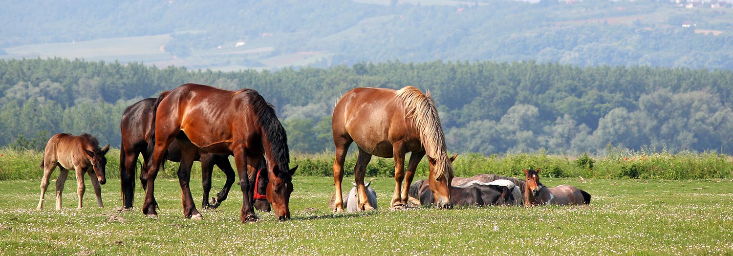
- Lyme disease in livestock
- GENERAL INTRODUCTION: SPIROCHAETES
- Swine dysentery
- Borrelia theileri infection
- Borrelia suilla infection
- Lyme disease in livestock
- Leptospirosis
- GENERAL INTRODUCTION: AEROBIC ⁄ MICRO-AEROPHILIC, MOTILE, HELICAL ⁄ VIBROID GRAM-NEGATIVE BACTERIA
- Genital campylobacteriosis in cattle
- Proliferative enteropathies of pigs
- Campylobacter jejuni infection
- GENERAL INTRODUCTION: GRAM-NEGATIVE AEROBIC OR CAPNOPHILIC RODS AND COCCI
- Moraxella spp. infections
- Bordetella bronchiseptica infections
- Pseudomonas spp. infections
- Glanders
- Melioidosis
- Brucella spp. infections
- Bovine brucellosis
- Brucella ovis infection
- Brucella melitensis infection
- Brucella suis infection
- Brucella infections in terrestrial wildlife
- GENERAL INTRODUCTION: FACULTATIVELY ANAEROBIC GRAM NEGATIVE RODS
- Klebsiella spp. infections
- Escherichia coli infections
- Salmonella spp. infections
- Bovine salmonellosis
- Ovine and caprine salmonellosis
- Porcine salmonellosis
- Equine salmonellosis
- Yersinia spp. infections
- Haemophilus and Histophilus spp. infections
- Haemophilus parasuis infection
- Histophilus somni disease complex in cattle
- Actinobacillus spp. infections
- Actinobacillus equuli infections
- Gram-negative pleomorphic infections: Actinobacillus seminis, Histophilus ovis and Histophilus somni
- Porcine pleuropneumonia
- Actinobacillus suis infections
- Pasteurella and Mannheimia spp. infections
- Pneumonic mannheimiosis and pasteurellosis of cattle
- Haemorrhagic septicaemia
- Pasteurellosis in sheep and goats
- Porcine pasteurellosis
- Progressive atrophic rhinitis
- GENERAL INTRODUCTION: ANAEROBIC GRAM-NEGATIVE, IRREGULAR RODS
- Fusobacterium necrophorum, Dichelobacter (Bacteroides) nodosus and Bacteroides spp. infections
- GENERAL INTRODUCTION: GRAM-POSITIVE COCCI
- Staphylococcus spp. infections
- Staphylococcus aureus infections
- Exudative epidermitis
- Other Staphylococcus spp. infections
- Streptococcus spp. infections
- Strangles
- Streptococcus suis infections
- Streptococcus porcinus infections
- Other Streptococcus spp. infections
- GENERAL INTRODUCTION: ENDOSPORE-FORMING GRAM-POSITIVE RODS AND COCCI
- Anthrax
- Clostridium perfringens group infections
- Clostridium perfringens type A infections
- Clostridium perfringens type B infections
- Clostridium perfringens type C infections
- Clostridium perfringens type D infections
- Malignant oedema⁄gas gangrene group of Clostridium spp.
- Clostridium chauvoei infections
- Clostridium novyi infections
- Clostridium septicum infections
- Other clostridial infections
- Tetanus
- Botulism
- GENERAL INTRODUCTION: REGULAR, NON-SPORING, GRAM-POSITIVE RODS
- Listeriosis
- Erysipelothrix rhusiopathiae infections
- GENERAL INTRODUCTION: IRREGULAR, NON-SPORING, GRAM-POSITIVE RODS
- Corynebacterium pseudotuberculosis infections
- Corynebacterium renale group infections
- Bolo disease
- Actinomyces bovis infections
- Trueperella pyogenes infections
- Actinobaculum suis infections
- Actinomyces hyovaginalis infections
- GENERAL INTRODUCTION: MYCOBACTERIA
- Tuberculosis
- Paratuberculosis
- GENERAL INTRODUCTION: ACTINOMYCETES
- Nocardiosis
- Rhodococcus equi infections
- Dermatophilosis
- GENERAL INTRODUCTION: MOLLICUTES
- Contagious bovine pleuropneumonia
- Contagious caprine pleuropneumonia
- Mycoplasmal pneumonia of pigs
- Mycoplasmal polyserositis and arthritis of pigs
- Mycoplasmal arthritis of pigs
- Bovine genital mycoplasmosis
- Neurotoxin-producing group of Clostridium spp.
- Contagious equine metritis
- Tyzzer's disease
- MYCOTIC AND ALGAL DISEASES: Mycoses
- MYCOTIC AND ALGAL DISEASES: Pneumocystosis
- MYCOTIC AND ALGAL DISEASES: Protothecosis and other algal diseases
- DISEASE COMPLEXES / UNKNOWN AETIOLOGY: Epivag
- DISEASE COMPLEXES / UNKNOWN AETIOLOGY: Ulcerative balanoposthitis and vulvovaginitis of sheep
- DISEASE COMPLEXES / UNKNOWN AETIOLOGY: Ill thrift
- Eperythrozoonosis
- Bovine haemobartonellosis
Lyme disease in livestock
This content is distributed under the following licence: Attribution-NonCommercial CC BY-NC  View Creative Commons Licence details here
Lyme disease in livestock
Previous authors: E HODZIC AND S W BARTHOLD
Current authors:
D M IMAI - Assistant, Clinical Health Sciences Professor, Comparative Pathology Laboratory, University of California, California, 95616, USA
E HODZIC - Director of Core Facility, DVM, MSci, PhD, Davis School of Veterinary Medicine, University of California, One Shields Avenue, California, 95616, USA
Introduction
Lyme disease, or Lyme borreliosis, was first described in 1977 as a distinctive entity in a cluster of children from Lyme, Connecticut, USA. These children had symptoms resembling juvenile rheumatoid arthritis, a disease that does not cluster and is rare in children.107 Based upon similarities with recognized human clinical syndromes in Europe that were of unknown aetiology, but associated with the bite of Ixodes ricinus ticks, the causative agent was isolated from North American Ixodes scapularis ticks in 1981,20 and subsequently named Borrelia burgdorferi.53 Once the connection was made between vector, agent, and the human disease, Lyme disease has been diagnosed throughout the world, but its significance is greatest in North America and Europe.47, 89, 98, 100 Borrelia burgdorferi has become an important public health problem and the most prevalent tick-borne disease in the United States.1, 64 It emerges across much of the northern hemisphere, causing considerable morbidity and in some cases mortality in humans, domestic animals, and occasionally wildlife.
The wide geographic distribution and the broad host range of both the vector and the bacterium provide ample opportunity for livestock species to be infected with B. burgdorferi. Most of what is known about Lyme disease, however, is based upon clinical studies in humans, and experimental studies in laboratory rodents. The amount of well-documented information on Lyme disease in livestock is limited.
Aetiology
Borrelia burgdorferi is a Gram-negative spiral or corkscrew shaped diderm bacterium, measuring 10 to 30 µm in length and 0,2 to 0,3 µm in diameter, with a lipoprotein-rich outer cell membrane and periplasmic flagella.53, 88, 114 For culture in vitro, this micro-aerophilic slow-growing organism requires a complex liquid medium, and an optimal temperature of 33 to 35 °C. 3
The genome of B. burgdorferi is small (~1.5 Mb) but complex, composed of 1 large, highly conserved linear chromosome and many small, highly variable linear and circular plasmids.24
Based on 16S ribosomal DNA gene sequences, the genus Borrelia belongs to the order Spirochaetales, family Spirochetaceae, but genomically unique and not closely related to any other bacteria, including the other spirochaetes. Other related genera include Spirochaeta, Cristispira, Treponema and Brachyspira (formerly Serpulina). It was first believed that B. burgdorferi was the only bacterium that could cause Lyme disease, but differences in morphology among isolates from diverse geographic locations suggested that additional spirochaetes may be involved in the aetiology.41
A major effort has been undertaken to analyse the phenotypic and genotypic diversity of B. burgdorferi isolates from around the world, using polymerase chain reaction (PCR) techniques that target 16S and 23S ribosomal DNA, flagellin, OspA, bdr genes, and intergenic spacers, as well as complete genome sequencing. It is now apparent that B. burgdorferi is genetically diverse, and is represented by more than 20 different genospecies grouped within the B. burgdorferi sensu lato (s.l.) complex. Borrelia burgdorferi sensu stricto (s.s.) is present in the USA and Europe (but not in Eurasia and Asia); B. afzelii, B. garinii, B. valaisiana, B. spielmanii, and B. lusitaniae are present in Eurasia; B. japonica, B. turdae, and B. tanukii in Japan; and B. bissettii and B. andersoni in the USA.34, 51, 80, 89 The causative genospecies of Lyme disease in humans and livestock are B. burgdorferi s.s., B. afzelii, and B. garinii. B. valaisiana has also been proposed as a causative agent of the human disease.59, 62, 116
Borrelia burgdorferi s.l. belongs to a guild of pathogens, including Ehrlichia and Babesia spp. and tick-borne encephalitis viruses, that are maintained within the same vector reservoir niches.112 Thus, co-infection with one or more of these agents can occur and may be responsible for ‘paraLyme disease’ syndromes. Since these other agents have a wide host range, they also infect livestock.
Epidemiology
Borrelia burgdorferi s.l. is geographically distributed in temperate regions of the northern hemispheres. These areas have the environmental conditions and reservoir hosts conducive to ixodid ticks. The range of B. burgdorferi s.l. was previously limited to northern Africa at its most southern extreme.2 Nevertheless, migratory birds, particularly seabirds, likely contributed to the expansion of its geographic distribution, with documentation of B. burgdorferi s.l. DNA in ixodid ticks or seropositivity in livestock, as far south as the Falkland Islands, Crozet Islands, and off-shore islands of New Zealand,76 and now, in Chile, Brazil, and Uruguay.4
Borrela burgdorferi and its vector ticks tend to be nonselective in their host range, and B. burgdorferi has been isolated from a wide variety of birds and mammals.2 Most hosts harbour subclinical infections. For example, in hyperendemic areas such as New York and Connecticut states in the USA, a high percentage of dogs are seropositive (and probably infected), but only a small fraction manifest clinical signs.61 A hallmark of Lyme borreliosis is persistent infection (even without antimicrobial agents), which is the rule in its many hosts. This has been experimentally proven in Peromyscus mice,95 laboratory mice,9 rats,68 hamsters,40 gerbils,85 guinea pigs,102 dogs,108 and non-human primates,90 and in reported confirmed spontaneous cases in humans based on culture,3, 57, 63, 65, 101, 104, 109 and PCR.17, 70, 71, 117
Among a large number of hard tick species, B. burgdorferi has been primarily detected in Ixodes ricinus, which is prevalent in Europe, Ixodes persulcatus, which is prevalent in Eastern Europe and Asia, and Ixodes scapularis and Ixodes pacificus in Northern America.81, 103 Clinical cases of Lyme disease could therefore be expected in localities where these Ixodes ticks are common. During their life cycle, which could take from two to six years to complete, ticks feed as larvae on small rodent reservoir hosts, thereby acquiring infection. After the larvae moult and harden, infected nymphs attach and feed on a variety of birds, reptiles and mammals, including livestock. Finally, adult I. scapularis, I. ricinus, and I. persulcatus ticks seek and parasitize large mammals, including deer or livestock, and are considered to be the most common vectors of Lyme disease for large animals.60, 78, 94 Borrelia burgdorferi s.l. has been identified in other ixodid ticks that parasitize livestock, such as Haemaphysalis spp., Hyalomma spp., Amblyomma spp., and Rhipicephalus spp.13, 28, 32, 37, 119, 121
Serosurveys for B. burgdorferi antibody in horses have revealed that the endemic and seasonal distribution of exposure closely corresponds to the prevalence of B. burgdorferi infection in humans in the same regions. Seroprevalence of B. burgdorferi antibodies was demonstrated in 10 per cent of horses in the New Jersey-Pennsylvania area,29 and in 62 per cent of horses in Wisconsin.22 In Connecticut, where Lyme disease is endemic, 45,1 per cent of horses had detectable antibodies to B. burgdorferi.62 In the UK the percentage of seropositive horses was low, but increased in areas that had a high prevalence of human and canine borreliosis,23 while in the region of München, Germany, 48 per cent of all tested horses were found to be seropositive.38 A study conducted in Sweden demonstrated that 16,8 per cent of horses had seroreactivity to B. burgdorferi s.l.31 Thus, like dogs within endemic areas, horses frequently seroconvert, indicating exposure and probable infection, but most do not manifest clinical signs.
Lyme disease in cattle often occurs as a herd problem with a geographic distribution similar to that among horses.22 In eastern Minnesota, a study revealed that higher prevalence rates of Lyme disease in lactating dairy cows were present during summer than in spring. This study also revealed a positive association between antibody titres and clinical lameness in the cows.120 Clinical Lyme disease has also been diagnosed in cows in Japan,50, 110 and in cows in Switzerland.32 A report from Norway showed that 10 per cent of tested sheep were seropositive to B. burgdorferi, mainly in areas where ticks were prevalent.35 Seropositive sheep and goats were reported in Italy.27 In Bolivia, goats were reported seropositive, but not sheep.26 In Tunisia, borrelial DNA was most prevalently detected in goats (30.4 per cent), and less commonly in sheep (6.2 per cent), cattle (1.3 per cent), and camels (1.8 per cent).13 A report from Egypt identified PCR-positive blood samples from 16 per cent of cattle tested and was able to successfully culture spirochaetes from one cow.32 Furthermore, the prevalence of seroconversion was higher than the appearance of clinical signs in these species.
Pathogenesis
Transmission by the vector (nymphal or adult ticks) to the host is a complex three-way interaction between the tick, the host, and the pathogen. Hosts mount local inflammatory, haemostatic and immune responses to the feeding tick, while tick saliva contains substances that counter the host response, thereby facilitating a successful blood meal and transmission of the pathogens.74 After tick attachment, B. burgdorferi spirochaetes undergo striking variations in the expression of antigens, and migrate from the tick midgut to its salivary glands.30, 96 After being transmitted, spirochaetes remain and multiply in the skin at the attachment site for several days, and then disseminate to many organs and tissues throughout the host.45, 97 Motility plays a major role in the spread of spirochaetes from the site of tick attachment, and allow contact with the host vasculature.69, 72 As a result of spirochaete multiplication at the tick attachment site, the host reacts with a localized skin rash (erythema migrans), which generally persists for weeks after detachment of the tick. The rash is observed in the majority of human cases, and has been reported occasionally in livestock. 19, 29, 59, 84
Clinical signs of illness are generally most apparent during the early stages of disseminated infection when spirochaetes are believed to elaborate pro-inflammatory lipoproteins that appear to facilitate the process of dissemination through host tissues. Despite the highly immunogenic nature of these lipoproteins, spirochaetes effectively evade host immune clearance by yet-to-be understood mechanisms.11 It should be emphasized that Lyme borreliosis in untreated humans (and experimental animals) is ephemeral, with “spontaneous” resolution (without antimicrobial treatment) of erythema migrans, myocarditis, arthritis, and other signs.105, 106 Studies in animal models have shown that resolution of arthritis and myocarditis is mediated by the acquired humoral immune response of the host. Under these conditions, anatomically defined inflammation resolves, but infection persists.7, 8, 10
Whatever the mechanism, it is clear that spirochaetes undergo dramatic shifts in protein expression. For example, outer surface protein A (OspA) is a highly immunogenic lipoprotein that is expressed in the midgut of ticks, but is rapidly down-regulated upon onset of feeding, and is minimally expressed in the mammalian host. In contrast, OspC is up-regulated under similar circumstances.12, 66, 96 Up- and down-regulation have also been documented for several other lipoproteins, some of which are expressed exclusively in vivo, but their role is still unclear.11 Other possible explanations of immune evasion by B. burgdorferi spirochaetes have been proposed. These include sequestration in extracellular matrix,49 intracellular localization,67, 44 formation of cystic-like forms found in vivo 46 and in vitro,18 and the existence of complement-resistant strains that regulate and control complement activation.56 It has also been suggested that during the course of infection, B. burgdorferi proliferates and incidentally generates an increasingly heterogeneous population of replication-attenuated spirochaetes that emerge as a result of genetic alterations, including plasmid loss or formation of biofilm, and/or that suboptimal immune responses induced by the infection make persistence and resurgence possible.6, 16, 42, 43, 44, 92, 93, 115 Once spirochaetes have disseminated, some animals and humans develop clinical signs of Lyme disease, including fever, lethargy, weight loss, stiffness, swollen joints, laminitis, lameness, abortion, uveitis and, sometimes, encephalitis. In livestock clinical signs have been observed in cattle, horses, and sheep.21, 22, 27, 36, 48, 52, 54, 86, 120
The mechanisms by which B. burgdorferi spirochaetes cause disease in infected hosts are being studied in animal models. Borrelia burgdorferi spirochaetes produce no known toxins yet are capable of invading virtually any mammalian tissue and causing infection and disease manifestations for months to years. It is suspected that the approximately 150 lipoproteins that are encoded by the B. burgdorferi genome play an important role in not only disease pathogenesis, but also host immunity, and are considered to be virulence determinants.73 An important first step in the establishment of infection is adhesion of spirochaetes to host cells and extracellular matrix components.33
Differential expression of pro-inflammatory lipoproteins, of which the majority are outer surface proteins, in various tissues and at different times during persistent infection appear to be critical determinants of disease.83 The genome has 12 linear and nine circular plasmids, and loss of plasmids has been correlated with decreased infectivity and pathogenicity.58, 87, 113
Clinical signs
The incubation period for Lyme disease in livestock has not been determined. Observed clinical signs in naturally infected horses and cows correlated within a month of the seasonal emergence of adult ticks, indicating a long incubation period.22, 38 No significant clinical signs were observed in experimentally infected cattle with cultured B. burgdorferi,116 nor in ponies that were exposed to ticks.25 Experimental infection of a pony with pulverized B. burgdorferi-infected ticks resulted in seroconversion on day nine, which continued for a three-week period, then waned. Re-challenge with cultured B. burgdorferi six months later led to seroconversion within four days.29 Infection efficiency has been found to depend upon the infectious dose, the strain of Borrelia, and the age of the host. Experimental infection of cattle with different doses and different strains of cultured organisms demonstrated that the mean infectious dose was 106 to 107 organisms, with some variations in infectivity among different strains.116
Clinical signs in horses and cattle, when present, are varied and often vague and non-specific. A skin rash resembling erythema migrans in humans is not a prominent feature of Lyme borreliosis in livestock though there are rare reports of erythema rashes in cattle and horses at the site of tick attachment, which can be under the lower jaw, on the abdomen and ventral chest, on the legs, in the region of the mane of horses, on the udder of cows, and on the tail.59, 78 In horses, clinical signs may include depression, low-grade fever (38,2 to 38,8 °C), swollen joints, shifting lameness, muscle tenderness, hyperaesthesia, ataxia, behavioural changes and uveitis.19, 21, 22, 38, 48, 52, 54, 78, 80, 86
Animals with disseminated disease can develop single or multiple swollen joints (arthritis), laminitis, stiffness, hyperaesthesia, and sporadic lameness, which are the most common clinical signs. Untreated cases can persist for years with chronic weight loss, neurologic dysfunction and polyarthritis.48 In pregnant mares, there is a suggested association with borreliosis and reproductive failure due to foetal resorption or abortion.25 In cattle, stiffness, swollen joints, and/or lameness are the most commonly reported clinical signs although reproductive failure, reduced milk production, fever and chronic weight loss are also observed.22, 50, 59, 78, 120 Arthritis has been most notable in the carpal-metacarpal, tibiotarsal and/or stifle joints. Other clinical signs in cattle include abortion and decreased milk production.
In small ruminants, clinical signs are not often observed or reported. Lameness, poor condition and poor appetite have been reported in seropositive lambs,36 but other studies have found that all seropositive sheep were healthy at the time of serum sampling.27, 35 Sheep experimentally exposed by tick bite did not develop systemic infection.75 In the few reports involving goats, a high prevalence of B. burgdorferi antibodies have been found,26, 35 but clinical signs were not evaluated. It has been suggested that goats might develop clinical signs similar to those in cattle.84
Pathology
It appears that Lyme disease in livestock is not fatal, signs are often ephemeral, and disease is mild, if present at all, so the pathology of disease in these animals is not well characterized.
Post-mortem examination and histopathologic finding associated with Lyme borreliosis in livestock in general include arthritis, synovitis, and myocarditis.48, 50, 59, 80 In spontaneous infections in horses, suppurative uveitis, necrotizing to non-suppurative meningitis and radiculoneuritis and non-suppurative myocarditis with intralesional argyrophilic spirochaetes have been documented.21, 48, 52, 54, 86 In cases of uveitis, spirochetaes have been visualized within the intravitreal suppurative exudate using silver stain.54, 86 The neurologic manifestation of Lyme borreliosis in the horse recapitulates the human disease and the equid is the only non-human host to develop neurologic disease after natural infection or after a biologically relevant route of experimental infection is used.48 A cow diagnosed with Lyme disease showed distention of the tibiotarsal joints. The synovial fluid collected from the joints was copious, watery, clear and amber. Endometritis and a few abnormalities in the abdomen were also found.59 In experimentally infected equids, lymphohistiocytic dermatitis at the tick attachment site with reactive regional lymphadenopathy were the only lessions identified.25
Although descriptions of abortion and metritis have been included in some reports, reproductive tract and foetal lesions have not been characterized and the direct role of B. burgdorferi is as yet undetermined but transplacental transmission of spirochetes has been documented.22
Diagnosis and differential diagnosis
The clinical signs of Lyme disease in domestic animals are very diverse, making an accurate clinical diagnosis difficult. An awareness of the tick vectors of the disease in the region and the opportunity for exposure to such ticks are important for diagnosis. For seroepidemiological studies, assays such as immunofluorescence assay, enzyme-linked immunosorbent assay and immunoblot are commonly used.14, 23, 35, 62, 118 The ‘gold standard’ for the diagnosis of Lyme disease, however, is the isolation of the organism from blood, skin biopsy, milk, vitreous humour, and synovial or cerebrospinal fluid. Culture requirements are stringent as this microaerophilic, slow-growing organism requires a complex liquid medium and incubation at 33 to 35ºC.5 A valuable ancillary procedure is PCR, especially real-time quantitative PCR (qPCR), which detects spirochaetal DNA, whether or not the organisms are alive. This is especially valuable when joint fluid, which tends not to contain viable organisms, is analysed.15, 25, 39, 101 Direct cytologic examination with identification of spirochaetes in vitreous humor has been successful in cases of uveitis.86
The differential diagnosis of Lyme disease must include, among numerous other possibilities, traumatically induced carpal hygromas and chronic carpal and hock swellings, interdigital dermatitis, white line disease, rabies, spinal compression, brucellosis, leptospirosis, equine protozoal myeloencephalitis, and vitamin E/selenium deficiency.21, 23, 60, 84, 120 In horses, negative serology and normal CSF or joint fluid analysis does not exclude the diagnosis.48, 54 For equine neuroborreliosis, histopathology is considered the most reliable diagnostic test.48, 54
Control
Chemotherapy of Lyme disease in animals is based upon that used for the treatment of the disease in humans. Antimicrobials such as doxycycline, amoxicillin, penicillin, ceftriaxone and cefotaxime have been shown to be effective against B. burgdorferi. Early treatment is desirable, as treatment is most effective when given during the early stages of infection. Chronic cases require prolonged treatment, which is often less successful.60, 78, 82 A bacterin and a recombinant vaccine based on OspA are available for veterinary use, but there is no evidence of their efficacy in livestock. The best way of controlling Lyme borreliosis in livestock is tick control. This can be achieved by biological control of ticks, by application of organophosphates or pyrethroids in the ticks’ habitat, or by the use of ectoparasiticides applied directly on animals.79, 91, 111
References
- ADRION, E.R., AUCOTT, J., LEMKE, K.W. & WEINER, J.P., 2015. Health care costs, utilization and patterns of care following Lyme disease. PLoS One, 10, e0116767.
- ANDERSON, J.F. & MAGNARELLI, L.A., 1993. Epizootiology of Lyme disease-causing Borreliae. Clinical Dermatology, 11, 339–351.
- ASBRINK, E. & HOVMARK, A., 1985. Successful cultivation of spirochetes from skin lesions of patients with erythema chronicum migrans Afzelius and acrodermatitis chronica atrophicans. Acta Pathologica, Microbiologica, et Immunologica Scandinavica, Section B, Microbiology, 93, 161-163.
- BARBIERI, A.M., VENZAL, J.M., MARCILI, A., ALMEIDA, A.P., GONZALEZ, E.M. & LABRUNA, M.B., 2013. Borrelia burgdorferi sensu lato infecting ticks of the Ixodes ricinus complex in Uruguay: first report for the Southern Hemisphere. Vector Borne and Zoonotic Diseases, 13, 147-153.
- BARBOUR, A.G., 1984. Isolation and cultivation of Lyme disease spirochetes. Yale Journal of Biology and Medicine, 57, 521-525.
- BARBOUR, A.G., 1984. Isolation and cultivation of Lyme disease spirochetes. Yale Journal of Biology and Medicine (New Haven CT), 57, 521–525.
- BARTHOLD, S.W., HODZIC, E., IMAI, D.M., FENG, S., YANG, X. & LUFT, B.J., 2010. Ineffectiveness of tigecycline against persistent Borrelia burgdorferi. Antimicrobial Agents and Chemotherapy, 54, 643-651.
- BARTHOLD, S.W., HODZIC, E., TUNEV, S. & FENG, S., 2006. Antibody-mediated disease remission in the mouse model of lyme borreliosis. Infection and Immunity, 74, 4817-4825.
- BARTHOLD, S W., DESOUZA, M. & FENG, S., 1996. Serum-mediated resolution of Lyme arthritis in mice. Laboratory Investigation, 74, 57-67.
- BARTHOLD, S. W., DE SOUZA, M.S., JANOTKA, J.L., SMITH, A.L. & PERSING, D.H., 1993. Chronic Lyme borreliosis in the laboratory mouse. American Journal of Pathology, 143, 959-971.
- BARTHOLD, S. W., FENG, S., BOCKENSTEDT, L.K., FIKRIG, E. & FEEN, K., 1997. Protective and arthritis-resolving activity in sera of mice infected with Borrelia burgdorferi. Clinical Infectious Diseases, 25, S9-17.
- BARTHOLD, S.W., 2000. Lyme Borreliosis. In: NATARO, J.P., BLASER, M.J. & CUNNINGHAM-RUNDLES, S., (eds). Persistent Bacterial Infection. Vol. 14. Washington, D.C.: ASM Press, 281–304.
- BARTHOLD, S.W., FIKRIG, E., BOCKENSTEDT, L.K. & PERSING, D.H., 1995. Circumvention of outer surface protein A immunity by host-adapted Borrelia burgdorferi. Infection and Immunity, 63, 2255–2261.
- BEN SAID, M., BELKAHIA, H., ALBERTI, A., ABDI, K., ZHIOUA, M., DAALOUL-JEDIDI, M. & MESSADI., L., 2016. First molecular evidence of [i]Borrelia burgdorferi[/i] sensu lato in goats, sheep, cattle and camels in Tunisia. Annals of Agricultural and Environmental Medicine, 23, 442-447.
- BERNARD, W.V., COHEN, D., BOSLER, E. & ZAMOS, D., 1990. Serologic survey for Borrelia burgdorferi antibody in horses referred to a mid-Atlantic veterinary teaching hospital. Journal of the American Veterinary Medical Association, 196, 1255–1258.
- BIL-LULA, I., MATUSZEK, P., PFEIFFER, T. & WOZNIAK, M., 2015. Lyme Borreliosis--the Utility of Improved Real-Time PCR Assay in the Detection of Borrelia burgdorferi Infections. Advances in Clinical and Experimental Medicine, 24, 663-670.
- BOCKENSTEDT, L. K., MAO, J., HODZIC, E., BARTHOLD, S.W. & FISH, D., 2002. Detection of attenuated, noninfectious spirochetes in Borrelia burgdorferi-infected mice after antibiotic treatment. Journal of Infectious Diseases, 186, 1430-1437.
- BRADLEY, J. F., JOHNSON, R.C. & GOODMAN, J.L., 1994. The persistence of spirochetal nucleic acids in active Lyme arthritis. Annals of Internal Medicine, 120, 487-489.
- BRORSON, O. & BRORSON, S.H., 1998. In vitro conversion of Borrelia burgdorferi to cystic forms in spinal fluid, and transformation to mobile spirochetes by incubation in BSK-H medium. Infection, 26, 144–150.
- BROWNING, A., CARTER, S.D., BARNES, A., MAY, C. & BENNETT, D., 1993. Lameness associated with Borrelia burgdorferi infection in the horse. The Veterinary Record, 132, 610–611.
- BURGDORFER, W., BARBOUR, A.G., HAYES, S.F., BENACH, J.L., GRUNWALT, E. & DAVIS, J.P., 1982. Lyme disease — a tickborne spirochaetosis? Science, 216, 1317–1319.
- BURGESS, E.C. & MATTISON, M., 1987. Encephalitis associated with Borrelia burgdorferi infection in a horse. Journal of the American Veterinary Medical Association, 191, 1457–1458.
- BURGESS, E.C., 1988. Borrelia burgdorferi infection in Wisconsin horses and cows. Annals of the New York Academy of Sciences, 539, 233–243.
- CARTER, S.D., MAY, C., BARNES, A. & BENNETT, D., 1994. Borrelia burgdorferi infection in UK horses. Equine Veterinary Journal, 26, 187–190.
- CASJENS, S. R., EGGERS, C.H. & SCHWARTZ, I., 2010. Borrelia Genomics: Chromosome, Plasmids, Bacteriphages and Genetic Variation. In S. D. Samuels and J. Radolf (ed.), Borrelia; Molecular Biology, Host Interaction and Pathogenesis. Caister Academic Press, Norfolk, UK, 21-47.
- CHANG, Y-F., NOVOSOL, V., MCDONOUGH, S.P., CHANG, C-F., JACOBSON, R.H., DIVERS, T., QUIMBY, F.W., SHIN, S. & LEIN, D.H., 2000. Experimental infection of ponies with Borrelia burgdorferi by exposure to Ixodid ticks. Veterinary Pathology, 37, 68–76.
- CHANG, Y-F., NOVOSOL, V., MCDONOUGH, S.P., CHANG, C-F., JACOBSON, R.H., DIVERS, T., QUIMBY, F.W., SHIN, S. & LEIN, D.H., 2000. Experimental infection of ponies with Borrelia burgdorferi by exposure to Ixodid ticks. Veterinary Pathology, 37, 68–76.
- CICERONI, L., BARTOLONI, A., CIARROCCHI, S., PINTO, A., GUGLIELMETTI, P., VALDEZ VASQUEZ, C., GAMBOA BARAHONA, H., ROSELLI, M. & PARADISI, F., 1997. Serologic survey for antibodies to Borrelia burgdorferi in sheep, goats and dogs in Cordillera Province, Bolivia. Zentralblatt für Veterinärmedizin, [B], 44, 133–137.
- CICERONI, L., SIMEONI, J., PACETTI, A.I., CIARROCCHI, S. & CACCIAPUOTI, B., 1996. Antibodies to Borrelia burgdorferi in sheep and goats. Alto Adige-South Tyrol, Italy. New Microbiology, 19, 171–174.
- CLARK, K. L., LEYDET, B. & HARTMAN, S., 2013. Lyme borreliosis in human patients in Florida and Georgia, USA. International Journal of Medical Sciences, 10, 915-931.
- COHEN, D., BOSLER, E.M., BERNARD, W., MEIRS, DN., EISNER, R. & SCHULZE, T.L., 1988. Epidemiologic studies of Lyme disease in horses and their public health significance. Annals of the New York Academy of Sciences, 539, 244–257
- DE SILVA, A.M. & FIKRIG, E., 1995. Growth and migration of Borrelia burgdorferi in Ixodes ticks during blood feeding. American Journal of Tropical Medicine and Hygiene, 53, 397–404.
- EGENVALL, A., FRANZEN, P., GUNNARSSON, A., OLSSON ENGVALL, E., VAGSHOLM, I., WIKSTROM, U-B. & ARTURSSON, K., 2001. Cross-sectional study of the seroprevalence to Borrelia burgdorferi sensu lato and granulocytic Ehrlichia spp. and demographic, clinical and tick-exposure factors in Swedish horses. Preventive Veterinary Medicine, 49, 191–208.
- ELHELW, R.A., EL-ENBAAWY, M.I. & SAMIR, A., 2014. Lyme borreliosis: A neglected zoonosis in Egypt. Acta Tropica, 140, 188-192.
- FINLAY, B.B. & FALKOW, S., 1997. Common themes in microbial pathogenicity revisited. Microbiology and Molecular Biology, 61, 136-169.
- FRASER, C. M., CASJENS, S., HUANG, W.M., SUTTON, G.G., CLAYTON, R., LATHIGRA, R., WHITE, O., KETCHUM, K.A., DODSON, R., HICKEY, E.K., GWINN, M., DOUGHERTY, B., TOMB, J.F., FLEISCHMANN, R.D., RICHARDSON, D., PETERSON, J., KERLAVAGE, A.R., QUACKENBUSH, J., SALZBERG, S., HANSON, M., VAN VUGT, R., PALMER, N., ADAMS, M.D., GOCAYNE, J., VENTER, J.C., UTTERBACK, T., WEIDMAN, J., WATTHEY, L., MCDONALD, L., ARTIACH, P., BOWMAN, C., GARLAND, S., FUJI, C., COTTON, M.D., HORST, K., ROBERTS, K., HATCH, B., SMITH, H.O., 1997. Genomic sequence of a Lyme disease spirochaete, Borrelia burgdorferi. Nature, 390, 580-586.
- FRIDRIKSDOTTIR, V., NESSE, L.L. & GUDDING, R., 1992. Seroepidemiological studies of Borrelia burgdorferi infection in sheep in Norway. Journal of Clinical Microbiology, 30, 1271–1277.
- FRIDRIKSDOTTIR, V., OVERNES, G. & STUEN, S., 1992. Suspected Lyme borreliosis in sheep. The Veterinary Record, 130, 323–324.
- FU, Y., LIU, Z., GUAN, G., NIU, Q., LI, Y., YANG, J., REN, Q., MA, M., LIU, A., PENG, Y., LUO, J. & YIN, H., 2012. Development of real-time polymerase chain reaction for detection of Borrelia burgdorferi sensu lato in China. Vector Borne and Zoonotic Diseases, 12, 341-345.
- GERHARDS, H. & WOLLANKE, B., 1996. Antibody titers against Borrelia in horses in serum and in eyes and occurrence of equine recurrent uveitis. Berliner und Münchener Tierärztliche Wochenschrift, 109, 273–278.
- GERN, L., ROUVINEZ, E., TOUTOUNGI, L.N. & GODFROID, E., 1997. Transmission cycles of Borrelia burgdorferi sensu lato involving Ixodes ricinus and/or I. hexagonus ticks and the European hedgehog, Erinaceus europaeus, in suburban and urban areas in Switzerland. Folia Parasitologica, 44, 309–314.
- GOODMAN, J. L., JURKOVICH, P., KODNER, C. & JOHNSON, R.C., 1991. Persistent cardiac and urinary tract infections with Borrelia burgdorferi in experimentally infected Syrian hamsters. Journal of Clinical Microbiology, 29, 894-896.
- HAYES, S.F. & BURGDORFER, W., 1993. Characteristic of Borrelia burgdorferi. 3 Ultrastructure of Borrelia burgdorferi. In: WEBER, K. & BURGDORFER, W., (eds). Aspects of Lyme borreliosis. Vol. 1. Berlin, Heidelberg, New York, London, Paris, Tokyo, Hong Kong, Barcelona, Budapest: Springer-verlag, 29–43.
- HODZIC, E., 2015. Lyme Borreliosis: Is there a preexisting (natural) variation in antimicrobial susceptibility among Borrelia burgdorferi strains? Bosnian Journal of Basic Medical Sciences, 15, 1-13.
- HODZIC, E., IMAI, D., FENG, S. & BARTHOLD, S.W., 2014. Resurgence of persisting non-cultivable Borrelia burgdorferi following antibiotic treatment in mice. PLoS One, 9, e86907.
- HODZIC, E., FENG, S., HOLDEN, K., FREET, K.J. & BARTHOLD, S.W., 2008. Persistence of Borrelia burgdorferi following antibiotic treatment in mice. Antimicrobial Agents and Chemotherapy, 52, 1728-1736.
- HODZIC, E., FENG, S., FREET, K.J., BORJESSON, D.L. & BARTHOLD, S.W., 2002. Borrelia burgdorferi population kinetics and selected gene expression at the host-vector interface. Infection and Immunity, 70, 3382-3388.
- HULINSKA, D., BARTAK, P., HERCEGOVA, J., HANCIL, J., BASTA, J. & SCHRAMLOVA, J., 1994. Electron microscopy of Langerhans cells and Borrelia burgdorferi in Lyme disease patients. Zentralblatt für Mikrobiologie, 280, 348–359.
- HUMAIR, P.F. & GERN, L., 2000. The wild hidden face of Lyme borreliosis in Europe. Microbes and Infection, 2, 915–922.
- IMAI, D. M., BARR, B.C., DAFT, B., BERTONE, J.J., FENG, S., HODZIC, E., JOHNSTON, J.M., OLSEN, K.J. & BARTHOLD, S.W., 2011. Lyme neuroborreliosis in 2 horses. Veterinary Pathology, 48, 1151-1157.
- IMAI, D. M., FENG, S., HODZIC, E. & BARTHOLD, S.W., 2013. Dynamics of connective-tissue localization during chronic Borrelia burgdorferi infection. Laboratory Investigation, 93, 900-910.
- ISOGAI, H., ISOGAI, E., MASUZAWA, T., YANAGIHARA, Y., MATSUBARA, M., SHIMANUKI, M., SETA, T., FUKAI, K., KUROSAWA, N., ENOKIDANI, M., NAKAMURA, T., TAJIMA, M., TAKAHASHI, K., TAKAHASHI, K. & FUJII, N., 1992. Seroepidemiological survey for antibody to Borrelia burgdorferi in cows. Microbiology and Immunology, 36, 1029–1039.
- IVANOVA, L. B., TOMOVA, A., GONZALEZ-ACUNA, D., MURUA, R., MORENO, C.X., HERNANDEZ, C., CABELLO, J., CABELLO, C., DANIELS, T.J., GODFREY, H.P. & CABELLO, F.C., 2014. Borrelia chilensis, a new member of the Borrelia burgdorferi sensu lato complex that extends the range of this genospecies in the Southern Hemisphere. Environmental Microbiology, 16, 1069-1080.
- JAMES, F. M., ENGILES, J.B. & BEECH, J., 2010. Meningitis, cranial neuritis, and radiculoneuritis associated with Borrelia burgdorferi infection in a horse. Journal of the American Veterinary Medical Association, 237, 1180-1185.
- JOHNSON, R.C., SCHMID, G.P., HYDE, F.W., STEIGERWALT, A.G. & BRENNER, D.J., 1984. Borrelia burgdorferi sp. nov.: Etiologic agent of Lyme disease. International Journal of Systematic Bacteriology, 34, 496–497.
- JOHNSTONE, L.K., ENGILES, J.B., ACETO, H., BUECHNER-MAXWELL, V., DIVERS, T., GARDNER, R., LEVINE, R., SCHERRER, N., TEWARI, D., TOMLINSON, J. & JOHNSON, A.L., 2016. Retrospective Evaluation of Horses Diagnosed with Neuroborreliosis on Postmortem Examination: 16 Cases (2004-2015). Journal of Veterinary Internal Medicine, 30, 1305-1312.
- KASBOHRER, A. & SCHONBERG, A., 1990. [Serologic studies of the occurrence of Borrelia burgdorferi in domestic animals in Berlin (West)]. Berliner und Münchener Tierärztliche Wochenschrift, 103, 374–378.
- KRAICZY, P., SKERKA, C., KIRSCHFINK, M., ZIPFEL, P.F. & BRADE, V., 2001. Mechanism of complement resistance of pathogenic Borrelia burgdorferi isolates. International Immunopharmacology, 1, 393–401.
- KUIPER, H., VAN DAM, A.P., SPANJAARD, L., DE JONGH, B.M., WIDJOJOKUSUMO, A., RAMSELAAR, T.C., CAIRO, I., VOS, K. & DANKERT, J., 1994. Isolation of Borrelia burgdorferi from biopsy specimens taken from healthy-looking skin of patients with Lyme borreliosis. Journal of Clinical Microbiology, 32, 715-720.
- LABANDEIRA-REY, M. & SKARE, J.T., 2001. Decreased infectivity in Borrelia burgdorferi strain B31 is associated with loss of linear plasmid 25 or 28-1. Infection and Immunity, 69, 446–455.
- LISCHER, C.J., LEUTENEGGER, C.M., BRAUN, U. & LUTZ, H., 2000. Diagnosis of Lyme disease in two cows by the detection of Borrelia burgdorferi DNA. The Veterinary Record, 146, 497–499.
- MADIGAN, J.E., 1993. Lyme disease (Lyme borreliosis) in horses. Veterinary Clinics of North America: Equine Practitioner, 9, 429–435
- MAGNARELLI, L.A., ANDERSON, J.F., SCHREIER, A.B. & FICKE, C.M., 1987. Clinical and serologic studies of canine borreliosis. Journal of the American Veterinary Medical Association, 191, 1089–1094.
- MAGNARELLI, L.A., IJDO, J.W., VAN ANDEL, A.E., WU, C., PADULA, S.J. & FIKRIG, E., 2000. Serologic confirmation of Ehrlichia equi and Borrelia burgdorferi infections in horses from the northeastern United States. Journal of the American Veterinary Medical Association, 217, 1045–1050
- MARASPIN, V., OGRINC, K., RUZIC-SABLJIC, E., LOTRIC-FURLAN, S. & STRLE, F., 2011. Isolation of Borrelia burgdorferi sensu lato from blood of adult patients with borrelial lymphocytoma, Lyme neuroborreliosis, Lyme arthritis and acrodermatitis chronica atrophicans. Infection, 39, 35-40.
- MEAD, P.S. 2015. Epidemiology of Lyme disease. Infectious Disease Clinics of North America, 29, 187-210.
- MIKLOSSY, J., KHALILI, K., GERN, L., ERICSON, R.L., DAREKAR, P., BOLLE, L., HURLIMANN, J. & PASTER, B.J., 2004. Borrelia burgdorferi persists in the brain in chronic lyme neuroborreliosis and may be associated with Alzheimer disease. Journal of Alzheimer's Disease, 6, 639-649; discussion 673-681.
- MONTGOMERY, R.R., MALAWISTA, S.E., FEEN, K.J.M. & BOCKENSTEDT, L.K., 1996. Direct demonstration of antigenic substitution of Borrelia burgdorferi ex vivo: Exploration of the paradox of the early immune response to outer surface proteins A and C in Lyme disease. Journal of Experimental Medicine, 183, 261–269.
- MONTGOMERY, R.R., NATHANSON, M.H. & MALAWISTA, S.E., 1993. The fate of Borrelia burgdorferi, the agent for Lyme disease, in mouse macrophages: Destruction, survival, recovery. Journal of Immunology, 150, 909–915.
- MOODY, K. D., BARTHOLD, S.W., TERWILLIGER, G.A., BECK, D.S., HANSEN, G.M. & JACOBY, R.O., 1990. Experimental chronic Lyme borreliosis in Lewis rats. Journal of Alzheimer's Disease, 42, 165-174.
- MORIARTY, T. J., NORMAN, M.U., COLARUSSO, P., BANKHEAD, T., KUBES, P. & CHACONAS, G., 2008. Real-time high resolution 3D imaging of the lyme disease spirochete adhering to and escaping from the vasculature of a living host. PLoS Pathogens, 4, e1000090.
- MOTER, S. E., HOFMANN, H., WALLICH, R., SIMON, M.M. & KRAMER, M.D., 1994. Detection of Borrelia burgdorferi sensu lato in lesional skin of patients with erythema migrans and acrodermatitis chronica atrophicans by ospA-specific PCR. Journal of Clinical Microbiology, 32, 2980-2988.
- NOCTON, J. J., DRESSLER, F., RUTLEDGE, B.J., RYS, P.N., PERSING, D.H. & STEERE, A.C., 1994. Detection of Borrelia burgdorferi DNA by polymerase chain reaction in synovial fluid from patients with Lyme arthritis. New England Journal of Medicine, 330, 229-234.
- NORMAN, M. U., MORIARTY, T.J., DRESSER, A.R., MILLEN, B., KUBES, P. & CHACONAS, G., 2008. Molecular mechanisms involved in vascular interactions of the Lyme disease pathogen in a living host. PLoS Pathogens, 4, e1000169.
- NORRIS, S. J., COBURN, J., LEONG, J.M., HU, L.T. & HOOK, M., 2010. Pathobiology of Lyme Disease Borrelia. In: SAMUELS, S.D. & RADOLF, J., (ed.), Borrelia; Molecular Biology, Host Interaction and Pathogenesis. Caister Academic Press, Norfolk, UK, 293-325.
- NUTTALL, P.A., 1998. Displaced tick-parasite interactions at the host interface. Parasitology, 116, S65–S72
- OGDEN, N.H., NUTTALL, P.A. & RANDOLPH, S.E., 1997. Natural Lyme disease cycles maintained via sheep by cofeeding ticks. Parasitology, 115, 591–599
- OLSEN, B., DUFFY, D.C., JAENSON, T.G.T., GYLFE, A., BONNEDAHL, J. & BERGSTRO, S., 1995. Transhemispheric exchange of Lyme disease spirochetes by seabirds. Journal of Clinical Microbiology, 33, 3270–3274.
- PACHNER, A.R., BASTA, J., DELANEY, E. & HULINSKA, D., 1995. Localization of Borrelia burgdorferi in murine Lyme borreliosis by electron microscopy. American Journal of Tropical Medicine, 52, 128–133.
- PARKER, J.L. & WHITE, K.K., 1992. Lyme borreliosis in cattle and horses: A review of the literature. Cornell Veterinarian, 82, 153–174.
- PAROLA, P. & RAOULT, D., 2001. Ticks and tickborne bacterial diseases in humans: An emerging infectious threat. Clinical Infectious Diseases, 32, 897–928.
- PASSAMONTI, F., VERONESI, F., CAPPELLI, K., CAPOMACCIO, S., REGINATO, A., MIGLIO, A., VARDI, D.M., STEFANETTI, V., COLETTI, M., BAZZICA, C. & PEPE, M., 2015. Polysynovitis in a horse due to Borrelia burgdorferi sensu lato infection--Case study. Annals of Agricultural and Environmental Medicine, 22, 247-250.
- PEAVEY, C.A. & LANE, R.S., 1995. Transmission of Borrelia burgdorferi by Ixodes pacificus nymphs and reservoir competence of deer mice (Peromyscus maniculatus) infected by tick-bite. Journal of Parasitology, 81, 175–178.
- POPOVIC, N., DJURICIC, B. & VALCIC, M., 1993. [The importance of Lyme borreliosis veterinary medicine]. Glasnik Srpske Akademije Nauka i Umetnosti [Med], 43, 277–285.
- PORCELLA, S.F. & SCHWAN, T.G., 2001. Borrelia burgdorferi and Treponema pallidum: A comparison of functional genomics, environmental adaptations, and pathogenic mechanisms. Journal of Clinical Investigation, 107, 651–656.
- POST, J.E., 1990. Lyme disease in large animals. New Jersey Medicine, 87, 575–577
- PREAC MURSIC, V., PATSOURIS, E., WILSKE, B., REINHARDT, S., GROSS, B. & MEHRAEIN, P., 1990. Persistence of Borrelia burgdorferi and histopathological alterations in experimentally infected animals. A comparison with histopathological findings in human Lyme disease. Infection, 18, 332-341.
- PRIEST, H. L., IRBY, N.L., SCHLAFER, D.H., DIVERS, T.J., WAGNER, B., GLASER, A.L., CHANG, Y.F. & SMITH, M.C., 2012. Diagnosis of Borrelia-associated uveitis in two horses. Veterinary Ophthalmology, 15, 398-405.
- PURSER, J.E. & NORRIS, S.J., 2000. Correlation between plasmid content and infectivity in Borrelia burgdorferi. Proceedings of the National Academy of Science, USA, 97, 13865–13870.
- RADOLF, J. D., SALAZAR, J.C. & DATTWYLER, R., 2010. Lyme Disease in Humans. In: SAMUEL, D.S. & RADOLF, J,D.,(ed.), Borrelia. Molecular Biology, Host Interaction and Pathogenesis. Caister Academic Press, Norfolk, UK, 481-527.
- ROBERTS, D.M., CARLYON, J.A., THEISEN, M. & MARCONI, R.T., 2000. The bdr gene families of the Lyme disease and relapsing fever spirochetes: Potential influence on biology, pathogenesis, and evaluation. Emerging Infectious Diseases, 6, 110–122.
- ROBERTS, E. D., BOHM JR, R.P., COGSWELL, F.B., LANNERS, H.N., LOWRIE JR, R.C., POVINELLI, L., PIESMAN, J. & PHILIPP, M.T., 1995. Chronic lyme disease in the rhesus monkey. Laboratory Investigation, 72, 146-160.
- SAMISH, M. & REHECAK, J., 1999. Pathogens and predators of ticks and their potential in biological control. Annual Review of Entomology, 44, 159–182
- SAPI, E., BALASUBRAMANIAN, K., PORURI, A., MAGHSOUDLOU, J.S., SOCARRAS, K.M., TIMMARAJU, A.V., FILUSH, K.R., GUPTA, K., SHAIKH, S., THEOPHILUS, P.A., LUECKE, D.F., MACDONALD, A. & ZELGER, B., 2016. Evidence of In Vivo Existence of Borrelia Biofilm in Borrelial Lymphocytomas. European Journal of Microbiology and Immunology. (Bp), 6, 9-24.
- SAPI, E., BASTIAN, S.L., MPOY, C.M., SCOTT, S., RATTELLE, A., PABBATI, N., PORURI, A., BURUGU, D., THEOPHILUS, P.A., PHAM, T.V., DATAR, A., DHALIWAL, N.K., MACDONALD, A., ROSSI, M.J., SINHA, S.K. & LUECKE, D.F., 2012. Characterization of biofilm formation by Borrelia burgdorferi in vitro. PLoS One, 7, e48277.
- SCHMIDTMANN, E.T., SCHLATER, J.L., MAUPIN, G.O. & MERTINS, J.W., 1998. Vegetational associations of host-seeking adult blacklegged ticks, Ixodes scapularis Say (Acari: Ixodidae), on dairy farms in northwestern Wisconsin. Journal of Dairy Science, 81, 718–721.
- SCHWAN, T. G., BURGDORFER, W., SCHRUMPF, M.E. & KARSTENS, R.H., 1988. The urinary bladder, a consistent source of Borrelia burgdorferi in experimentally infected white-footed mice (Peromyscus leucopus). Journal of Clinical Microbiology, 26, 893-895.
- SCHWAN, T.G., PIESMAN, J., GOLDE, W.T., DOLAN, M.C. & ROSA, P.A., 1995. Induction of an outer surface protein on Borrelia burgdorferi during tick feeding. Proceedings of the National Academy of Science, USA, 92, 2909–2913.
- SHIH, C.M., POLLACK, R.J., TELFORD, S.R.I. & SPIELMAN, A., 1992. Delayed dissemination of Lyme-disease spirochetes from the site of deposition in the skin of mice. Journal of Infectious Diseases, 166, 827–831
- SIGAL, L.H., 1994. Summary of the fifth international congress on Lyme borreliosis. Arthritis and Rheumatism, 37, 10–14.
- SMITH, M., GRAY, J., GRANSTROM, M., REVIE, C. & GETTINBY, G., 2000/2001. European Union concerted action on Lyme borreliosis. http://www.dis.strath.ac.uk/LymeEU/disease—overview—index—html. Last accessed July 28.
- SMITH, R., O’CONNELL, S. & PALMER, S., 2000. Lyme disease surveillence in England and Wales, 1986–1998. Emerging Infectious Diseases, 6, 404– 407
- SNYDMAN, D. R., SCHENKEIN, D.P., BERARDI, V.P., LASTAVICA, C.C. & PARISER, K.M., 1986. Borrelia burgdorferi in joint fluid in chronic Lyme arthritis. Annals of Internal Medicine, 104, 798-800.
- SONNESYN, S. W., MANIVEL, J.C., JOHNSON, R.C. & GOODMAN, J.L., 1993. A guinea pig model for Lyme disease. Infection and Immunity, 61, 4777-4784.
- SPIELMAN, A., LEVINE, J.F. & WILSON, M.L., 1984. Vectorial capacity of North American Ixodes ticks. Yale Journal of Biology and Medicine, 57, 507– 513
- STANEK, G., KLEIN, J., BITTNER, R. & GLOGAR, D., 1990. Isolation of Borrelia burgdorferi from the myocardium of a patient with longstanding cardiomyopathy. New England Journal of Medicine, 322, 249-252.
- STEERE, A.C., 2001. Lyme disease. New England Journal of Medicine, 345, 115-125.
- STEERE, A. C., SCHOEN, R.T. & TAYLOR, E., 1987. The clinical evolution of Lyme arthritis. Annals of Internal Medicine, 107, 725-731.
- STEERE, A.C., MALAWISTA, S.E., SNYDMAN, D.R., SHOPE, R.E., ANDIMAN, W.A., ROSS, M.R. & STEELE, F.M., 1977. Lyme arthritis: An epidemic of oligoarticular arthritis in children and adults in three Connecticut communities. Arthritis and Rheumatism, 20, 7–17.
- STRAUBINGER, R. K., SUMMERS, B.A., CHANG, Y.F. & APPEL, M.J., 1997. Persistence of Borrelia burgdorferi in experimentally infected dogs after antibiotic treatment. Journal of Clinical Microbiology, 35, 111-116.
- STRLE, F., CHENG, Y., CIMPERMAN, J., MARASPIN, V., LOTRIC-FURLAN, S., NELSON, J.A., PICKEN, M.M., RUZIC-SABLJIC, E. & PICKEN, R.N., 1995. Persistence of Borrelia burgdorferi sensu lato in resolved erythema migrans lesions. Clinical Infectious Diseases, 21, 380-389.
- TAKAHASHI, K., ISOGAI, E., ISOGAI, H., TAKAGI, T., SASAKI, K., FUJII, N. & KIMURA, K., 1993. Serological survey for Borrelia burgdorferi infection in cattle in southern Hokkaido. Journal of Veterinary Medical Science, 55, 921–924.
- TAYLOR, M.A., 2001. Recent developments in ectoparasiticides. Veterinary Journal, 161, 253–268.
- TELFORD, III S.R., ARMSTRONG, P.M., KATAVALOS, P., FOPPA, I., GARCIA, A.S.O., WILSON, M.L. & SPIELMAN, A., 1997. A new tick-borne encephalitis-like virus infecting New England deer ticks, Ixodes dammini. Emerging Infectious Diseases, 3, 165–170.
- THOMAS, V., ANGUITA, J., SAMANTA, S., ROSA, P.A., STEWART, P., BARTHOLD, S.W. & FIKRIG, E., 2001. Dissociation of infectivity and pathogenicity in Borrelia burgdorferi. Infection and Immunity, 69, 3507–3509.
- TILLY, K., ROSA, P.A. & STEWART, P.E., 2008. Biology of infection with Borrelia burgdorferi. Infectious Disease Clinics of North America, 22, 217-234, v.
- TUNEV, S. S., HASTEY, C.J., HODZIC, E., FENG, S., BARTHOLD, S.W. & BAUMGARTH, N., 2011. Lymphoadenopathy during lyme borreliosis is caused by spirochete migration-induced specific B cell activation. PLoS Pathogens, 7, e1002066.
- TUOMI, J., RANTAMAKI, L.K. & TANSKANEN, R., 1998. Experimental infection of cattle with several Borrelia burgdorferi sensu lato strains; immunological heterogeneity of strains as revealed in serological tests. Veterinary Microbiology, 60, 27–43.
- VON STEDINGK, L. V., OLSSON, I., HANSON, H.S., ASBRINK, E. & HOVMARK, A., 1995. Polymerase chain reaction for detection of Borrelia burgdorferi DNA in skin lesions of early and late Lyme borreliosis. European Journal of Clinical Microbiology & Infectious Diseases, 14, 1-5.
- WANG, G., AGUERO-ROSENFELD, M., WORMSER, G. & SCHWARTZ, I., 2010. Detection of Borrelia burgdorferi, p. 437-460. In S. D. Samuels and J. Radolf (ed.), Borrelia. Molecular Biology, Host Interaction and Pathogenesis. Caister Academic Press, Norfolk, UK.
- WANG, Y. Z., MU, L.M., ZHANG, K., YANG, M.H., ZHANG, L., DU, J.Y., LIU, Z.Q., LI, Y.X., LU, W.H., CHEN, C.F., WANG, Y., CHEN, R.G., XU, J., YUAN, L., ZHANG, W.J., ZUO, W.Z. & SHAO, R.F., 2015. A broad-range survey of ticks from livestock in Northern Xinjiang: changes in tick distribution and the isolation of Borrelia burgdorferi sensu stricto. Parasites & Vectors, 8, 449.
- WELLS, S.J., TRENT, A.M., ROBINSON, R.A., KNUTSON, K.S. & BEY, R.F., 1993. Association between clinical lameness and Borrelia burgdorferi antibody in dairy cows. American Journal of Veterinary Research, 54, 398–405.
- YBANEZ, A. P., SATO, F., NAMBO, Y., FUKUI, T., MASUZAWA, T., OHASHI, N., MATSUMOTO, K., KISHIMOTO, T., & INOKUMA, H., 2013. Survey on tick-borne pathogens in thoroughbred horses in the Hidaka district, Hokkaido, Japan. Journal of Veterinary Medical Science, 75, 11-15.


