- Infectious Diseases of Livestock
- Part 3
- MYCOTIC AND ALGAL DISEASES: Protothecosis and other algal diseases
- GENERAL INTRODUCTION: SPIROCHAETES
- Swine dysentery
- Borrelia theileri infection
- Borrelia suilla infection
- Lyme disease in livestock
- Leptospirosis
- GENERAL INTRODUCTION: AEROBIC ⁄ MICRO-AEROPHILIC, MOTILE, HELICAL ⁄ VIBROID GRAM-NEGATIVE BACTERIA
- Genital campylobacteriosis in cattle
- Proliferative enteropathies of pigs
- Campylobacter jejuni infection
- GENERAL INTRODUCTION: GRAM-NEGATIVE AEROBIC OR CAPNOPHILIC RODS AND COCCI
- Moraxella spp. infections
- Bordetella bronchiseptica infections
- Pseudomonas spp. infections
- Glanders
- Melioidosis
- Brucella spp. infections
- Bovine brucellosis
- Brucella ovis infection
- Brucella melitensis infection
- Brucella suis infection
- Brucella infections in terrestrial wildlife
- GENERAL INTRODUCTION: FACULTATIVELY ANAEROBIC GRAM NEGATIVE RODS
- Klebsiella spp. infections
- Escherichia coli infections
- Salmonella spp. infections
- Bovine salmonellosis
- Ovine and caprine salmonellosis
- Porcine salmonellosis
- Equine salmonellosis
- Yersinia spp. infections
- Haemophilus and Histophilus spp. infections
- Haemophilus parasuis infection
- Histophilus somni disease complex in cattle
- Actinobacillus spp. infections
- Actinobacillus equuli infections
- Gram-negative pleomorphic infections: Actinobacillus seminis, Histophilus ovis and Histophilus somni
- Porcine pleuropneumonia
- Actinobacillus suis infections
- Pasteurella and Mannheimia spp. infections
- Pneumonic mannheimiosis and pasteurellosis of cattle
- Haemorrhagic septicaemia
- Pasteurellosis in sheep and goats
- Porcine pasteurellosis
- Progressive atrophic rhinitis
- GENERAL INTRODUCTION: ANAEROBIC GRAM-NEGATIVE, IRREGULAR RODS
- Fusobacterium necrophorum, Dichelobacter (Bacteroides) nodosus and Bacteroides spp. infections
- GENERAL INTRODUCTION: GRAM-POSITIVE COCCI
- Staphylococcus spp. infections
- Staphylococcus aureus infections
- Exudative epidermitis
- Other Staphylococcus spp. infections
- Streptococcus spp. infections
- Strangles
- Streptococcus suis infections
- Streptococcus porcinus infections
- Other Streptococcus spp. infections
- GENERAL INTRODUCTION: ENDOSPORE-FORMING GRAM-POSITIVE RODS AND COCCI
- Anthrax
- Clostridium perfringens group infections
- Clostridium perfringens type A infections
- Clostridium perfringens type B infections
- Clostridium perfringens type C infections
- Clostridium perfringens type D infections
- Malignant oedema⁄gas gangrene group of Clostridium spp.
- Clostridium chauvoei infections
- Clostridium novyi infections
- Clostridium septicum infections
- Other clostridial infections
- Tetanus
- Botulism
- GENERAL INTRODUCTION: REGULAR, NON-SPORING, GRAM-POSITIVE RODS
- Listeriosis
- Erysipelothrix rhusiopathiae infections
- GENERAL INTRODUCTION: IRREGULAR, NON-SPORING, GRAM-POSITIVE RODS
- Corynebacterium pseudotuberculosis infections
- Corynebacterium renale group infections
- Bolo disease
- Actinomyces bovis infections
- Trueperella pyogenes infections
- Actinobaculum suis infections
- Actinomyces hyovaginalis infections
- GENERAL INTRODUCTION: MYCOBACTERIA
- Tuberculosis
- Paratuberculosis
- GENERAL INTRODUCTION: ACTINOMYCETES
- Nocardiosis
- Rhodococcus equi infections
- Dermatophilosis
- GENERAL INTRODUCTION: MOLLICUTES
- Contagious bovine pleuropneumonia
- Contagious caprine pleuropneumonia
- Mycoplasmal pneumonia of pigs
- Mycoplasmal polyserositis and arthritis of pigs
- Mycoplasmal arthritis of pigs
- Bovine genital mycoplasmosis
- Neurotoxin-producing group of Clostridium spp.
- Contagious equine metritis
- Tyzzer's disease
- MYCOTIC AND ALGAL DISEASES: Mycoses
- MYCOTIC AND ALGAL DISEASES: Pneumocystosis
- MYCOTIC AND ALGAL DISEASES: Protothecosis and other algal diseases
- DISEASE COMPLEXES / UNKNOWN AETIOLOGY: Epivag
- DISEASE COMPLEXES / UNKNOWN AETIOLOGY: Ulcerative balanoposthitis and vulvovaginitis of sheep
- DISEASE COMPLEXES / UNKNOWN AETIOLOGY: Ill thrift
- Eperythrozoonosis
- Bovine haemobartonellosis
MYCOTIC AND ALGAL DISEASES: Protothecosis and other algal diseases
This content is distributed under the following licence: Attribution-NonCommercial CC BY-NC  View Creative Commons Licence details here
Protothecosis and other algal diseases
H F VISMER AND J A PICARD
Protothecosis
Introduction
Protothecosis is a rare algal infection caused by chlorophyllfree (alchloric) Prototheca spp. that affect both healthy and immunocompromised humans6, 7, 51, 74 and animals.56, 67 These organisms are the cause of chronic, non-treatable mastitis in cows,1, 18, 21, 30, 33, 63 cutaneous, ocular, enteric or systemic disease in dogs,31, 32, 40, 42, 64, 67 and cutaneous disease in domestic cats.12, 22, 24 Rare infections have been recorded in a sheep,69 deer,25 fruit bat (Pteropus lylei),44 a platypus (Ornithorhynchus anatinus),48 Atlantic salmon (Salmo salar),26 and a corn snake (Elaphe guttata guttata).19
Protothecosis is most probably found throughout the world, although most reports of disease in animals have originated from Europe, the USA, New Zealand and Australia. 1, 18, 30, 63 Two cases of protothecosis in domestic animals in South Africa have been described: one in a dog with a disseminated infection,32 and the other in a cow with mastitis caused by a mixed infection of a Prototheca sp., Staphylococcus aureus and Streptococcus agalactiae.28
Aetiology
The genus Prototheca is closely related to the genus Chlorella and contains five species,2, 52, 57 but protothecosis in animals is only caused by Prototheca zopfii Krüger and P. wickerhamii Tubaki and Soneda, with P. moriformis, P. stagnora and P. ulmea not being incriminated as pathological agents.2, 13, 57 Most outbreaks of protothecal mastitis in cattle are caused by P. zopfii, although P. wickerhamii has occasionally been isolated.16, 23 In dogs and cats P. zopfii has been incriminated in disseminated disease and P. wickerhamii in cutaneous disease.22, 27 Most cases in humans have been attributed to P. wickerhamii.6, 74 The species typically produce thick-walled cells (sporangia) that, at maturity, divide by irregular cleavage, forming 2 to 15 aplanospores (endospores) that enlarge on rupture of the sporangia.10, 33 One to 3 per cent of the sporangia will cleave to form thick-walled resting cells (hypnospores).16
Generally, the sporangia of P. zopfii are round and 14 to 15 μm in diameter, and those of P. wickerhamii are oval to cylindrical and 7 to 13 μm in diameter, but this characteristic can be influenced by environmental conditions.72
The organisms are cultured using Sabouraud’s dextrose agar or potato dextrose agar plus chloramphenicol (0,1 g/l ) and incubated aerobically at room temperature for up to seven days. The optimum incubation temperature is 30 °C and growth is inhibited by the presence of cycloheximide,72 tetracycline, 45 and aminoglycosides.61 Colonies of the organisms are pale, friable, and cream-coloured and must be distinguished from those of yeasts.30 On blood agar the colonies are grey.16 Cultured organisms appear as ovoid to spherical nonbudding cells 4 to 20 μm in diameter, containing variable numbers of endospores.30 They often have a mulberry-like appearance. Species identification is done using the API 20C49 and API ZYM (bioMérieux, France) sugar assimilation tests.8 Both P. zopfii and P. wickerhamii assimilate glucose and galactose, and, additionally, P wickerhamii assimilates trehalose.72 A selective medium known as Prototheca enrichment medium (PEM) allows the isolation of these algae from the environment or milk samples.55 Dull, creamish-white colonies (1 to 2mmin diameter) develop after 48 to 72 hours of incubation.1
Epidemiology
Prototheca spp. are common in aqueous habitats and have frequently been isolated from the soil, plants and the slime flux of trees.1, 32 Other sites from which these algae have been isolated include domestic or municipal sewage, fresh and marine water, animal faeces, human fingernails, sputum and skin scrapings, potato skins, clams, crabs, beef and pork products.38 Epidemiological studies following outbreaks of protothecal mastitis in dairy herds indicate that P. zopfii and P. wickerhamii are associated with wetness and the presence of organic material.32 In the dairy herd environment these algae are found in organically rich soil, demanuring and feeding aisles, drinking troughs, slurry basins and sewage.1, 17, 30, 32
Humansand monogastric animals such as rats and pigs are thought to play a role in spreading protothecal organisms by ingesting them in their feed, and then passing them intact in their faeces for them to continue their saprophytic cycle.1, 54 Prototheca zopfii is also transmitted in the milk and faeces of affected animals and in this manner contaminates the environment. 1, 17, 55 The organisms are fairly resistant to milk pasteurization. 43 They can be harboured in a non-lactating bovine udder and shed during the subsequent lactation.17, 23, 30
Transmission is thought to be from a contaminated environment and there is no evidence that there is direct transmission from animal to animal.47 Although there is no evidence of animal to human transmission, some fear that the drinking of raw milk could create faecal shedding or disease in humans.43 Improper chlorination of, and the presence of organic debris in milking equipment may result in them playing a role in the transmission of these algae.1 Wet, hot weather conditions and the geographic location of dairies may also influence the prevalence of mastitis.18, 53 In one study in Brazil it was found that the occurrence of Prototheca spp. mastitis was higher in large dairy herds in cows kept in free stalls, cows not receiving dry cow therapy, cows that were not fed immediately after milking and cows that were subjected to post-milking teat-dipping.18
Pathogenesis
It is uncertain why the disease is so rare in animals since the organisms are common in the environment and as little as 40 to 480 colony-forming units of P. zopfii has experimentally caused infectionwhenintroduced by the intramammary route into the udders of cows.41 It is thought not to be highly invasive, but will invade through damaged skin, e.g. via a teat damaged by a malfunctioning milking machine or, in the case of dogs, via the intestines, especially the colon.1, 30, 67 Dissemination via the blood and lymphatic systems can occur, especially in dogs and humans, with granulomatous lesions being found particularly in the eyes, brain or kidneys.27, 42, 67, 73, 74 In cows lesions tend to be limited to the mammary glands and supramammary lymph nodes.11
Immunity to Prototheca infections is primarily cellular, with phagocytosis and destruction of the organism by macrophages and extracellular killing by neutrophils.11 Macrophages seem to be able to inhibit replication and lyze internal structures of the algae without destroying their cell walls.11 It was found that serum factors enhance the oxidative metabolism and degranulation of neutrophils, and it has been shown that a Collie dog and a human patient which were infected had defects in neutrophil function.50, 56, 68
Clinical signs
Clinically, protothecosis in humans can be observed mainly as a localized cutaneous, olecranon bursitis or systemic disease disease, 6 while in cattle it most often presents as sporadic or serious outbreaks of mastitis involving both the mammary glands and the supramammary lymph nodes.1, 18, 30, 33, 63
Protothecal mastitis is a chronic disease that reduces milk production and is characterized by swollen udder quarter(s) and the presence of watery milk containing flakes or small white clots.16, 30, 37, 63 As many as one-third of cows in a herd may be affected.11 In addition, many of the other cows in the herd may be subclinically infected.17, 30, 37 Although rare, disseminated protothecosis has been described in cows with involvement of the udder, kidneys, pulmonary artery and large intestine.66
There has been only one report on disseminated protothecosis in a ewe, with the liver, kidneys, spleen and lymph nodes of the abdomen being affected.69
In dogs, protothecosis presents either as disseminated disease, with the intestines, eyes, brain and kidneys being most frequently affected, or as cutaneous disease, presenting as nodules or crusty ulcerative lesions.27, 42, 67, 73 Clinical signs include chronic, haemorrhagic diarrhoea, progressive weight loss, sudden blindness, and deafness.4, 14, 42, 65, 67, 73
Pathology
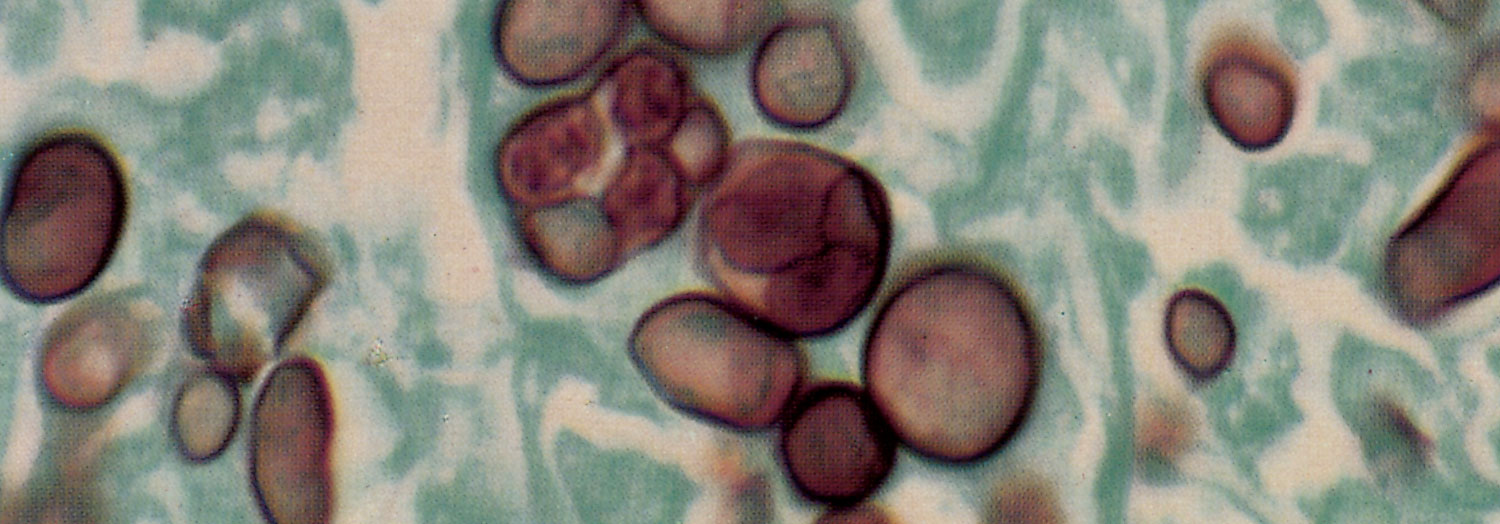
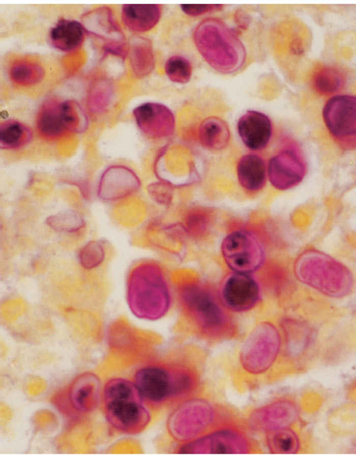
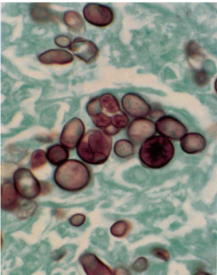
Macroscopically, affected bovine udder tissue is swollen and dense, with cords and nodules apparent on deep palpation.41 Glandular tissue appears a light pinkish-grey without the normal white cast of healthy tissue.41 Microscopically, changes present in affected lobules include diffuse infiltration of lymphocytes, epithelioid cells and neutrophils, and fibroblast cell proliferation in the interstitium. Granulomatous foci consisting of epithelioid cells and a few Langerhans’ giant cells which are encapsulated by fibrous tissue are present.30 The alveolar epithelium appears hyperplastic and vacuolated and is infiltrated by large numbers of neutrophils and laminated concretions. 30 In early lesions abundant Prototheca sp. organisms, 10 to 30 μm in diameter, containing 2 to 20 endospores (average two to eight) (Figure 211.1) are found mainly intracytoplasmically in macrophages and occasionally extracellularly within alveolar lumens.5, 11, 30 In chronic disease the infection is interstitial, with large numbers of algae-bearing macrophages sequestered between epithelial cells of the mammary acini and periacinar connective tissue.11 The cell wall of the endospores stain well with periodic acid-Schiff (PAS) and Gomori’s methenamine silver (GMS) stains (Figure 211.2), and is highly refractile when thick, unstained sections are viewed under polarized light.32
In skin lesions, typical protothecal sporangia may be surrounded by a mixed inflammatory cell infiltrate and necrotizing granulomas.71
The typical morphology of the algae helps to distinguish them histologically from pathogenic fungi such as Blastomyces dermatitidis, Cryptococcus neoformans, Coccidioides immitis, Paracoccidioides brasiliensis and Rhinosporidium seeberi, and other organisms such as Pneumocystis carinii.
Chlorella spp. can be distinguished from Prototheca spp. by the presence of a green colour in unfixed tissues, culture and the presence of cytoplasmic granules (chloroplasts) that stain positive with either PAS or GMS.10
Diagnosis
Protothecal infections should be considered when cows with chronic mastitis do not respond to routine antimicrobial therapy.1, 5, 28, 33, 58, 59 Appropriate samples for laboratory culture are milk from affected quarters, and tissue or fine needle aspirates from lesions. Should swabs be used, it is essential that they be placed in transport medium as it is believed that desiccation will deletariously affect the viability of the organism.40 If available, formalin-fixed mammary tissues or other affected tissue should be submitted for histopathology. A fluorescent antibody test can be used on tissue impression smears to distinguish the species involved.4, 64 More recently, an indirect enzyme-linked immunosorbent assay (ELISA) has been developed to identify not only infected cows but also infected cows at various stages of the clinical disease.58
Control
Protothecosis in humans, including those with acquired immunodeficiency syndrome (AIDS), has successfully been treated with amphotericin B in combination with tetracycline or ketoconazole, or with fluconazole.7, 9, 29, 36 Most cutaneous cases in animals have a good to fair prognosis, whereas systemic and mastitic infections tend to be refractory to treatment. Thus, therapy of protothecal mastitis is usually unsuccessful and not always desirable. Although the organism was not eliminated from infected mammary glands in one outbreak of mastitis in cows, the clinical signs improved in those treated by the intramammary administration of nystatin and dimethyl sulfoxide (DMSO).16 Cutaneous lesions in cats and dogs have been treated by excising the entire lesion and, where this was not possible, oral ketoconazole for four months or longer.22, 40
An important measure in the control of protothecal mastitis is the segregation and subsequent elimination of infected animals, as small numbers of algae are capable of causing infection.30, 37, 63 Furthermore, all objects or procedures that might give rise to teat trauma, such as milking machine malfunction, should be eliminated or modified.37 As for the control of many other pathogens causing mastitis, the implementation of proper sanitary measures in the dairy environment and equipment is essential to prevent this disease from occurring.37
Other algal diseases
Infections in livestock due to unicellular green (chlorophyllcontaining) algae have also been reported. Several cases have been noted in cattle and sheep,15, 35, 59 and a single case has been described in a dromedary camel (Camelus dromedarius).39 Single cases of subcutaneous infections have been reported in a Canadian beaver (Castor canadensis) 62 and a human.34 The responsible green algae are microscopically very similar to the chlorophyll-free algae that cause protothecosis. Their cultural morphology, however, indicates that they fall within the Chlorella spp. in the order Chloroccales, hence the clinical name, chlorellosis.
It has been maintained that Chlorella spp. produce green lesions in internal organs due to their ability to produce chlorophyll in the absence of light, a characteristic that is maintained when they are kept or grown in the dark.35 Enlarged, green lymph nodes (retropharyngeal, mandibular and rarely mediastinal) have been detected in cattle slaughtered in abattoirs.47, 59, 60 Similar disseminated infections due to Chlorella spp. have been described in sheep and lambs which manifested as green necrotic foci in the liver and hepatic lymph nodes.15, 35, 75 In the camel, a granulomatous enteritis affecting the distal ileum, caecum and proximal part of the colon as well as involvement of the mesenteric lymph nodes was noted.39 Contaminated water is thought to have been the source of the infection in all these cases.15, 39, 60, 75 The mouth was considered to be the possible portal of entry to the internal organs.15, 39, 60, 70
Lesions have been experimentally induced in Wistar rats by intraperitoneal inoculation of a Chlorella sp. They remained limited to the inoculation site. A mixed cellular inflammatory response was provoked which is in contrast to the reaction in cattle where an extensive proliferation of macrophages is the most striking feature.60
A presumptive diagnosis can be made when green coloured lesions are noted in unfixed specimens, endosporulating spherical or ellipsoidal organisms approximately 9 μmin diameter are present in the tissues, and large abundant, strongly PAS and GMS-positive granules are detected in the cytoplasm of individual endospores.10, 60 The chloroplasts are easily identifiable on transmission electron microscopy examination.39, 60 Chlorellosis is further confirmed or distinguished from protothecosis by culture. After 48 hours of incubation at room temperature, small round light to dark green pasty colonies are noted.75
References
- ANDERSON, K.L. & WALKER, R.L., 1988. Sources of Prototheca spp. in a dairy herd environment. Journal of the American Veterinary Medical Association, 193, 553–556.
- ARNOLD, P. & AHEARN, D.G., 1972. The systematics of the genus Prototheca with a description of a new species, P. filamenta. Mycologica, 64, 265–276.
- BAUMGARTNER, B., 1997. Vorkommen und Bekampfung der Prototheken mastitis des Rindes im Einzugsgebiet des Staatlichen Veterinar- und Lebensmitteluntersuchungsamtes Potsdam. Der Praktische Tierarzt, 78, 406, 412–414.
- BLOGG, J.R. & SYKES, J.E., 1995. Sudden blindness associated with protothecosis in a dog. Australian Veterinary Journal, 72, 147–149.
- BODENHOFF, J. & MADSEN, P.S., 1978. Bovine protothecosis: A brief report of ten cases. Acta Pathologica et Microbiologica Scandinavica, Section B, Microbiology, 86, 51–52.
- BOYD, A.S., LANGLEY, M. & KING, L.E., JR., 1995. Cutaneous manifestations of Prototheca infections. Journal of the American Academy of Dermatology, 32, 758–764.
- CAREY, W.P., KAYKOVA, Y., BANDRES, J.C., SIDHU, G.S. & BRAU, N., 1997. Cutaneous protothecosis in a patient with AIDS and a severe functional neutrophil defect: Successful therapy with amphotericin B. Clinical Infectious Diseases, 25, 1265–1266.
- CASAL, M., LINARES, M.J. & MORALES, M.M., 1985. Enzymatic profile of Prototheca species. Mycopathologia, 92, 81–82.
- CHAN, J.C., JEFFERS, L.J., GOULD, E.W., HUTSON, D., MARTINEZ, O.V., REDDY, K.R., HASSAN, F. & SCHIFF, E.R., 1990. Visceral protothecosis mimicking sclerosing cholangitis in an immunocompetent host: Successful antifungal therapy. Reviews of Infectious Diseases, 12, 802–807.
- CHANDLER, F.W., KAPLAN, W. & CALLAWAY, C.S., 1978. Differentiation between Prototheca and morphologically similar green algae in tissue. Archives of Pathology and Laboratory Medicine, 102, 353–356.
- CHEVILLE, N.F., MCDONALD, J. & RICHARD, J., 1984. Ultrastructure of Prototheca zopfii in bovine granulomatous mastitis. Veterinary Pathology, 21, 341–348.
- COLOE, P.J. & ALLISON, J.F., 1982. Protothecosis in a cat. Journal of the American Veterinary Medical Association, 180, 78–79.
- CONNOLE, M.D., 1990. Review of animal mycoses in Australia. Mycopathologica, 111, 133–164.
- COOK, J.R., TYLER, D.E., COULTER, D.B. & CHANDLER, F.W., 1984. Disemminated protothecosis causing acute blindness and deafness in a dog. Journal of the American Veterinary Medical Association, 184, 1266–1272.
- CORDY, D.R., 1973. Chlorellosis in a lamb. Veterinary Pathology, 10, 171–176.
- COSTA, E.O., CARCIOFI, A.C., MELVILLE, P.A., PRADA, M.S. & SCHALCH, U., 1996. Prototheca sp. outbreak of bovine mastitis. Journal of Veterinary Medicine, Series B, 43, 321–324.
- COSTA, E.O., MELVILLE, P.A., RIBEIRO, A.R., WATANABE, E.T. & PAROLARI, M.C.F.F., 1997. Epidemiologic study of environmental sources in a Prototheca zopfii outbreak of bovine mastitis. Mycopathologia, 137, 33–36.
- COSTA, E.O., RIBEIRO, A.R., WATANABE, E.T. & MELVILLE, P.A., 1998. Infectious bovine mastitis caused by environmental organisms. Journal of Veterinary Medicine, Series B, 45, 65–71.
- CRISPENS, C.G. & MARION, K.R., 1975. Algal infection in a corn snake (Elaphe guttata guttata). Laboratory Animal Science, 25, 788–789.
- DE CAMARGO, Z.P. & FISHMAN O., 1979. Prototheca stagnora, an encapsulated organism. Sabouraudia, 17, 197–200.
- DE VARGAS, A.C., LAZZARI, A., SANTURIO, J.M., ALVES, S.H., FERREIRA, G. & KREUTZ, L.C. 1998. Isolation of Prothoteca zopfii from a case of bovine mastitis in Brazil. Mycopathologia, 142, 135–137.
- DILLBERGER, J.E., HOMER, B., DAUBERT, D. & ALTMAN, N.H., 1988. Protothecosis in two cats. Journal of the American Veterinary Medical Association, 192, 1557–1559.
- DION, W.M., 1982. Bovine mastitis due to Prototheca zopfi. II. Canadian Veterinary Journal, 23, 272–275.
- FINNIE, J.W. & COLOE, P.J., 1981. Cutaneous protohecosis in a cat. Australian Veterinary Journal, 57, 307–308.
- FRESE, K. & GEDEK, B., 1968. Ein Fall von Protothecosis beim Reh. Berliner und Münchener Tierärztliche Wochenschrift, 81, 174–178.
- GENTLES, J.C. & BOND, P.M., 1977. Protothecosis of Atlantic salmon. Sabouraudia, 15: 2, 133–139.
- GINEL, P.J., PÉREZ, J., MOLLEDA, J.M., LUCENA, R. & MOZOS, E., 1997. Cutaneous protothecosis in a dog. The Veterinary Record, 140, 651–653.
- GIESECKE, W.H., NEL, E.E. & VAN DEN HEEVER, L.W., 1968. Blastomycotic mastitis in South Africa. Journal of the South African Veterinary Medical Association, 39, 69–70.
- HEITZMAN, H.B., BROOKS, T.J. & PHILLIPS, B.J., 1984. Protothecosis. Southern Medical Journal, 77, 1477–1478.
- HODGES, R.T., HOLLAND, J.T.S., NEILSON, F.J.A. & WALLACE, N.M., 1985. Prototheca zopfii mastitis in a herd of dairy cows. New Zealand Veterinary Journal, 33, 108–111.
- HOLLINGSWORTH, S.R., 2000. Canine protothecosis. The Veterinary Clinics of North America. Small Animal Practice, 30, 1091–1101.
- IMES, G.D., LLOYD, J.C. & BRIGHTMAN, M.P., 1977. Disseminated protothecosis in a dog. Onderstepoort Journal of Veterinary Research, 44, 1–6.
- JENSEN, H.E., AALBAEK, B., BLOCH, B. & HUDA, A., 1998. Bovine mammary protothecosis due to Prototheca zopfii. Medical Mycology, 36, 89–95.
- JONES, J.W., MCFADDEN, H.W., CHANDLER, F.W., KAPLAN, W. & CONNOR, D.H., 1983. Green algal infections in a human. American Journal of Clinical Pathology, 80, 102–107.
- KAPLAN, W., CHANDLER, F.W., CHOUDARY, C. & RAMACHANDRAN, P.K., 1983. Disseminated unicellular green algal infection in two sheep in India. American Journal of Tropical Medicine and Hygiene, 32, 405–411.
- KIM, S.T., SUH, K.S., CHAE, Y.S. & KIM, Y.J., 1996. Successful treatment with fluconazole of protothecosis developing at the site of an intralesional corticosteroid injection. British Journal of Dermatology, 135, 803–806.
- KIRK, J.H., 1991. Part 2. Diagnosis and treatment of difficult mastitis cases. Agri Practice, 12, 15–20.
- KWONG-CHUNG, K.J. & BENNETT, J.E., 1992. Protothecosis. Infections caused by algae. Medical Mycology, 29, 785.
- LE-NET, J.L., AHMED, M.F., SAINT-MARTIN, G., MASSON, M.T., MONTOIS, C., LONGEART, L. & FADL-AHMED, M., 1993. Granulomatous enteritis in a dromedary (Camelus dromedarius) due to green algal infection. Veterinary Pathology, 30, 370–373.
- MACARTNEY, L., RYCROFT, A.N. & HAMMIL, J., 1988. Cutaneous protothecosis in the dog: First confirmed case in Britain. The Veterinary Record, 123, 494–496.
- MCDONALD, J.S., RICHARD, J.L. & CHEVILLE, N.F., 1984. Natural and experimental bovine intramammary infection with Prototheca zopfii. American Journal of Veterinary Research, 45, 592–595.
- MEEHAN, C. & POLLOCK, J., 1996. Prototheca zopfii infection in a Cocker spaniel. Australiam Veterinary Practioner, 26, 146–147.
- MELVILLE, P.A., WATANABE, E.T., BENITES, N.R., RIBEIRO, A.R., SILVA, J.A., GARINO JUNIOR, F. & COSTA, E.O., 1999. Evaluation of the susceptibility of Prototheca zopfii to milk pasteurization. Mycopathologia, 146, 79–82.
- METTLER, F., 1975. Generalisierte Protothecose bei einem Flughund (Pteropus lylei). Veterinary Pathology, 12, 118–124.
- MEZGER, E., EISSES, J.F. & SMITH, M.J., 1981. Protothecal cellulitis in a renal transplant patient [abstract]. Laboratory Investigation, 44, 81A.
- MIGAKI, G., GARNER, F.M. & IMES, G.D., JR., 1969. Bovine protothecosis. A report of three cases. Pathological Veterinaria, 6, 444–453.
- MIGAKI, G., FONT, R.L., SAUER, R.M., KAPLAN, W. & MILLER, R.L., 1982. Canine protothecosis: Review of the literature and report of an additional case. Journal of the American Veterinary Medical Association, 181, 794–797.
- MUNDAY, B.L. & PEEL, B.F., 1983. Severe ulcerative dermatitis in platypus (Ornithorhynchus anatinus). Journal of Wildlife Diseases, 19, 363–365.
- PADHYE, A.A., BAKER, J.G. & D’AMATO, R.F., 1979. Rapid identification of Prototheca species by the API 20C system. Journal of Clinical Microbiology, 10, 579–582.
- PHAIR, J.P., WILLIAMS, J.E., BASSARIS, H.P., ZEISS, C.R. & MORLOCK, B.A., 1981. Phagocytosis and algicidal activity of human polymorphonuclear neutrophils against Prototheca wickerhamii. Journal of Infectious Diseases, 144, 72–76,
- POLK, P. & SAUNDERS, D.Y., 1997. Cutaneous protothecosis in association with the acquired immunodeficiency syndrome. Southern Medical Journal, 90, 831–832.
- PORE, R.S., 1998. Chapter 33. Prototheca and Chlorella. In: ajello, l. & hay, r.j., (eds). Topley & Wilson’s Microbiology and Microbial Infections. Vol. 4, 9th edn. London: Arnold LTD, Publications.
- PORE, R.S., BARNETT, E.A., BARNES, W.C., & WALKER, J.D., 1983. Prototheca ecology. Mycopathologica, 81, 49–62.
- PORE, R.S. & SHAHAN, T.A., 1988. Prototheca zopfii: Natural, transient, occurrence in pigs and rats. Mycopathologia, 101, 85–88.
- PORE, R.S., SHAHAN, T.A., PORE, M.D. & BLAUWIEKEL, R., 1987. Occurrence of Prototheca zopfii, a mastitis pathogen, in milk. Veterinary Microbiology. 15, 315–323.
- RAKICH, P.M. & LATIMER, K.S., 1984. Altered immune function in a dog with disseminated protothecosis. Journal of the American Veterinary Medical Association, 185, 681–683.
- RIPPON, J.W., 1988. Medical Mycology. The Pathogenic Fungi and Pathogenic Actinomycetes. 3rd edn. Philadelphia: W.B. Saunders Company. pp. 723–728.
- ROESLER, U., SCHOLZ, H. & HENSEL, A., 2001. Immunodiagnostic identification of dairy cows infected with Prototheca zopfii at various clinical stages and discrimination between infected and uninfected cows. Journal of Clinical Microbiology, 39, 539–543.
- ROGERS, R.J., 1974. Protothecal lymphadenitits in an ox. Australian Veterinary Journal, 50, 281–282.
- ROGERS, R.J., CONNOLE, M.D., NORTON, J., THOMAS, A., LADDS, P.W. & DICKSON, J., 1980. Lymphadenitis of cattle due to infection with green algae. Journal of Comparative Pathology, 90, 1–9.
- SHAHAN, T.A. & PORE, R.S., 1991. In vitro susceptibility of Prototheca spp. to gentamicin. Antimicrobial Agents and Chemotherapy, 35, 2434–2435.
- SILEO, L. & PALMER, N.C., 1973. Probable cutaneous protothecosis in a beaver. Journal of Wildlife Diseases, 9, 320–322.
- SPALTON, D.E., 1985. Bovine mastitis caused by Prototheca zopfii: A case study. The Veterinary Record, 116, 347–349.
- SUDMAN, M.S. & KAPLAN, W., 1973. Identification of Prototheca species by immunofluorescence. Applied Microbiology, 25, 981–990.
- SUDMAN, M.S., MAJKA, J.A. & KAPLAN, W., 1973. Primary mucocutaneous protothecosis in a dog. Journal of the American Veterinary Medical Association, 163, 1372–1374.
- TANIYAMA, H., OKAMOTO, F., KUROSAWA, T., FURUOKA, H., KAJI, Y., OKADA, H. & MATSUKAWA, K., 1994. Disseminated protothecosis caused by Prototheca zopfii in a cow. Veterinary Pathology, 31, 123–125.
- THOMAS J.B. & PRESTON, N., 1990. Generalized protothecosis in a Collie dog. Australian Veterinary Journal, 67, 25–27.
- VENEZIO, F.R., LAVOO, E., WILLIAMS, J.E., ZEISS, C.R., CARO, W.A., MANGKORNKANSK, M.M. & PHAIR, J.P., 1982. Progressive cutaneous protothecosis, American Journal of Clinical Pathology, 77, 485–493.
- VON MATSCHULLAT, G. & DAHLE, J., 1984. Protothekose bei einem Schaf. Der Praktische Tierartz, 4, 318, 320–321.
- VON METTLER, F., 1983. Algeninfektionen bei Mensch und Tier. Schweizer Archiv für Tierheilkunde, 125, 433–442.
- WALSH, S.V., JOHNSON, R.A. & TAHAN, S.R., 1998. Protothecosis: An unusual case of chronic subcutaneous and soft tissue infection. American Journal of Dermatology, 20, 379–382.
- WARREN, N.G. & HAZEN, K.C., 1995. Chapter 61. Candida, Cryptococcus, and other yeasts of medical importance. In: MURRAY, P.R., BARON, E.J., PFALLER, M.A., TENOVER, F.C. & YOLKEN, R.H., (eds) Manual of Clinical Microbiology. 6th edn. Washington, D.C.: American Society for Microbiology Press. pp. 723–737.
- WILKINSON, G.T. & LEONG, G., 1988. Protothecosis in a dog. Australian Veterinary Practioner, 18, 47–49.
- WIRTH, F.A., PASSALACQUA, J.A. & KAO, G., 1999. Disseminated cutaneous protothecosis in an immunocompromised host: A case report and literature review. Cutis, 63, 185–188.
- ZAKIA, A.M., OSHEIK, A.A. & HALIMA, M.O., 1989. Ovine chlorellosis in the Sudan. The Veterinary Record, 125, 625–626.


