- Infectious Diseases of Livestock
- Part 3
- GENERAL INTRODUCTION: SPIROCHAETES
- Swine dysentery
- Borrelia theileri infection
- Borrelia suilla infection
- Lyme disease in livestock
- Leptospirosis
- GENERAL INTRODUCTION: AEROBIC ⁄ MICRO-AEROPHILIC, MOTILE, HELICAL ⁄ VIBROID GRAM-NEGATIVE BACTERIA
- Genital campylobacteriosis in cattle
- Proliferative enteropathies of pigs
- Campylobacter jejuni infection
- GENERAL INTRODUCTION: GRAM-NEGATIVE AEROBIC OR CAPNOPHILIC RODS AND COCCI
- Moraxella spp. infections
- Bordetella bronchiseptica infections
- Pseudomonas spp. infections
- Glanders
- Melioidosis
- Brucella spp. infections
- Bovine brucellosis
- Brucella ovis infection
- Brucella melitensis infection
- Brucella suis infection
- Brucella infections in terrestrial wildlife
- GENERAL INTRODUCTION: FACULTATIVELY ANAEROBIC GRAM NEGATIVE RODS
- Klebsiella spp. infections
- Escherichia coli infections
- Salmonella spp. infections
- Bovine salmonellosis
- Ovine and caprine salmonellosis
- Porcine salmonellosis
- Equine salmonellosis
- Yersinia spp. infections
- Haemophilus and Histophilus spp. infections
- Haemophilus parasuis infection
- Histophilus somni disease complex in cattle
- Actinobacillus spp. infections
- Actinobacillus equuli infections
- Gram-negative pleomorphic infections: Actinobacillus seminis, Histophilus ovis and Histophilus somni
- Porcine pleuropneumonia
- Actinobacillus suis infections
- Pasteurella and Mannheimia spp. infections
- Pneumonic mannheimiosis and pasteurellosis of cattle
- Haemorrhagic septicaemia
- Pasteurellosis in sheep and goats
- Porcine pasteurellosis
- Progressive atrophic rhinitis
- GENERAL INTRODUCTION: ANAEROBIC GRAM-NEGATIVE, IRREGULAR RODS
- Fusobacterium necrophorum, Dichelobacter (Bacteroides) nodosus and Bacteroides spp. infections
- GENERAL INTRODUCTION: GRAM-POSITIVE COCCI
- Staphylococcus spp. infections
- Staphylococcus aureus infections
- Exudative epidermitis
- Other Staphylococcus spp. infections
- Streptococcus spp. infections
- Strangles
- Streptococcus suis infections
- Streptococcus porcinus infections
- Other Streptococcus spp. infections
- GENERAL INTRODUCTION: ENDOSPORE-FORMING GRAM-POSITIVE RODS AND COCCI
- Anthrax
- Clostridium perfringens group infections
- Clostridium perfringens type A infections
- Clostridium perfringens type B infections
- Clostridium perfringens type C infections
- Clostridium perfringens type D infections
- Malignant oedema⁄gas gangrene group of Clostridium spp.
- Clostridium chauvoei infections
- Clostridium novyi infections
- Clostridium septicum infections
- Other clostridial infections
- Tetanus
- Botulism
- GENERAL INTRODUCTION: REGULAR, NON-SPORING, GRAM-POSITIVE RODS
- Listeriosis
- Erysipelothrix rhusiopathiae infections
- GENERAL INTRODUCTION: IRREGULAR, NON-SPORING, GRAM-POSITIVE RODS
- Corynebacterium pseudotuberculosis infections
- Corynebacterium renale group infections
- Bolo disease
- Actinomyces bovis infections
- Trueperella pyogenes infections
- Actinobaculum suis infections
- Actinomyces hyovaginalis infections
- GENERAL INTRODUCTION: MYCOBACTERIA
- Tuberculosis
- Paratuberculosis
- GENERAL INTRODUCTION: ACTINOMYCETES
- Nocardiosis
- Rhodococcus equi infections
- Dermatophilosis
- GENERAL INTRODUCTION: MOLLICUTES
- Contagious bovine pleuropneumonia
- Contagious caprine pleuropneumonia
- Mycoplasmal pneumonia of pigs
- Mycoplasmal polyserositis and arthritis of pigs
- Mycoplasmal arthritis of pigs
- Bovine genital mycoplasmosis
- Neurotoxin-producing group of Clostridium spp.
- Contagious equine metritis
- Tyzzer's disease
- MYCOTIC AND ALGAL DISEASES: Mycoses
- MYCOTIC AND ALGAL DISEASES: Pneumocystosis
- MYCOTIC AND ALGAL DISEASES: Protothecosis and other algal diseases
- DISEASE COMPLEXES / UNKNOWN AETIOLOGY: Epivag
- DISEASE COMPLEXES / UNKNOWN AETIOLOGY: Ulcerative balanoposthitis and vulvovaginitis of sheep
- DISEASE COMPLEXES / UNKNOWN AETIOLOGY: Ill thrift
- Eperythrozoonosis
- Bovine haemobartonellosis
infections
This content is distributed under the following licence: Attribution-NonCommercial CC BY-NC  View Creative Commons Licence details here
Actinobacillus lignieresii infections
M M HENTON AND J J VAN DER LUGT
Introduction and aetiology
Actinobacillosis is caused by Actinobacillus lignieresii and is an ubiquitous, but usually sporadic, disease of cattle, sheep and goats. The disease is generally chronic and is characterized by pyogranulomatous inflammation, particularly of the skin and other soft tissues of the head and mouth, and of the regional lymph nodes. Infection in the tongue of cattle, a common site in this species, induces a marked fibroplastic response resulting in its common name, ‘wooden tongue’. The bacterium occasionally causes abscesses or granulomas in the teats and udders of sows.10 In horses, lower airway disease and abcesses have also been associated with A. lignieresii and related bacteria infections,9, 15 but these are rare.
The cultural, morphological and other characteristics of A. lignieresii are outlined in the introduction to Actinobacillus spp. infections.
Epidemiology, pathogenesis, clinical signs and pathology
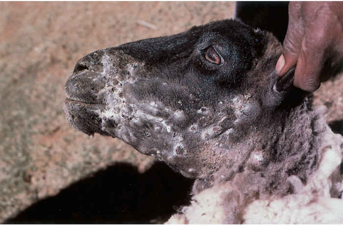
Actinobacillus lignieresii occurs as a commensal of the oral mucosae of ruminants in particular. Disease may follow injury to the mucous membranes in cattle, sheep and goats. Such injuries might be caused by coarse feeds, grass awns, or teeth abrasions or eruptions. Injuries to the skin, such as those inflicted by thorns and tick bites, may also lead to disease. In cattle the disease tends to occur sporadically, with only single cases being encountered, but in sheep it may occur as outbreaks involving several or many animals in a flock are involved. Because of the differences in prehension of feed between cattle and sheep (cattle use their tongues, while sheep use their lips and generally feed with their heads closer to the ground), the lesions in cattle occur most frequently in the tissues of the tongue. The site of entry of the organisms is often the lingual groove because of injuries to the tongue epithelium that have followed the entrapment there of sharp objects, such as the awns of grasses. Lesions in sheep, on the other hand, tend to occur initially in the cutaneous and subcutaneous tissues of the lips and cheeks, with multiple cases occurring in sheep as a result of the nature of the feed.13 In one outbreak, sheep that were fed the leaves of prickly pears (Opuntia sp.) developed lesions in the mouth, pharynx, and forestomachs.12
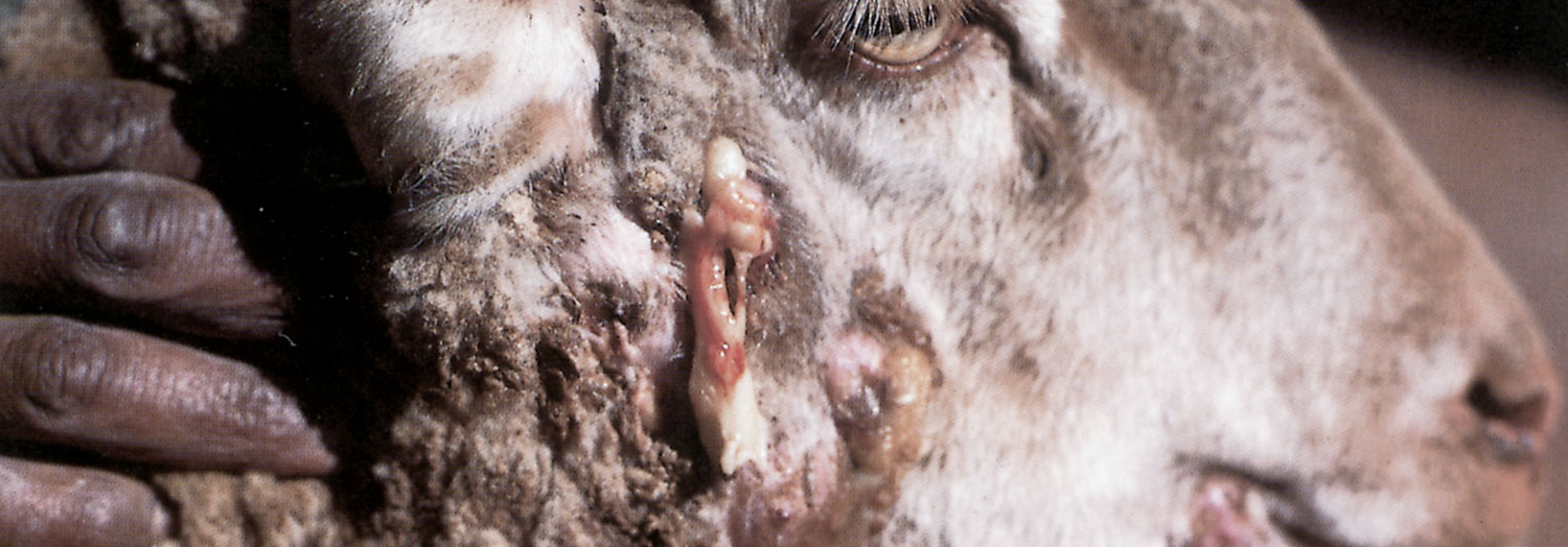
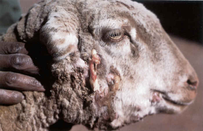
In cattle, sheep and goats, pyogranulomas caused by A. lignieresii most commonly in the soft tissues of the head and oral cavity and less commonly in other sites such as the forestomachs, lungs, uterus 5 and skin. Lesions spread by extension, as well as by the lymphatic system to regional lymph nodes which are frequently affected and become enlarged and firm. The lesions are intitially hard, circumscribed nodules, varying in size from a few to several millimetres in diameter. Some eventually become soft and fluctuate, and discharge their contents through the skin or mucous membrane, leaving deep ulcers or fistulae (Figure 158.1 and Figure 158.2). These lesions may expand and extend into surrounding tissues. When the tongue is involved, part of the organ becomes indurated, enlarged and immobile. This interferes with mastication and deglutition, and results in salivation and loss of weight. In some, the surface of the tongue becomes unevenly elevated. In severe cases the enlarged tongue may protrude from the mouth. Similarly, when the skin and subcutaneous tissues of the lips, cheeks and submandibular region of the head of sheep are affected, the thickened indurated lesions interfere progressively with prehension and mastication of feed, and animals gradually starve to death.
Abscesses and granulomas may occur in the teats and udders of sows as a result of small wounds inflicted by the sharp teeth of suckling pigs.
Other bacteria such as Trueperella (Corynebacterium) pyogenes, Streptococcus spp. and Pseudomonas aeruginosa are commonly found together with A. lignieresii in lesions, and it is likely that they gain entry to tissues in the same way.
The lesion is pyogranulomatous and contains barely visible colonies of the organism. In some lesions, or parts of lesions, the granulation tissue is hard and firm and somewhat resembles a fibroma,4 the colonies standing out as yellowish- white specks which are referred to as ‘sulphur granules’;in others the affected tissue has a nodular appearance and has more numerous granules dispersed in the fibromatous connective tissue. Lesions may coalesce to form large nodules which may be of a softer consistency and show evidence, on cut surface, of suppuration and consequently abscessation. The pus is thick, mucoid, greenish-yellow and odourless, and contains numerous granules which have the appearance of grains of sand. Ulcers and discharging fistulae and sinuses are sometimes formed when lesions break out into the lumen of hollow organs or to the externum. Frank abscessation does not often occur in tongue lesions.4 Affected lymph nodes may contain only a few nodules or they may be considerably enlarged and oedematous. In long- standing cases the nodes are fibrous and nodular, the nodules containing greenish-yellow pus and characteristic granules.4 Affected nodes, if superficially located, may adhere to the skin and some may form sinuses and discharge pus. Histologically, the sulphur granules consist of masses of cocco-bacilli which are embedded in a homogenous eosinophilic material that has a palisaded outer circumference consisting of radiating finger-like or club-shaped structures. The cocco-bacilli stain negatively by Gram’s method. Around the granules is a zone containing mainly neutrophils but also macrophages and a few giant cells. Lymphocytes and plasma cells occur peripherally in the encompassing fibrous connective tissue, which may be very dense in parts and replace normal tissue structures in the vicinity.
Diagnosis and differential diagnosis
Actinobacillus lignieresii may be difficult to culture from pus and only a few colonies may grow in primary cultures prepared from fresh lesions.8 If sulphur granules are present, the pus should be vigorously shaken in sterile physiological saline in a test tube in order to liquify it. This enables the granules to sink to the bottom of the tube, where they may be collected and utilized either for the preparation of smears or for culturing.
For smears, granules are placed on a glass slide, crushed by firm application of a cover-slip and examined microscopically, either directly or after staining. The peripheral club-shaped structures, which are thought to be comprised of immune complexes, may be seen in both stained and unstained preparations. Gram’s staining reveals the presence of the negatively staining cocco-bacilli in the centres of the granules, which are surrounded by radiating red-staining clubs.3, 11 Granules used for the preparation of bacteriological cultures should also be crushed prior to the inoculation of media.
Actinomycosis, caused by Actinomyces bovis, botryomycosis, caused by Staphylococcus aureus, and tuberculosis are the most important differential diagnoses to be considered. Granules resembling those of actinobacillosis may be encountered in the pus of both actinomycotic and botryomycotic lesions. Actinomyces bovis infection in cattle frequently causes a chronic purulent and proliferative inflammation of bony structures (particularly the mandible), the resultant disease being known as ‘lumpy jaw’. In addition, in contrast to actinobacillosis, the regional lymph nodes tend not to be affected in actinomycosis. The granules in actinomycosis are larger than those in actinobacillosis, in which they are generally less than 1 mm in diameter. They also tend to be harder in the former disease, their centres consisting of Gram- positive filamentous organisms. Botryomycosis is a chronic disease characterized by the formation of granulomas that contain suppurative foci.4 The essential feature is the presence in the pus of the so-called botryomycotic granules, which in histological sections are seen to consist of closely packed Gram-positive cocci embedded in a homogenous matrix, although the latter may not be discernible in younger granules. Numerous cocci are present when granules are placed on a glass slide, crushed with a cover-slip and then stained and examined.
The macroscopic lesions caused by A. lignieresii may also be confused with those caused by other pyogenic bacteria such as T. pyogenes, Corynebacterium pseudotuberculosis, Staphylococcus spp. and Streptococcus spp.
Control
Disease caused by A. lignieresii occurs sporadically and may be controlled to some extent by the application of sound managemental procedures, such as the timely and appropriate treatment of wounds and the minimization of injuries to the lips and oral mucosa.2, 6, 14 Cases or even outbreaks can be anticipated and early preventive measures and treatment adopted when animals are, for example, fed prickly pear (the thorns of which should be burned off with a blowtorch before feeding) or other material (such as barley) likely to injure soft tissues in and around the mouth.
Iodine is a satisfactory disinfectant. Treatment is more successful when applied during the initial stages of infection than in the chronic stage.1
Lesions that are well circumscribed and accessible can be surgically excised under local anaesthesia and the wound cavity packed with gauze tampons soaked in tincture of iodine. If surgical excision cannot be carried out, fistulae, if present, should be curetted and packed with gauze saturated with tincture of iodine. In some cases it may be advisable to make an incision into a lesion which is then irrigated with iodine. Lugol’s iodine can be injected directly into lesions.7
Prior to the advent of modern antimicrobial drugs, the oral administration of iodine preparations was widely recommended, whether surgical procedures had been performed or not.7 Potassium iodide, at a dosage level of 6 to 10 g/day, should be administered orally or in the drinking water for two to four weeks. During or shortly after this form of therapy an animal may develop signs of catarrh of the nose and eyes, dandruff, loss of hair or wool, cutaneous eruptions, and loss of appetite. These are indicative of iodism — the maximum systemic level of iodine that can be tolerated has been reached. The administration of iodine should then be suspended for about a week and then resumed if the full course of iodine has not been given.1 Potassium iodide should never be administered parenterally.
Actinobacillus lignieresii is usually sensitive to the sulphonamides or trimethoprim-sulphanomide combinations, ampicillin, streptomycin, tetracylines and chloramphenicol. Antibiotic treatment should be chosen with due regard to the sensitivity of the isolate. As abscesses and purulent foci are generally enclosed in a thick, fibrous tissue capsule, antimicrobial preparations may not be able to penetrate them to reach the causative organisms, and prolonged (longer than five days) therapy may be necessary to achieve this.16
References
- BLOOD, D.C., HENDERSON, J.A. & RADOSTITS, O.M., 1979. Veterinary Medicine, 5th edn. London: Bailiére Tindall.
- CAMPBELL, S.G., WHITLOCK, R.H., TOMONEY, J.F. & UNDERWOOD, A.M., 1975. An unusual epizootic of actinobacillosis in dairy heifers. Journal of the American Veterinary Medical Association, 166, 604–606.
- CARTER, G.R., 1984. Diagnostic Procedures in Veterinary Bacteriology and Mycology, 4th edn. Springfield: Charles C. Thomas. 122–125.
- DAVIES, G.O., 1947. Gaiger and Davies Veterinary Pathology and Bacteriology, 3rd edn. London: Baillière, Tindall.
- DE KUIF, A., MIJTEN, P., HAESEBROUCK, F., HOORENS, J. & DEVRIESE, L., 1992. Actinobacillosis in bovine caesarian sections. The Veterinary Record, 131, 414–415.
- HEBELER, H.F., LINTON, A.H. & OSBORNE, A.D., 1961. A typical actinobacillus in a dairy herd. The Veterinary Record, 73, 517–521.
- HENNING, M.W., 1956. Animal Diseases in South Africa, 3rd edn. South Africa: Central News Agency Ltd.
- PHILLIPS, J.E., 1984. Actinobacillus. In: KRIEG, N.R. & HOLT, J.G., (eds). Bergey’s Manual of Systematic Bacteriology, Vol. I. Baltimore, London: Williams and Wilkins.
- SAMITZ, E.M. & BIBERSTEIN, E.L., 1991. Actinobacillus suis-like organisms and evidence of haemolytic strains of Actinobacillus lignieresii in horses. American Journal of Veterinary Research, 52, 1245–1251.
- SANDERS, D.A. & RISTIC, M., 1956. Actinobacillus of cattle. Journal of the American Veterinary Medical Association, 129, 478–481.
- SMITH, H.A & JONES, T.C., 1966. Veterinary Pathology, Philadelphia: Lea and Febiger. 500–501.
- THOMAS, A.D., 1931. Actinobacillosis and other complications in sheep which may arise from the feeding of prickly pear (Opuntia sp.). Seventeenth Report of the Director of Veterinary Services and Animal Industry of the Union of South Africa, 215–229.
- TUSTIN, R.C., 1993. Faculty of Veterinary Science, University of Pretoria. Personal observations.
- WALKER, R.D., 1993. Actinobacillosis and actinomycosis. In: howard, j.l., (ed.). Current Veterinary Therapy 3: Food Animal Practice, Philadelphia: W.B. Saunders.
- WARD, C.L., WOOD, J.L., HOUGHTON, S.B., MUMFORD, J.A. & CHANTER, N., 1998. Actinobacillus and Pasteurella sp. isolated from horses with lower airway disease. The Veterinary Record, 143, 277–279.
- WILLSON, P.J., 1990. Haemophilus, Actinobacillus, Pasteurella: Mechanisms of resistance and antibiotic therapy. Canadian Journal of Veterinary Research, 54, S73–S77.


