- Infectious Diseases of Livestock
- Part 3
- Clostridium chauvoei infections
- GENERAL INTRODUCTION: SPIROCHAETES
- Swine dysentery
- Borrelia theileri infection
- Borrelia suilla infection
- Lyme disease in livestock
- Leptospirosis
- GENERAL INTRODUCTION: AEROBIC ⁄ MICRO-AEROPHILIC, MOTILE, HELICAL ⁄ VIBROID GRAM-NEGATIVE BACTERIA
- Genital campylobacteriosis in cattle
- Proliferative enteropathies of pigs
- Campylobacter jejuni infection
- GENERAL INTRODUCTION: GRAM-NEGATIVE AEROBIC OR CAPNOPHILIC RODS AND COCCI
- Moraxella spp. infections
- Bordetella bronchiseptica infections
- Pseudomonas spp. infections
- Glanders
- Melioidosis
- Brucella spp. infections
- Bovine brucellosis
- Brucella ovis infection
- Brucella melitensis infection
- Brucella suis infection
- Brucella infections in terrestrial wildlife
- GENERAL INTRODUCTION: FACULTATIVELY ANAEROBIC GRAM NEGATIVE RODS
- Klebsiella spp. infections
- Escherichia coli infections
- Salmonella spp. infections
- Bovine salmonellosis
- Ovine and caprine salmonellosis
- Porcine salmonellosis
- Equine salmonellosis
- Yersinia spp. infections
- Haemophilus and Histophilus spp. infections
- Haemophilus parasuis infection
- Histophilus somni disease complex in cattle
- Actinobacillus spp. infections
- Actinobacillus equuli infections
- Gram-negative pleomorphic infections: Actinobacillus seminis, Histophilus ovis and Histophilus somni
- Porcine pleuropneumonia
- Actinobacillus suis infections
- Pasteurella and Mannheimia spp. infections
- Pneumonic mannheimiosis and pasteurellosis of cattle
- Haemorrhagic septicaemia
- Pasteurellosis in sheep and goats
- Porcine pasteurellosis
- Progressive atrophic rhinitis
- GENERAL INTRODUCTION: ANAEROBIC GRAM-NEGATIVE, IRREGULAR RODS
- Fusobacterium necrophorum, Dichelobacter (Bacteroides) nodosus and Bacteroides spp. infections
- GENERAL INTRODUCTION: GRAM-POSITIVE COCCI
- Staphylococcus spp. infections
- Staphylococcus aureus infections
- Exudative epidermitis
- Other Staphylococcus spp. infections
- Streptococcus spp. infections
- Strangles
- Streptococcus suis infections
- Streptococcus porcinus infections
- Other Streptococcus spp. infections
- GENERAL INTRODUCTION: ENDOSPORE-FORMING GRAM-POSITIVE RODS AND COCCI
- Anthrax
- Clostridium perfringens group infections
- Clostridium perfringens type A infections
- Clostridium perfringens type B infections
- Clostridium perfringens type C infections
- Clostridium perfringens type D infections
- Malignant oedema⁄gas gangrene group of Clostridium spp.
- Clostridium chauvoei infections
- Clostridium novyi infections
- Clostridium septicum infections
- Other clostridial infections
- Tetanus
- Botulism
- GENERAL INTRODUCTION: REGULAR, NON-SPORING, GRAM-POSITIVE RODS
- Listeriosis
- Erysipelothrix rhusiopathiae infections
- GENERAL INTRODUCTION: IRREGULAR, NON-SPORING, GRAM-POSITIVE RODS
- Corynebacterium pseudotuberculosis infections
- Corynebacterium renale group infections
- Bolo disease
- Actinomyces bovis infections
- Trueperella pyogenes infections
- Actinobaculum suis infections
- Actinomyces hyovaginalis infections
- GENERAL INTRODUCTION: MYCOBACTERIA
- Tuberculosis
- Paratuberculosis
- GENERAL INTRODUCTION: ACTINOMYCETES
- Nocardiosis
- Rhodococcus equi infections
- Dermatophilosis
- GENERAL INTRODUCTION: MOLLICUTES
- Contagious bovine pleuropneumonia
- Contagious caprine pleuropneumonia
- Mycoplasmal pneumonia of pigs
- Mycoplasmal polyserositis and arthritis of pigs
- Mycoplasmal arthritis of pigs
- Bovine genital mycoplasmosis
- Neurotoxin-producing group of Clostridium spp.
- Contagious equine metritis
- Tyzzer's disease
- MYCOTIC AND ALGAL DISEASES: Mycoses
- MYCOTIC AND ALGAL DISEASES: Pneumocystosis
- MYCOTIC AND ALGAL DISEASES: Protothecosis and other algal diseases
- DISEASE COMPLEXES / UNKNOWN AETIOLOGY: Epivag
- DISEASE COMPLEXES / UNKNOWN AETIOLOGY: Ulcerative balanoposthitis and vulvovaginitis of sheep
- DISEASE COMPLEXES / UNKNOWN AETIOLOGY: Ill thrift
- Eperythrozoonosis
- Bovine haemobartonellosis
Clostridium chauvoei infections
This content is distributed under the following licence: Attribution-NonCommercial CC BY-NC  View Creative Commons Licence details here

Clostridium chauvoei infections
N P J KRIEK AND M W ODENDAAL
Introduction
Blackquarter in cattle arises as a consequence of the activation of spores of Clostridium chauvoei latent in the musculature. Clostridium chavoei may also cause gas gangrene in cattle, sheep, and goats, and rarely in horses and pigs following wound infection.21, 33, 36, 42 These infections cause a peracute or acute, usually fatal, non-contagious disease which is characterized by focal, gangrenous myositis and associated localized cellulitis. Death is caused by the local and systemic effects of the toxins elaborated by C. chauvoei. In this chapter the use of the term ‘blackquarter’ is restricted to that condition in cattle which arises as a result of endogenous C. chauvoei infection, whereas the lesions that develop as a consequence of wound infections by this organism in various animal species, including cattle, are referred to as gas gangrene.
Blackquarter is a universal disease of cattle. It was recognized as a distinct disease in 1782 by Chabert who named it ‘charbon symptomatique’ (quoted by Henning25) and distinguished it from anthrax with which it was often confused. Later the disease and the properties of the causal organisms were studied by Arloing, Cornevin and Thomas,2 who also developed the first practical method of prophylactic immunization. 37
It would appear that in South Africa blackquarter was one of the most prevalent diseases of cattle from the time of the earliest European settlement of the Cape. Thus, as early as 1780, Le Vaillant31 described the disease as a ‘terrible scourge, spons-siekte (literally ‘‘sponge disease’’), which causes speedy destruction of more than half the herd’. The first official reference to the occurrence of blackquarter in South Africa was made by Commissioner De Mist in 1805.48
Immunization against blackquarter has been practised since before the turn of the eighteenth century. As early as 1883 blackquarter powder vaccine was imported into Natal by Wiltshire. The first effective blackquarter vaccine in South Africa was prepared in 1887 at the Grahamstown Laboratory in the Cape Province.25
In spite of the effective vaccines available today, sporadic outbreaks and individual cases of the disease are still regularly encountered in livestock in southern Africa and blackquarter remains one of the important bacterial diseases of cattle under three years of age, particularly those in feedlots. For this reason cattle should be regularly immunized against this infection.
Aetiology
Clostridium chauvoei is a Gram-positive anaerobic rod, 3 to 8 μm long and 0,5 to 1 μm wide. When grown in fluid media the organisms are most commonly found as single cells, but sometimes occur in pairs and, rarely, in short chains. The cells are motile and have peritrichous flagella. In older cultures they are pleomorphic, showing irregular staining; citron, barred and spindle shapes occur frequently. Spores, which are are formed when the organism is cultured on solid media and in broth, are oval, occur in central or subterminal positions, and distort the shape of the cell. They are resistant to the effects of being boiled in water as well as to phenolic and quaternary disinfectants at concentrations used to sterilize contaminated instruments.5, 43
Clostridium chauvoei grows well in peptone-yeast-glucose broth, in which, after four days of incubation, the pH decreases to 5,0 to 5,4. The optimum temperature for growth is 37 °C; poor growth is obtained at between 25 and 30 °C, but no growth occurs at 45 °C. The addition of liver extract to the medium favours growth which is also stimulated by fermentable carbohydrates. Growth is inhibited by concentrations of NaCl above 6,5 per cent as well as by bile levels of 20 per cent or higher; pH values of 8,5 or higher also inhibit growth.
Surface colonies on blood agar are circular, 0,5 to 3 mm in diameter, haemolytic, slightly raised or low convex, whitish-grey, translucent or opaque, and granular with a glossy surface and an entire margin. Red blood cells of cattle, sheep, pigs, rabbits and dogs are readily haemolysed by C. chauvoei on blood agar, whilst those of guinea pigs, horses, humans, and chickens are more resistant.
Clostridium chauvoei has fastidious anaerobic growth requirements. It is saccharolytic, non-proteolytic and does not produce lecithinase and lipase. Lactose, galactose, mannose, glucose, maltose and sucrose are fermented, esculin is hydrolysed by 90 to 100 per cent of strains, and ribose by 40 to 60 per cent of strains, while nitrate is reduced by 61 to 89 per cent of strains. No starch is hydrolysed nor is acid produced from amygdalin, arabinose, cellobiose, fructose, glycogen, inositol, mannitol, melezitose, melibiose, raffinose, rhamnose, salicin, sorbitol, starch, trehalose and xylose. Meat is not digested by 90 to 100 per cent of strains.10, 35 Large volumes of gas (carbon dioxide and hydrogen) are produced in liquid media. The organism has a high demand for cysteine, biotin, nicotinic acid, pantothenic acid, pyridoxamine, thiamin and para-aminobenzoic acid.41
Clostridium chauvoei produces protein toxins and other protective antigens in amounts which vary with the strain. Both bacteria and the filtrate of fluid media in which they have been grown are immunogenic.13, 14, 46 Whole cultures of C. chauvoei are more immunogenic than washed bacterial cells alone. Immunization with formalinized whole cultures produces high levels of protective immunity which are probably induced by the heat-labile protective antigen that occurs in the cell wall of the organism.11, 12
The alpha toxin produced by the bacterium is lethal, necrotizing, and haemolytic, the beta toxin behaves as a deoxyribonuclease, the gamma toxin is a hyaluronidase, and the delta toxin is an oxygen-labile haemolysin. However, the role of these toxins in the pathogenesis of blackquarter is poorly defined.
The alpha toxin is formed as part of a soluble immunogenic complex from which it dissociates spontaneously, and has a molecular mass of 27 000 Daltons. Beta toxin is produced in an active form and is relatively heat-stable, being able to withstand exposure to a temperature of 95 °C for 10 minutes without appreciable loss of activity. The oxygenlabile haemolysin (delta toxin) is serologically related but not identical to streptolysin O, Clostridium perfringens theta toxin, tetanolysin and the haemolysin produced by some of the other clostridia.10, 16, 17, 41 Failure of some commercial C. chauvoei vaccines to protect cattle during outbreaks of blackquarter has led to the belief that cross-protection between different strains may not be absolute.51
Epidemiology
Blackquarter occurs throughout the world in association with livestock and is primarily a disease of cattle and to a lesser extent of sheep. Goats, pigs, horses, camels, deer and mink are only very rarely affected while birds, dogs, cats, rabbits and humans are resistant to infection with C. chauvoei.42
Outbreaks of blackquarter tend to be seasonal; they occur most commonly during summer and autumn, especially after heavy rains. There appears to be a direct relationship between the prevalence of blackquarter and rainfall;the higher the rainfall the greater the number of outbreaks during a season.3
Distribution of outbreaks is often regional and patchy, and its prevalence in some localities, which may be portions of one farm, may be high while adjoining areas are apparently free from infection.25 It appears that regional differences in the soil composition play a role in determining the survival of the bacteria in the areas of high prevalence.
The mode of infection in blackquarter has not yet been accurately established. It is generally believed that infection follows ingestion of spores from the soil or in contaminated drinking water, particularly during the period when the eruption of permanent teeth takes place. The ingested spores or those that have formed after vegetative bacteria have germinated and sporulated in the gut lumen, cross the intestinal epithelium and enter the blood and lymph streams to be distributed in tissues and organs throughout the body, including the musculature, in which they can remain dormant, probably in phagocytic cells, for prolonged periods.28, 41
Though the spores of C. chauvoei are highly resistant to environmental conditions and remain viable for a number of years outside the body, there is no experimental evidence that these organisms actually multiply in the soil; C. chauvoei apparently does not live as a saprophyte.32 The principle habitat of C. chauvoei is the animal body and it may be isolated from the intestinal contents. Contamination of vegetation and water by faeces containing the spores of the bacterium ensures animal-to-animal passage. Infected carcasses of animals that have died of the disease, however, appear to be the main source of the bacteria by contaminating the surroundings such as stables, sheds, kraals, pastures and drinking pools, which then become reservoirs of infection. Shearing sheds usually become contaminated by spores when animals that have died from blackquarter or gas gangrene caused by C. chauvoei are skinned in them. Once an area is contaminated, spores persist for years. Direct transmission from animal to animal does not occur.40, 41
In cattle, blackquarter is mainly confined to young stock between the ages of nine months and two years. Rapidly growing animals in good condition and on a high plane of nutrition are more susceptible to the disease than underfed animals or those in poor condition. Exercise, cold and heat do not appear to influence animals’ susceptibility to blackquarter. 41
Cattle, sheep, goats, horses and pigs of all ages may suffer from localized gas gangrene following the infection by C. chauvoei of wounds contracted, for example, during shearing, dipping, parturition, and castration or tail-docking operations, or of the umbilicus in new-born animals.
Wound infection of vaccination sites, particularly those induced by vaccines containing an irritant substance (e.g. saponin) may precipitate blackquarter within a matter of two hours. Goats are much more resistant than sheep to infection, and only isolated cases of gas gangrene have been recorded in pigs.7, 36, 42
Pathogenesis
The pathogenesis of C. chauvoei infections is essentially unknown. It is believed that blackquarter in cattle develops when latent spores within larger muscle groups germinate and multiply when these muscles are traumatized, resulting in localized areas of low redox potential. The vegetative cells grow, ferment muscle glycogen, digest protein, and produce exotoxins and gas. The alpha, beta, delta and gamma toxins of C. chauvoei are produced and released into the tissues, resulting in muscle necrosis.23 The lesions, which usually remain localized and in most animals are situated in one of the large muscle groups, expand along fascial planes but bacteraemia also develops, particularly terminally. Occasionally, latent spores in the myocardium are stimulated to germinate and multiply with the production of typical lesions in the myocardium; the stimulus to germinate possibly being the consequence of changes in the myocardium caused by increased blood levels of cortisol and catecholamines as a response to stress.19 The latter form of the disease that is sometimes referred to as the visceral form of blackquarter in contrast to the classical form that is much more common and easily diagnosed.24 The exotoxins and certain metabolites produced by the bacteria during their multiplication and those which arise from tissue damage, are absorbed from the affected muscles into the systemic circulation, causing toxaemia and death. Stevens45 reviewed the current hypothesis proposing the mechanisms of the pathogenesis of clostridial myonecrosis and the systemic disease caused by the toxaemia.
Clinical signs
As the course of blackquarter is so rapid, generally being less than 24 hours, clinical signs are not often observed in affected animals prior to death. Most affected cattle do not recover, but a small proportion of sheep and other animals that contracted the disease due to wound infection, may survive the infection.25, 36, 39, 42, 49
In cattle the first clinical signs are a rise in the body temperature, loss of appetite, ruminal stasis, and if the primary lesion is situated in one of the larger muscle groups of a limb, as is most often the case, there is initial stiffness followed by lameness which rapidly progresses in severity in the limb concerned. The muscles or muscle groups most commonly affected are those situated in the shoulder, buttock or loin regions, but those in the chest and neck regions and, more rarely, muscles such as the intrinsic and extrinsic muscles of the tongue, and the diaphragmatic and sublumbar muscles or myocardium may be primarily involved. The clinical signs, in the few cases in which they are evident, therefore vary according to the site of the lesion(s). If the muscles affected are superficial, there is subcutaneous swelling and crepitation at the site. The swelling increases rapidly in severity and size, and initially is hot and painful but terminally becomes cold and painless. Lesions situated in deeper body sites are not clinically detectable.
Rarely, lesions may develop simultaneously in more than one location. 39 Affected animals soon become recumbent, and manifest signs of dyspnoea and accelerated pulse rates. The clinical signs of visceral blackquarter are very similar to those of the classical form. Affected animals are depressed, reluctant to move, stiff and become recumbent.24
The clinical signs of localized gas gangrene that develop following wound infection by C. chauvoei are very similar to those of blackquarter and are also dependent on the site of the infection. In sheep and occasionally in cattle and goats, the vagina, cervix and the uterus may be infected following parturition. In such cases the perineal and adjacent tissues are severely swollen and emphysematous, and bloodstained droplets of serous fluid may ooze from the surface of the affected parts.42 In the case of contaminated injection sites, lesions are localized to the site of injection, and are oedematous and crepitant. This localized reaction expands rapidly, causing swelling of the tissues and the exudation of drops of blood-stained serous fluid through the skin, the exudate having a rancid smell. Affected animals may die within 18 hours of being injected with a contaminated needle.42 The course of the infection in pigs is usually less acute, the infection being localized.42
Localized gas gangrene as a consequence of wound infection with C. chauvoei rarely occurs in horses.21, 33, 36, 49 However, in cases in which this does occur, the infection may spread and involve large areas adjacent to the initial lesion. Affected horses either die without being seen to be ill, or are severely ill, in which case they manifest depression, signs of shock, and an unwillingness to move the affected part, if it is in a limb or the neck. The swelling, which is initially hot and painful, becomes cold, oedematous and crepitant. Most affected horses die but in those that do recover following intensive antibiotic treatment, the affected tissues may slough, leaving large cavities a few centrimetres deep which heal slowly.49
Pathology
Clinical pathological changes in animals suffering from blackquarter are not characteristic. Elevated levels of enzymes associated with muscle damage such as creatine kinase, may be detectable, while the results of haemograms are indicative of toxic shock.34, 36
The carcasses of animals that have died of blackquarter undergo rapid putrefaction and bloat, and blood-stained, frothy fluid may ooze from the nose and anus.6 When the lesions are localized in a hind limb, that limb may be extended at an awkward angle (Figure 181.1). The subcutaneous, intermuscular and other interstitial tissues in the vicinity of the affected musculature are distended by a yellow oedematous fluid which may be partially blood-stained and contain bubbles of gas. On cutting into an affected muscle, a reddish fluid exudes and there is a characteristic sweetish smell which resembles that of rancid butter.25
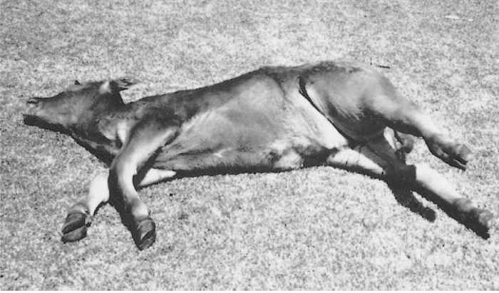
Figure 181.1 Note the swollen hind leg extended at an awkward angle due to blackquarter lesions in the large muscle groups
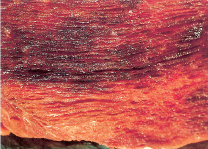
Lesions in muscles are characteristically well-circumscribed, spongy due to the accumulation of gas bubbles between muscle fibres, friable, and either uniformly dark-red or have alternating streaks varying in colour from a mottled greyish-red to pale yellowish or blackish (Figure 181.2). Affected muscles tend to be dryish towards the centre of the lesions. The spleen is usually not enlarged, but regional lymph nodes are hyperaemic and oedematous.25
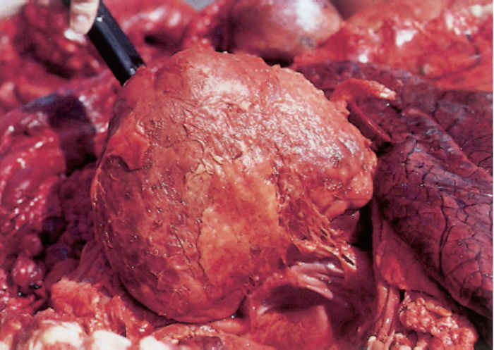
Localized crepitating lesions similar to those in the skeletal muscles are occasionally confined to the muscles of the tongue or diaphragm, or even the myocardium. In the latter case, the disease is known as visceral blackquarter or in South Africa, as ‘harsslag sponssiekte’ (literal translation from the Afrikaans is ‘heart-pluck blackquarter’). The myocardial lesions are often accompanied by a fibrinohaemorrhagic pericarditis (Figure 181.3), and occasionally by one or more mural thrombi in the chambers of the heart. A reddish serous fluid is often found in the thoracic and peritoneal cavities and the lungs are generally severely hyperaemic and oedematous. A localized serofibrinous pleuritis may occur adjacent to the lesions in the pericardium.24
Histologically the muscle lesions are characterized by extensive necrosis thought to be caused by both the effect of the toxins elaborated by the bacteria, and as a result of vascular involvement. Haemorrhage, the presence of gas bubbles and numerous clostridial organisms as well as the lack of an inflammatory response are also characteristic features of blackquarter.27
The lesions of gas gangrene in sheep following wound infection resemble those in cattle that have died of blackquarter. They may be associated with the presence of a severe fibrinous, fibrinopurulent or fibrinohaemorrhagic pericarditis in association with locally extensive areas of necrosis in the myocardium.19 In lambs that have died of the disease following infection by the umbilical route, there is marked congestion of the subcutaneous tissues in the umbilical region and the visceral organs. This is accompanied by petechiation and the presence of a fibrinous exudate on the surface of the liver.26
The lesions in horses that have died from gas gangrene caused by C. chauvoei are similar in nature and appearance to those in cattle.21 In one case in which no wound was detected, there was a large hepatic infarct, blood-stained peritoneal exudation, numerous fibrinous tags on the peritoneal serosa, hydrothorax and hydropericardium.33
Diagnosis
In typical cases of blackquarter a presumptive diagnosis can be made on the basis of the history, clinical signs and lesions at necropsy. It should be borne in mind that in some of the more ‘atypical’ cases of blackquarter characterized by muscle lesions in, for example, the tongue, crura of the diaphragm or sublumbar region, their presence may have to be actively sought during the necropsy. The aetiology of the disease can only be confirmed microbiologically or by the application of immunological staining techniques, which are important if immunoprophylaxis is considered.
From the exudate emanating from lesions, approximately six, thin, air-dried smears should be prepared on microscope glass slides for diagnostic purposes. One should be stained by Gram’s method and the others (keeping some in reserve) are used for fluorescent antibody testing in order to distinguish between C. chauvoei, C. septicum and C. novyi.4 Specimens of affected muscles as well as liver should be collected using aseptic techniques, placed separately in sterile plastic or glass jars and submitted at 4 °C to a laboratory for isolation of the organism. For microbiological processing, each specimen should be divided into two parts, one of which is heated to 80 °C for 15 to 30 minutes before a portion is plated out on blood tryptose agar medium and another portion of it inoculated into Robertson’s meat broth containing glucose. The second part should be inoculated into another tube containing the same meat broth medium without it being heated. Clostridium chauvoei has fastidious anaerobic requirements for its growth on blood agar. Selective media may be compiled by incorporating antibiotics such as neomycin, kanamycin, polymyxin, oleandomycin, or compounds such as sodium azide, crystal violet or sulphadiazine. 10 The isolation and subculturing of suspect single colonies are required to obtain pure cultures for use in biochemical tests. For these purposes a single colony is inoculated into either Robertson’s meat broth, liver broth or thioglycollate broth, each of which contains 5 per cent glucose and is incubated for 24 to 48 hours before the bacteria are subjected to biochemical testing. The determination of the fatty acid composition of the bacteria by gas chromatography can also be used to differentiate between the different clostridia.39 An indirect immunoenzymatic test, the peroxidase-antiperoxidase (PAP) technique was developed and used to detect C. chauvoei in tissue sections prepared from experimental cases in sheep.18
An identification system for C. chauvoei using PCR amplification of the 16S rRNA gene (rrs) with specific oligonucleotide primers and subsequent restriction digestion of the amplification product was developed by Kuhnert.37 It has advantages over the traditional isolation and identification methods including the immunofluorescence method and in vivo testing procedures in laboratory animals.
Differential diagnosis
Blackquarter may be difficult to distinguish clinically from other peracute or acute infectious diseases, particularly anthrax. Because most affected animals die suddenly and rapidly become bloated and putrified with blood-stained fluid oozing from natural orifaces, blackquarter is difficult to distinguish from anthrax. For this reason, when confronted with animals that have died acutely, it is advisable to examine suitably stained blood smears in order to exclude anthrax before proceeding with a necropsy. Lesions at necropsy are generally easily distinguished from those caused by most other infectious diseases except those due to some of the other clostridial infections (C. perfringens, C. septicum, C. novyi, C. sordellii and C. carnis) that cause gas gangrene or malignant oedema, and to snake venoms such as of puff adders (Bitis arietans arietans) and Gaboon adders (Bitis gabonica). In cases where other clostridial infections are concerned, the identification of the causative species by application of fluorescent antibody techniques is a prerequisite to the institution of control measures.50
Control
Only a small percentage of animals receiving specific chemotherapy for blackquarter, even if this is instituted early in the course of the disease, recover.22, 36, 49 Necrotic muscle may slough in animals that do recover. Animals treated in the advanced stages of the disease have no chance of recovery.
Systemic treatment with large doses of penicillin for five days in addition to accepted surgical methods of wound treatment may result in the recovery of animals suffering from gas gangrene; they do, however, take several weeks to recover fully as lesions that result from sloughing of the necrotic tissue take a long time to heal. In horses it may be necessary to administer penicillin at doses of approximately 44 000 IU/kg intravenously every two to four hours until the animal has stabilized (which may take from one to five days), whereafter they should be maintained on eighthourly penicillin injections at the same dosage level for up to five weeks. Supportive fluid therapy and analgesics should be administered in addition to the antibiotic treatment.36
Control measures for the prevention of blackquarter in cattle and gas gangrene due to C. chauvoei infection in other livestock species include the destruction of the carcasses of animals that have died of these conditions by incineration or deep burial. Contamination of wounds and the transmission of spores (or even vegetative forms of the organism) by the use of contaminated hypodermic needles and surgical instruments should be prevented. All susceptible animals should be vaccinated. Sheep that have died from C. chauvoei infection should not be skinned in order to salvage the price of the skin. However, if they are skinned, the skins should not be stored in or near shearing sheds.
The occurrence of blackquarter and wound infections caused by C. chauvoei can be adequately controlled by immunization although the calves of immune dams have colostrum-derived immunity which lasts for up to three months and interferes with active immunization.38
Calves should be vaccinated when three to six months of age. The initial course of immunization should consist of two subcutaneous innoculations administered at an interval of approximately four weeks, followed by annual boosters until the animals are three years old. On heavily infected farms on which blackquarter occurs regularly, the period between booster vaccinations may have to be reduced to nine months.15 The vaccine should only be inoculated subcutaneously, and particularly when vaccine is administered during an actual outbreak of blackquarter, a separate, clean, sterile needle should be used to inoculate each animal to prevent transmission of the bacteria between them. Several combinations of clostridial antigens in vaccines are available. 8, 9, 29, 44 Such polyvalent vaccines may include one or more antigens of the following organisms: C. chauvoei, C. botulinum, C. tetani, C. septicum, C. novyi, C. sordellii, C. haemolyticum, and C. perfringens types C and D.
Although there are distinct advantages in using multivalent vaccines, their application may elicit a severe inflammatory response that leads to a decrease in feed consumption and the development of subcutaneous lesions detectable at slaughter.1, 47
On farms on which problems with C. chauvoei infections in sheep are consistently encountered, the animals should be vaccinated routinely, the time of administration being determined by the form of the disease likely to be encountered. In the case of pregnant ewes, those not previously vaccinated should be vaccinated twice at an interval of a month, the second inoculation being given at least one month before lambing; thereafter a booster inoculation given one month prior to lambing is sufficient to protect them from the genital disease. Not only will ewes vaccinated four weeks before parturition be protected from possible infection acquired by contamination of obstetric wounds, but their lambs will be protected by virtue of the passive immunity against infections of the umbilicus as well as wounds resulting from tail-docking and castration.20 If C. chauvoei gas gangrene following contaminated shearing wounds is anticipated, the booster vaccinations should be administered six to eight weeks before shearing. In exceptional instances sheep may for unknown reasons develop a shock reaction after administration of vaccine.15 It is therefore advisable to inoculate about 10 animals in a flock and then to observe them for clinical signs of shock for a period of 30 minutes. If no evidence of shock arises, the remaining animals may be injected with reasonable impunity. The same vaccines (mono- and polyvalent combinations) as for cattle can be used at the prescribed dosage levels to vaccinate sheep.
References
- APLEY, M., WRAY, M. & ARMSTRONG, D., 1994. Subcutaneous injection site comparison of two multivalent clostridial bacterin/toxoids in feedlot cattle. Agri-Practice, 15, 9–12.
- ARLOING, CORNEVIN, & THOMAS, 1887. Le Charbon Symptomatique de Boeuf, 2nd edn. Paris.
- BAGADI, H.O., 1978. The relationship between the annual rainfall and outbreaks of blackquarter of cattle in northern Nigeria. Tropical Animal Health and Production, 10, 124–126.
- BATTY, I. & WALKER, P.D., 1963. Differentiation of Clostridium septicum and Clostridium chauvoei by the use of fluorescent labelled antibodies. Journal of Pathology and Bacteriology, 85, 517–521.
- BISPING, W. & AMTSBERG, G., 1988. Colour Atlas for the Diagnosis of Bacterial Pathogens in Animals. Berlin & Hamburg: Paul Parey Scientific Publishers.
- BREWITT, J.M., 1966. A practitioner’s investigation of a possible outbreak of blackleg. Canadian Veterinary Journal, 7, 231–232.
- BROWN, C.M., KANEENE, J.B. & WALKER, R.D., 1988. Intramuscular injection techniques and the development of clostridial myositis or cellulitis in horses. Journal of the American Veterinary Medical Association, 193, 668–670.
- BROWN, K.K., PARIZEK, R.E. & STEWART, R.C., 1976. Prevention of clostridial disease in cattle and sheep by vaccination with a multivalent bacterin-toxoid. Veterinary Medicine/Small Animal Clinician, 71, 1717–1721.
- CAMERON, C.M., BOTHA, W.J.S. & SCHOEMAN, H., 1986. Immunization of guinea pigs and cattle with a reduced dose Clostridium chauvoei vaccine produced in a semi-synthetic medium. Onderstepoort Journal of Veterinary Research, 53, 51–53.
- CATO, E.P., GEORGE, W.L. & FINEGOLD, S.M., 1986. Genus Clostridium Prazmowski 1880. In: sneath, p.h.a., mair, n.s., sharpe, m.e. & holt, j.g., (eds). Bergey’s Manual of Systematic Bacteriology. Vol. II. Baltimore & London: Williams & Wilkens.
- CHANDLER, H.M. & GULASEKHARAM, J., 1970. An evaluation of characteristics of Clostridium chauvoei which possibly indicate a highly protective strain. Australian Journal of Experimental Medicine and Science, 48, 187–197.
- CHANDLER, H.M. & HAMILTON, R.C., 1975. The protective antigenicity of protoplasts and spheroplasts of a highly protective strain of Clostridium chauvoei. Journal of General Microbiology, 88, 179–183.
- CLAUS, K.D. & MACHEAK, M.E., 1972. Preparation of a C. chauvoei antigen and determination of protective immunity by plate agglutination test. American Journal of Veterinary Research, 33, 1045–1052.
- CLAUS, K.D. & MACHEAK, M.E., 1972b. Characteristics and immunizing properties of culture filtrates of Clostridium chauvoei. American Journal of Veterinary Research, 33, 1031–1038.
- ERASMUS, B.J., CAMERON, C.M., HUNTER, P., CILLIERS, J.A., OBEREM, P.T., STOLTSZ, W.H. & DE WAAL, D.T., 1990. Onderstepoort Vaccines. Booklet issued by the Department of Agriculture and Development, Private Bag X144, Pretoria 0001.
- FOEGEDING, P.M., 1988. Detection and quantitation of sporeforming pathogens and their toxins. In: pierson, m.d. & stern, n.j., (eds). Foodborne Microorganisms and their Toxins: Developing Methodology, New York & Basel: Marcel Dekker, Inc.
- GILL, M., 1987. Bacterial toxins. In: laskin, a.i. & lechevalier, h.a., (eds). CRC Handbook of Microbiology, 2nd edn. Vol. VIII. Toxins and Enzymes. Florida: CRC Press, Inc.
- GIRANDO CONTESA, L.C., VANELLI, S.A. & UZAL, F.A., 1995. Detection of Clostridium chauvoei in formaling-fixed, paraffin-embedded tissues of sheep by the peroxidase-antiperoxidase (PAP) technique. Veterinary Research Communications, 19, 451–456.
- GLASTONBURY, J.R.W., SEARSON, J.E., LINKS, I.J. & TUCKETT, L.M., 1988. Clostridial myocarditis in lambs. Australian Veterinary Journal, 65, 208–209.
- GREEN, D.S., GREEN, M.J., HILLYER, M.H. & MORGAN, K.L., 1987. Injection site reactions and antibody responses in sheep and goats after the use of multivalent clostridial vaccines. The Veterinary Record, 120, 435–439.
- HAGEMOSER, W.A., HOFFMAN, L.J. & LUNDVALL, R.L., 1980. Clostridium chauvoei infection in a horse. Journal of the American Veterinary Medical Association, 176, 631–633.
- HALL, K.E., 1989. Treatment of a calf with Clostridium chauvoei infection. Journal of the American Veterinary Medical Association, 194, 272.
- HATHAWAY, C.L., 1990. Toxigenic Clostridia. Journal of Clinical Microbiology, 3, 66–98.
- HELMAN, G., WELSH, R.D., STAIR, E.L. & ELY, R.W., 1997. Diagnosing visceral blackleg as a cause of sudden death in cattle. Veterinary Medicine, October, 914–918.
- HENNING, M.W., 1956. Animal Diseases in South Africa, 3rd edn. South Africa: Central News Agency.
- HUGHES, K.L., HAUGHEY, K.G. & HARTLEY, W.J., 1971. Perinatal lamb mortality: Infections occurring among lambs dying after parturition. Australian Veterinary Journal, 47, 472–476.
- HULLAND, T.J., 1993. Muscle and tendons. In: jubb, k.v.f., kennedy, p.c. & palmer, n., (eds). Pathology of Domestic Animals, 4th edn. San Diego: Academic Press.
- KERRY, J.B., 1964. A note on the occurrence of Clostridium chauvoei in the livers and spleens of normal cattle. The Veterinary Record, 76, 396.
- KNOTT, G.K.L., ERWIN, B.G. & CLASSICK, L.G., 1985. Benefits of a clostridial vaccination program in feedlot cattle. Veterinary Medicine, June, 95–97.
- KUHNERT, P., KRAMPE, M., CAPAUL, S.E., FREY, J. & NICOLET, J., 1997. Identification of Clostridium chauvoei in cultures and clinical material from blackleg using PCR. Veterinary Microbiology, 51, 291–298.
- LE VALLIANT, M., 1796. Travels into the Interior Parts of Africa. Vol. II.
- MINETT, F.C. & DHANDA, M.R., 1941. Multiplication of B. anthracis and Cl. chauvoei in soil and water. Indian Journal of Veterinary Science and Animal Husbandry, 11, 308–321.
- MURPHY, D.B., 1980. Clostridium chauvoei as the cause of malignant edema in a horse. Veterinary Medicine/Small Animal Clinician, 75, 1152–1154.
- PEMBERTON, J.R., BATES, F., MATSON, R., MACHEAK, M.E. & HIGBE, J., 1974. Changes in clinical values of cattle infected with Clostridium chauvoei: Clinical relationships during infection. American Journal of Veterinary Research, 35, 1041–1044.
- PHILLIPS, K.D., BRAZIER, J.S., LEVETT, P.N. & WILLIS, A.T., 1985. Clostridia. In: collins, c.h. & grange, j.m., (eds). Isolation and Identification of Micro-organisms of Medical and Veterinary Importance, London: Academic Press.
- REBHUN, W.C., SHIN, S.J., KING, J.M., BAUM, K.H. & PATTEN, V., 1985. Malignant edema in horses. Journal of the American Veterinary Medical Association, 187, 732–735.
- ROBERTSON, M., 1929. The organisms associated with gas gangrene. A System of Bacteriology, M.R.C., 3, 225–297.
- SCHIPPER, I.A., KELLING, C.L., MAYER, J. & PFEIFFER, N.W., 1978. Effects of passive immunity on immune response in calves vaccinated against Clostridium chauvoei infection (blackleg). Veterinary Medicine/Small Animal Clinician, December, 73, 1564–1566.
- SIPPEL, W.L., 1972. Diagnosis of clostridial diseases. Journal of the American Veterinary Medical Association, 161, 1299–1305.
- SMITH, L.D.S., 1975. The Pathogenic Anaerobic Bacteria, 2nd edn. Springfield, Illinois: Charles C. Thomas.
- SMITH, L.D.S. & WILLIAMS, B.L., 1984. The Pathogenic Anaerobic Bacteria, 3rd edn. Springfield, Illinois: Charles C. Thomas.
- STERNE, M., 1981. Clostridial infections. British Veterinary Journal, 137, 443–454.
- STERNE, M. & BATTY, I., 1975. Pathogenic Clostridia, Boston & London: Butterworths.
- STERNE, M., BATTY, I., THOMPSON, A. & ROBERTSON, J.M., 1962. Immunisation of sheep with multi-component clostridial vaccines. The Veterinary Record, 74, 909–913.
- STEVENS, D.L., 2000. The pathogenesis of clostridial myositis. International Journal of Medical Microbiology, 290, 497–502.
- STEVENSON, J.R. & STONGER, K.A., 1980. Protective cellular antigen of Clostridium chauvoei. American Journal of Veterinary Research, 41, 650–652.
- STOKKA, G.L., EDWARDS, A.J., SPIRE, M.F., BRANDT, R.T. & SMITH, J.E., 1994. Inflammatory response to clostridial vaccines in feedlot cattle. Journal of the American Veterinary Medical Association, 204, 415–419.
- THEAL, G.M., 1911. Belangryke Historische Dokumente over Zuid-Afrika. Reisen van Kommissaris de Mist.
- WESTMAN, C.W., TRAUB, J.L. & SCHROEDER, W.G., 1979. Clostridial infection in a horse. Journal of the American Veterinary Medical Association, 174, 725–726.
- WILLIAMS, B.M., 1977. Clostridial myositis in cattle: Bacteriology and gross pathology. The Veterinary Record, 100, 90–101.
- WOOLCOCK, J.B. & FROST, A.J., 1978. Failure of Clostridium chauvoei vaccines to protect against blackleg. Australian Veterinary Journal, 54, 319.


