- Infectious Diseases of Livestock
- Part 3
- Contagious caprine pleuropneumonia
- GENERAL INTRODUCTION: SPIROCHAETES
- Swine dysentery
- Borrelia theileri infection
- Borrelia suilla infection
- Lyme disease in livestock
- Leptospirosis
- GENERAL INTRODUCTION: AEROBIC ⁄ MICRO-AEROPHILIC, MOTILE, HELICAL ⁄ VIBROID GRAM-NEGATIVE BACTERIA
- Genital campylobacteriosis in cattle
- Proliferative enteropathies of pigs
- Campylobacter jejuni infection
- GENERAL INTRODUCTION: GRAM-NEGATIVE AEROBIC OR CAPNOPHILIC RODS AND COCCI
- Moraxella spp. infections
- Bordetella bronchiseptica infections
- Pseudomonas spp. infections
- Glanders
- Melioidosis
- Brucella spp. infections
- Bovine brucellosis
- Brucella ovis infection
- Brucella melitensis infection
- Brucella suis infection
- Brucella infections in terrestrial wildlife
- GENERAL INTRODUCTION: FACULTATIVELY ANAEROBIC GRAM NEGATIVE RODS
- Klebsiella spp. infections
- Escherichia coli infections
- Salmonella spp. infections
- Bovine salmonellosis
- Ovine and caprine salmonellosis
- Porcine salmonellosis
- Equine salmonellosis
- Yersinia spp. infections
- Haemophilus and Histophilus spp. infections
- Haemophilus parasuis infection
- Histophilus somni disease complex in cattle
- Actinobacillus spp. infections
- Actinobacillus equuli infections
- Gram-negative pleomorphic infections: Actinobacillus seminis, Histophilus ovis and Histophilus somni
- Porcine pleuropneumonia
- Actinobacillus suis infections
- Pasteurella and Mannheimia spp. infections
- Pneumonic mannheimiosis and pasteurellosis of cattle
- Haemorrhagic septicaemia
- Pasteurellosis in sheep and goats
- Porcine pasteurellosis
- Progressive atrophic rhinitis
- GENERAL INTRODUCTION: ANAEROBIC GRAM-NEGATIVE, IRREGULAR RODS
- Fusobacterium necrophorum, Dichelobacter (Bacteroides) nodosus and Bacteroides spp. infections
- GENERAL INTRODUCTION: GRAM-POSITIVE COCCI
- Staphylococcus spp. infections
- Staphylococcus aureus infections
- Exudative epidermitis
- Other Staphylococcus spp. infections
- Streptococcus spp. infections
- Strangles
- Streptococcus suis infections
- Streptococcus porcinus infections
- Other Streptococcus spp. infections
- GENERAL INTRODUCTION: ENDOSPORE-FORMING GRAM-POSITIVE RODS AND COCCI
- Anthrax
- Clostridium perfringens group infections
- Clostridium perfringens type A infections
- Clostridium perfringens type B infections
- Clostridium perfringens type C infections
- Clostridium perfringens type D infections
- Malignant oedema⁄gas gangrene group of Clostridium spp.
- Clostridium chauvoei infections
- Clostridium novyi infections
- Clostridium septicum infections
- Other clostridial infections
- Tetanus
- Botulism
- GENERAL INTRODUCTION: REGULAR, NON-SPORING, GRAM-POSITIVE RODS
- Listeriosis
- Erysipelothrix rhusiopathiae infections
- GENERAL INTRODUCTION: IRREGULAR, NON-SPORING, GRAM-POSITIVE RODS
- Corynebacterium pseudotuberculosis infections
- Corynebacterium renale group infections
- Bolo disease
- Actinomyces bovis infections
- Trueperella pyogenes infections
- Actinobaculum suis infections
- Actinomyces hyovaginalis infections
- GENERAL INTRODUCTION: MYCOBACTERIA
- Tuberculosis
- Paratuberculosis
- GENERAL INTRODUCTION: ACTINOMYCETES
- Nocardiosis
- Rhodococcus equi infections
- Dermatophilosis
- GENERAL INTRODUCTION: MOLLICUTES
- Contagious bovine pleuropneumonia
- Contagious caprine pleuropneumonia
- Mycoplasmal pneumonia of pigs
- Mycoplasmal polyserositis and arthritis of pigs
- Mycoplasmal arthritis of pigs
- Bovine genital mycoplasmosis
- Neurotoxin-producing group of Clostridium spp.
- Contagious equine metritis
- Tyzzer's disease
- MYCOTIC AND ALGAL DISEASES: Mycoses
- MYCOTIC AND ALGAL DISEASES: Pneumocystosis
- MYCOTIC AND ALGAL DISEASES: Protothecosis and other algal diseases
- DISEASE COMPLEXES / UNKNOWN AETIOLOGY: Epivag
- DISEASE COMPLEXES / UNKNOWN AETIOLOGY: Ulcerative balanoposthitis and vulvovaginitis of sheep
- DISEASE COMPLEXES / UNKNOWN AETIOLOGY: Ill thrift
- Eperythrozoonosis
- Bovine haemobartonellosis
Contagious caprine pleuropneumonia
This content is distributed under the following licence: Attribution-NonCommercial CC BY-NC  View Creative Commons Licence details here

Contagious caprine pleuropneumonia
Previous authors: P-C LEFÈVRE AND F THIAUCOURT
Current author:
F T H THIAUCOURT - OIE Expert and Head of CIRAD CBPP Reference Laboratory, Veterinarian, PhD, HDR, TA A117 Campus de Baillarguet, Montpellier, Occitanie, 34398, France
![]()
Introduction
Contagious caprine pleuropneumonia (CCPP) is one of the most severe diseases of goats. It presents as an acute, highly contagious disease characterized by fever, coughing, severe respiratory distress and high mortality, and is caused by Mycoplasma capricolum subsp. capripneumoniae (Mccp). It has also been described in certain wild ruminants.
The history of the disease is rather complex since there are several mycoplasmas that may produce pneumonia or pleuropneumonia in goats, and the causative agent was only isolated and characterized in 1976. Due to the fastidiousness of the agent and its antigenic relations with other mycoplasmas, it took many years before CCPP was recognized as a specific disease entity. Its history should therefore be reconsidered in the light of our present knowledge. The past literature on the disease was comprehensively reviewed by Thiaucourt in 1994.68
In 1873, in Algeria, a French military veterinary surgeon, Philippe Thomas, described a disease called Bou Frida with the characteristic clinical signs and lesions of what is now known as CCPP. This description seems to be the first one.73 A few years later in 1881 in South Africa, Duncan Hutcheon reported a disease in goats imported from Turkey that had spread to local goats and which appeared to be CCPP.22 He carried out experiments that gave valuable information on the length of the incubation period and the epidemiology of the disease.22, 23
From the beginning of the twentieth century, there were many reports of outbreaks of CCPP from different parts of the world, mainly the Mediterranean area or Africa, and attempts to reproduce it and isolate the causative organism were carried out. The latter, however, led to a growing confusion since several diseases caused by various Mycoplasma spp. were involved.5, 31, 37, 39, 49, 50, 55
It was only in 1976 that Macowan and Minette41 isolated a mycoplasma strain in Kenya that was called F38 and that was different in its growth inhibition test from the strains previously isolated from caprine pleuropneumonia, such as Mycoplasma mycoides subsp. capri or M. capricolum. Experimental reproduction of the disease, in which the clinical signs and lesions observed in natural cases were manifested, was obtained by inoculating pure cultures of this strain into goats, and F38-like strains were subsequently isolated from such experimentally induced cases.21, 42, 59 Since then CCPP has been considered a specific disease entity and this has been confirmed by the application of Koch postulates.18 In 1993 the F38 strain was given a definitive name.33
Aetiology
It has been established that the sole agent responsible for CCPP is Mycoplasma capricolum subsp. capripneumoniae (Mccp). Like all mycoplasmas, it is a small pleomorphic micro-organism lacking a cell wall. It shares serological and genetic properties with five Mycoplasma spp. that are pathogens of ruminants. It is included in the so-called Mycoides cluster (Table 1).7
Table 1 Evolution of the Mycoplasma mycoides cluster taxonomy from 198713 to 200948
| Mycoplasma mycoides cluster | ||||||
| mycoides sub-cluster | capricolum sub-cluster | |||||
| 1987 taxonomy Cottew et al. 198713 | M. mycoides subsp. capri (Mmc) | M. mycoides subsp. mycoides LC (MmmLC) | M. mycoides subsp. mycoides SC (MmmSC) | M. sp. Gr7 Leach (MGr7) | M. capricolum subsp. capricolum (Mcc) | M. capricolum subsp. capripneumoniae (Mccp) |
| 2017 taxonomy Manso-Silvan et al. 200948 | M. mycoides subsp. capri (Mmc) | M. mycoides subsp. mycoides (Mmm) | M. leachii (Ml) | M. capricolum subsp. capricolum (Mcc) | M. capricolum subsp. capripneumoniae (Mccp) | |
| Disease | MAKEPS* Contagious agalaxia | CBPP | MAKEPS* | MAKEPS* Contagious agalaxia | CCPP | |
| Main host | Goats | Cattle | Cattle | Goats | Goats | |
*MAKEPS: Mastitis, Arthritis, Keratoconjunctivitis, Pneumonia, Septicaemia
In vitro, Mccp is a slow-growing mycoplasma compared to the other species, and it does not hydrolyse arginine, while M. capricolum does. In addition, the use of substrates by the oxydative route, such as pyruvate, is typical of Mccp. 1 Molecular studies permit the F38-type strains to be classified as a subspecies of M. capricolum. A comparison of the genomic relationships between M. capricolum strains and F38 strains resulted in intra-group DNA-DNA relatedness values of 85 to 90 per cent, while the relatedness was only 70 per cent between the two groups. The M. capricolum species, therefore, had to be divided into two new subspecies.33
Mycoplasma capricolum subsp. capripneumoniae is a very vulnerable micro-organism and is not able to survive for long in the external environment.
Epidemiology
Mycoplasma capricolum subsp. capripneumoniae has been isolated in Kenya,41 Ethiopia,70 Eritrea, Sudan,19 Uganda,9 Chad,34 and Niger, but there is serological evidence of its wider presence in West Africa, for example in Mali.62 It has also been isolated in Turkey in the Middle-East74 and Oman24 and the United Arab Emirates60 in the Arabian Peninsula. More recently it has been isolated in East Turkey,11 Mauritius,66 Tajikistan,2 China (both from domestic goats and wildlife)78 and in Saudi Arabia.17 As a consequence CCPP distribution extends from Niger to China (see Figure 1). The presence of CCPP in the Thrace region of Turkey58 is threatening EU countries bordering this province.
Figure 1 CCPP distribution as known in 2017. It spans from West Niger in Africa to China in Asia. It occurred in Tunisia in 1982 and has a high prevalence in Turkey ( notably in the Thrace region). There is a good correlation between the genotypes and the geographical distribution of strains. The letters mentioned here refer to genogroups as defined by Dupuy et al. 2015.16 It is noteworthy that many groups co-exist in the Arabic Peninsula as a result of the constant influx of goats from various origins.
As the isolation of Mccp is difficult, it is probable that the distribution of CCPP was previously underestimated. With the development of new and more reliable diagnostic tests, such as PCR or cELISA, many more countries should be able to assess their status relative to CCPP infection. Recent advances in DNA sequencing technology have opened up new perspectives in molecular epidemiology in differentiating various Mccp strains. The complete sequencing of the 16S rRNA genes has revealed 11 polymorphisms leading to the construction of a phylogenetic tree with two major descent lines and seven groups.60
Similar studies were performed with multilocus sequence analysis using eight loci.45, 46 It notably confirmed the existence of two main lineages and five groups/clades. Group three contained three strains from China, Tajikistan and United Arab Emirates, an indication that CCPP has been present in Asia for a long time. The advent of whole genome sequencing resulted in studies including a much higher number of genes16 and a Bayesian analysis to determine the time of emergence of the most recent common ancestors to the various groups of strains. The analysis showed that Mccp is a recently emerged pathogen dating back about 270 years. The ancestor to Mccp strains circulating in Central Africa emerged about 40 years ago, which may be an indication that CCPP has been introduced recently into that part of Africa and that it may be expanding westwards. In East Africa there are two distant groups of Mccp strains that co-exist, a possible indication that CCPP had been introduced there on two occasions.
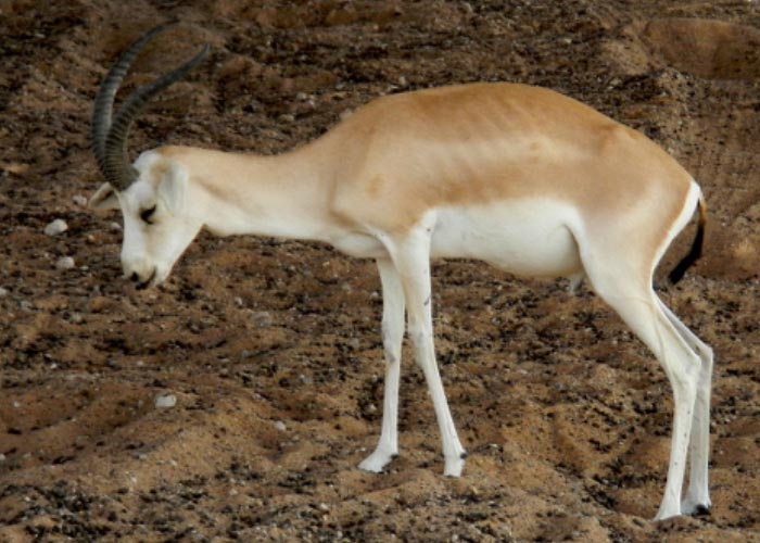
Goats were considered for a long time to be the only species affected by CCPP. Some Mccp isolates were obtained from sheep but usually in association with other pathogens that could explain the observed lesions. Since 2007, a number of CCPP outbreaks have been confirmed in wildlife species. (Figure 2) This was notably the case in a wildlife preservation centre in Qatar, where multiple species were affected, such as gerenuk, beira and Laristan mouflon.3 Oryx were also shown to be susceptible in the Arabic Peninsula12 as well as Tibetan antelopes in China.78 It is not known if these outbreaks result from increased opportunities for contact between wildlife and affected goats or, alternatively, simply the results of improved diagnostic capabilities.
The disease always appears in a herd after the introduction of an infected animal, the causative organism being transmitted to susceptible goats by inhalation of aerosols released by infected animals during bouts of coughing. The fragility of Mccp in the environment requires close contact between infected and susceptible animals for transmission to take place. The duration of this contact period need not be long; 24 hours is sufficient for transmission to occur. Sex and age do not seem to play any role in the susceptibility of the animals.20, 42, 59
The existence of latent carriers is still debated. Hutcheon in 1881 mentioned that the goats imported into the Cape Colony had been travelling for seven weeks. Unfortunately, unlike contagious bovine pleuropneumonia, the location of Mccp in latent carriers, if they do exist, is not known but several hypotheses can be suggested. The organism could be harboured in the ear canal of recovered animals as has been demonstrated in M. mycoides subsp. mycoides LC infections.14 It could also possibly be carried by another animal species without provoking clinical signs.67 Mycoplasma capricolum subsp. capripneumoniae has been found in the nares and the lungs of healthy sheep10 and the role of this in the epidemiology of CCPP should be investigated.
In countries where it exists, CCPP has a heavy toll on goats. In Kenya for example, in Narok, all the goat herds that were brought for vaccination had some positive animals. As the test used was highly specific (cELISA) this clearly showed the importance of the disease. The individual prevalence within each herd was variable (6 to 100 per cent). The case fatality rates induced by CCPP, PPR and mange were within a similar range (64 per cent) in Kenya in regions where vaccination was not practised.8
Pathogenesis
There are very few data available on the pathogenicity of Mccp and the pathogenesis of CCPP. Findings from other Mycoplasma infections are therefore used as a model. The disease is characterized by strict organ specificity: only the lungs are affected.37 The airborne route is the commonest for pulmonary infections with the upper respiratory tract serving as the portal of entry. Mycoplasmas reaching the lungs via infected aerosols become attached to the mucous membrane of the bronchial tree, reach the alveoli and are able to multiply and invade the alveolar cells.26
This strict organ specificity may be related to the loss of multiple genes from the Mccp genome,15, 21, 42 which could explain the fastidiousness of this organism as it lacks many functional genes enabling an efficient metabolism. In spite of a slow growth rate, Mccp remains pathogenic and it notably possesses the glycerol metabolism genes that were suspected to play a role in pathogenicity for other mycoplasmas through the production of H2O2 or reactive oxygen species. Mccp also produces and excretes polysaccharides that are of a different nature to those of Mmm7 and could play a role in the attachment to target cells or in protection from the host innate responses, such as phagocytosis or complement- mediated killing.
Clinical signs
The clinical signs have been described by many authors.21, 35, 41, 42, 52 During experimental infections induced by the intratracheal- endobronchial route, the incubation period is between six and ten days.21, 44 Hutcheon, during the first outbreaks in South Africa, observed that susceptible goats fell ill ten days after contact with infected animals. However, this period is often longer. In contact transmission experiments a mean duration for the incubation period of 26 ± 15 days was obtained.42 In the OIE International Animal Health Code, the incubation period has been set at 45 days.54
Pyrexia heralds the onset of clinical disease and persists for four to nine days. The peak of the temperature reaction — 40 °C or, in some, 41 °C — is reached 48 to 72 hours after the onset of fever. Initially, affected animals tend to lag behind the rest of the flock, but continue to eat and ruminate for some days. In the following days coughing and accelerated respiration develop and animals assume a typical posture with the forelimbs set wide apart, the neck extended and the head held down. Breathing becomes progressively more difficult and is accompanied by grunting, a sign of painful respiration, and a mucopurulent nasal discharge. When forced to move, affected animals may collapse, showing signs of respiratory distress before they die. In the terminal stage, the mouth is kept open, the lips are covered with froth, the tongue protrudes, and the animal bleats distressingly. In acute cases, which occur particularly in animals suffering from a primary infection, the duration of the disease is from two to seven days. Subacute forms are observed more often in areas where CCPP is endemic; the clinical signs are similar but less intense. Coughing occurs irregularly and generally follows a physical effort. The duration of the disease depends, to some extent, on the environmental conditions and the management system. When kept under favourable conditions, affected animals may survive for more than a month and may even recover. In the absence of treatment or control measures, morbidity can be as high as 100 per cent and mortality may reach 70 per cent.
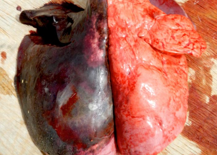
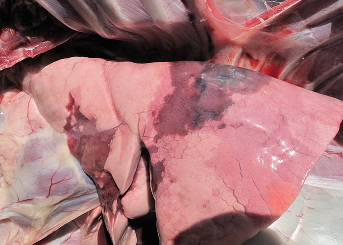
Pathology
Lesions are strictly confined to the thoracic cavity. Usually only one lung is affected, which shows severe hepatization involving all the lobes. The lung is enlarged, oedematous and variegated in colour due to the presence of red, yellow or grey foci (Figures 3 to 7). Incision into the affected part reveals a copious broncho-alveolar exudate. An extensive serofibrinous pleuritis leads to the accumulation of a large quantity of yellow pleural fluid exudate (Figure 4). Contrary to what may be found in cases of pleuropneumonia due to M. mycoides, subsp. capri there is no widening of the interlobular septa due to inflammatory oedema. In subacute cases, the gross pathology is not as clear-cut, as it is complicated by secondary bacterial infections. Numerous fibrinous adhesions between the parietal and visceral pleura may be present and numerous purulent abscesses may be present within the lung. Histologically, the pneumonia is characterized by fibrinopurulent exudate in dilated hyperplastic bronchi. Alveolar exudate is dominated by macrophages, neutrophils in acute cases and fibrosis in more chronic cases.76
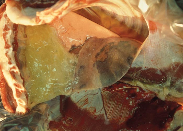
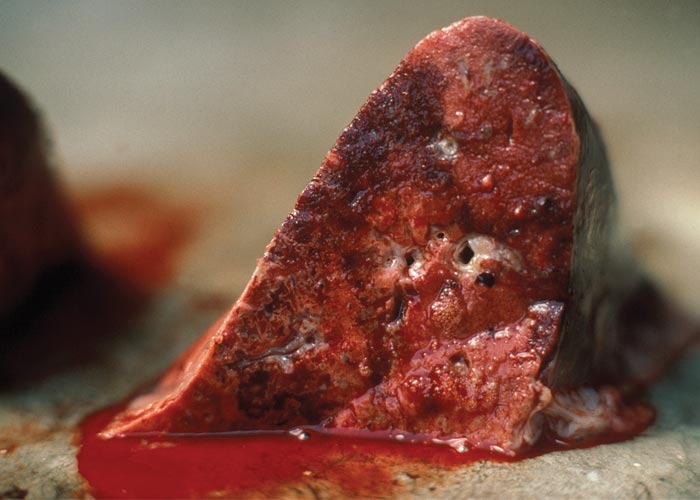
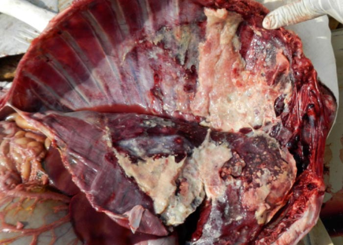
Figure 5 Goitered gazelle (Gazella marica) affected by CCPP (2013), United Arab Emirates. Severe serofibrinous pleuritis and pneumonia. (By courtesy of L Lignereux, Environment agency Abu Dhabi, United Arab Emirates)
Diagnosis and differential diagnosis
Reports on the occurrence of CCPP to international bodies, such as the OIE or FAO, should be considered with caution since there may still be misinterpretation in the name of the disease. Only outbreaks that have been confirmed by laboratory examination should be considered to be CCPP.
Under field conditions, the clinical signs and pathological lesions may lead to a suspected diagnosis of CCPP. Laboratory confirmation is essential in order to differentiate CCPP from pasteurellosis or other mycoplasmal infections. The difficulty in recognizing Mccp as the causal agent of CCPP arises from the fact that several other mycoplasmas may produce pleuropneumonia in goats, especially M. mycoides subsp. capri. The latter organism had, in fact, been considered for many years before 1976 to be the causative agent of CCPP, certainly because it coexists with Mccp strains in many countries and because it grows very easily.
The differences between Mccp and the other mycoplasmas involve not only its pathogenicity but also its in vitro behaviour. Infection of goats with Mccp will always, and only, produce pleuropneumonia, while after infection with the other mycoplasmas, induction of pleuropneumonia is only one of several possible lesions.
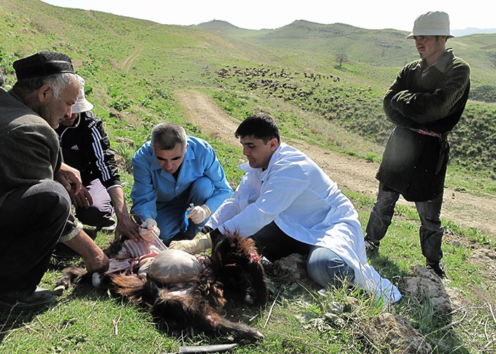
Samples for mycoplasmal isolation should consist of pleuritic fluid (Figure 8), when present, affected lung tissue especially from the interface between consolidated and more normal areas, and mediastinal lymph nodes.53 Samples for PCR may consist of several filter papers soaked in the pleural fluid and then air dried. Before being processed, the samples can be safely stored for months at −20 °C. If the tissue and exudate samples are to be transported, ampicillin can be added (1 mg/ml) to prevent bacterial contamination. Lyophilization is also possible and is used especially when samples are to be sent to reference laboratories. Filter papers should be sent in sealed plastic bags.
The diagnostic techniques that are used for the isolation (Figure 9) and identification of Mccp have been reviewed in several recent papers.25, 53, 72 Before isolation, the tissue samples should not be finely ground; it is preferable to dice them finely using scissors. Pleuritic fluid should be diluted by at least three ten-fold steps in the medium. The causative organism is not an easy mycoplasma to isolate and great care must be taken in the quality of the medium that is used for the purpose. Several types of media are routinely used, such as VFG (Viande Foie Goat),41 WJ or Newing’s tryptose.24, 27 Alternatively, using PPLO broth without crystal violet or Brain Heart Infusion (BHI) broth as a base, horse or donkey sera added to the media at a concentration of 20 to 30 per cent permit good growth of Mccp but should be tested, prior to use, for their ability to sustain an abundant growth.
Fresh yeast extract is required at a concentration of 5 to 10 per cent. Glucose is added at 0.2 per cent concentration and sodium pyruvate at 0.4 per cent. For solid medium, the final concentration of agar is 1 per cent but this could be adjusted to give a relatively soft agar. After four to five days of incubation at 37 °C in a humid atmosphere containing 5 per cent CO2, very small colonies (0,1 mm in diameter) appear on solid media. They should be visualized using a binocular microscope. In broth, the turbidity produced by growth of the organism is very faint; inoculated broth tubes should be compared with uninoculated tubes. Until recently, the identification of Mccp relied mainly on immunological tests such as the growth inhibition test using hyperimmune sera raised against the various Mycoplasma species found in goats. Biochemical tests, such as glucose breakdown, arginin hydrolysis, reduction of tetrazolium chloride (aerobically and anaerobically), and ‘film and spots’, do not permit complete and unevoquivocal characterization of Mccp. Nevertheless, they could be carried out as a preliminary screening system and in order to narrow down the likely identity of an isolate.25, 53
The biochemical and serological tests used for the identification of Mccp are now superseded by PCR tests, which can offer faster and more specific identification. One of them is based on the amplification of the gene coding for the 16S ribosomal RNA.4, 10 This gene, being well conserved amongst the Mycoides cluster species, makes it imperative that the PCR be followed by the digestion of the amplified products by an endonuclease to identify Mccp. Another PCR test yields a specific amplification if the sample contains Mccp DNA, as it exploits the presence of mutations that are present in Mccp and not in the genome of the closely related Mcc strains.77 The same primer set has also been used to build a qPCR test.40 Ultimately, the genome of the isolated Mccp strain can be fully sequenced and this will allow a precise identification of the strain and its positioning in the phylogenetic groups that have been defined, notably by extended multilocus sequence analysis.15 Molecular subtyping of the Mccp strains is particularly important when the disease is introduced into a naïve region, as this typing might provide clues to the origin of introduction.
Several tests have been used for serological diagnosis: complement fixation test, passive haemagglutination test,51 latex agglutination test64 and indirect ELISA.75 Each of these has different advantages, but their main common drawback is their lack of specificity due to the antigenic relationships of Mycoplasma spp. In addition, the specificty of these tests is reduced by the fact that joint or sequential infections caused by other mycoplasmal species is not infrequent and interpretation of the results may become very difficult. A competition enzyme-linked immunosorbent assay (c-ELISA) has been developed using a specific monoclonal antibody.69, 71 This assay permits the testing of large numbers of serum samples and should be recommended for sero-epidemiological surveys. However, as for contagious bovine pleuropneumonia, serological tests should never be used for individual diagnoses but only on a flock basis. The same cELISA could be used to monitor the outcome of vaccination campaigns as a vaccine that has been produced according to the OIE manual of standards is expected to yield consistent seroconversion for at least 3 months. The test is not able to differentiate antibodies resulting from vaccination from those arising from disease.61
Control
In many regions where CCPP exists, antibiotic treatment is the first and only means of control, especially since vaccines are not widely available and a stamping-out policy is unacceptable for socio-economic reasons, which include a lack of funds to compensate the owners and a general unwillingness by the owners to slaughter their animals.
The earlier treatment is implemented during the course of the disease, the better. It is important to remember, however, that samples for laboratory confirmation should be taken before inoculation of any antibiotic. Mycoplasma capricolum subsp. capripneumoniae, being deprived of a cell wall, is not susceptible to antibiotics of the penicillin group as their action depends on inhibition of cell wall synthesis. Some species are susceptible to streptomycin but its use is not advisable because of the rapid appearance of streptomycin-resistant strains. As a general rule, the antibiotics that give the best results are the tetracycline group at 5 to 10 mg/kg, the fluoroquinolones and the macrolide group or related compounds (tylosin, erythromycin and spiramycin) at 25 mg/kg.35, 56, 63 The duration of the treatment should always be at least five days, and all the animals that have been exposed should be treated. In remote areas and/or nomadic herds, long-acting formulations are preferred in order to achieve complete treatment. Despite antibiotic treatment being able to minimize the losses due to CCPP, it does not prevent the persistence of mycoplasmas in treated animals.57
In addition, failures of treatment and recurrence of the disease in the long term are not infrequent, especially when recommendations are not followed, as sometimes occurs under field situations.
In countries free of the disease, only a strict policy and control of animal movements will prevent CCPP from being introduced. Contagious caprine pleuropneumonia is a listed disease of the OIE and standards for trade have been established.54 When the disease is present, great care should be taken to avoid contact between different flocks. Animals introduced into a flock should always come from one that has been free of the disease for at least several months. Modern diagnostic tools permit access to information on the disease status of the flocks or of imported animals.
The best way to control CCPP should be to vaccinate the susceptible goat populations.
An experimental attenuated live vaccine has been tested with some encouraging results43 but has not been used in the field. The only vaccine available is a sonicated suspension of Mccp with saponin as an adjuvant.38, 65 It is theoretically produced in freeze-dried form and provides protection for over a year. Vaccination of kids should be practised after the age of ten weeks as antibodies of colostral origin persist for eight weeks.29 In the field, use of the vaccine reduces the morbidity and mortality rates in the vaccinated groups when compared to those unvaccinated control groups.31
Unfortunately, it appears that vaccines that are available in the field, although expensive, seldom meet the quality criteria that should be expected. Some so- called “CCPP vaccines” contain live mycoplasmas that are not Mccp. Other producers do not purify their Mccp antigen sufficiently and the proteins that are detected in the vaccine are linked to residual medium components. None of these low-quality products will yield the expected seroconversion by cELISA and therefore they should be easily detected if this test is performed.
The control tools that are available in the field are notably insufficient to obtain proper control of this disease, leaving little hope for eradication. It is therefore expected that the disease will continue its progressive geographical expansion. This expansion puts at risk a number of endangered wildlife species that have recently proved to be susceptible to CCPP. It also increases the antimicrobial resistance risk associated with antibiotic treatment. It may be expected that the actual campaigns aiming at controlling/eradicating PPR may indirectly help identify CCPP cases more easily and will help to establish its real impact. It is therefore important to provide good quality and affordable vaccines to goat owners and develop new products that should be effective but less costly.
References
- ABU-GROUN, E.A.M., TAYLOR, R.R., VARSANI, H., WADHER, B.J., LEACH, R.H. & MILES, R.J., 1994. Biochemical diversity within the ‘Mycoplasma mycoides cluster’. Microbiology, 140, 2033–2042.
- AMIRBEKOV, M., MURVATULLOEV, S., FERRARI, G., 2010. Contagious caprine pleuropneumonia detected for the first time in Tajikistan. EMPRES transboundary Animal Diseases Bulletin, 35, 20-22.
- ARIF, A., SCHULZ, J., THIAUCOURT, F., TAHA, A., HAMMER, S., 2007. Contagious caprine pleuropneumonia outbreak in captive wild ungulates at Al Wabra Wildlife Preservation, State of Qatar. Journal of Zoo and Wildlife Medicine, 38, 93-96.
- BASCUÑANA, C.R., MATTSON, J.G., BÖLSKE, G. & JOHANSSON, K.E., 1994. Characterization of the 16S rRNA genes from Mycoplasma sp. strain F38 and development of an identification system based on PCR. Journal of Bacteriology, 176, 2577–2586.
- BEATON, W.G., 1931. Diseases of goats in Nigeria. Journal of Comparative Pathology and Therapeutics, 44, 192–201.
- BÉRANGER, S., 1983. Contributions à l’étude de la sensibilité des mycoplasmes à quelques antibiotiques. Thèse de Doctorat en Pharmacie, University of Lyon, 118.
- BERTIN, C., PAU-ROBLOT, C., COURTOIS, J., MANSO-SILVÁN, L., TARDY, F., POUMARAT, F., CITTI, C., SIRAND-PUGNET, P., GAURIVAUD, P., THIAUCOURT, F., 2015. Highly Dynamic Genomic Loci Drive the Synthesis of Two Types of Capsular or Secreted Polysaccharides within the Mycoplasma mycoides Cluster. Applied and Environmental Microbiology, 81, 676-687.
- BETT, B., JOST, C., ALLPORT, R., MARINER, J., 2009. Using participatory epidemiological techniques to estimate the relative incidence and impact on livelihoods of livestock diseases amongst nomadic pastoralists in Turkana South District, Kenya. Preventive Veterinary Medicine, 90, 194-203.
- BÖLSKE, G., JOHANSSON, K.E., HEINONEN, R., PANVUGA, P.A. & TWINAMASIKO, E., 1995. Contagious caprine pleuropneumonia in Uganda and isolation of Mycoplasma capricolum subsp. capripneumoniae from goats and sheep. The Veterinary Record, 137, 594.
- BÖLSKE, G., MATTSON, J.G., BASCUÑANA, C.R., BERGSTRÖM, K., WESONGA, H. & JOHANSSON, K.E., 1996. Diagnosis of contagious caprine pleuropneumonia by detection and identification of Mycoplasma capricolum subsp. capripneumoniae by PCR and restriction analysis. Journal of Clinical Microbiology, 34, 785–791.
- ÇETINKAYA, B., KALIN, R., KARAHAN, M., ATIL, E., MANSO-SILVÁN, L., THIAUCOURT, F., 2009. Detection of contagious caprine pleuropneumonia in East Turkey. Revue scientifique et technique de l'OIE, 28, 1037-1044.
- CHABER, A., LIGNEREUX, L., QASSIMI, M.A., SAEGERMAN, C., MANSO-SILVAN, L., DUPUY, V., THIAUCOURT, F., 2014. Fatal transmission of contagious caprine pleuropneumonia to an Arabian oryx (Oryx leucoryx). Veterinary Microbiology, 173, 156-159.
- COTTEW, G.S., BRÉARD, A., DA MASSA, A.J., ERNØ, H., LEACH, R.H., LEFEVRE, P.-C., RODWELL, A.W., & SMITH, G., 1987. Taxonomy of the Mycoplasma mycoides cluster. Israel Journal of Medical Science, 23, 632–635.
- DA MASSA, A.J. & BROOKS, D.L., 1991. The external ear canal of goats and other animals as a mycoplasma habitat. Small Ruminant Research, 4, 85–93.
- DUPUY, V. & THIAUCOURT, F., 2014. Complete Genome Sequence of Mycoplasma capricolum subsp. capripneumoniae strain 9231-Abomsa. Genome Announcements, 2.
- DUPUY, V., VERDIER, A., THIAUCOURT, F. & MANSO-SILVÁN, L., 2015. A large-scale genomic approach affords unprecedented resolution for the molecular epidemiology and evolutionary history of contagious caprine pleuropneumonia. Veterinary Research, 46, 74.
- EL-DEEB, W., ALMUJALLI, A.A., ELJALII, I., ELMOSLEMANY, A. & FAYEZ, M., 2017. Contagious caprine pleuropneumonia: The first isolation and molecular characterization of Mycoplasma capricolum subsp. capripneumoniae in the Kingdom of Saudi Arabia. Acta Tropicana, 168, 74-79.
- EVANS, A.S., 1976. Causation and disease: The Henle-Koch postulates revisited. Yale Journal of Biology and Medicine, 49, 175–195.
- HARBI, M.S.M.A., EL TAHIR, M.S., MAC OWAN, K.J. & NAYIL, A.A., 1981. Mycoplasma strain F38 and contagious caprine pleuropneumonia in the Sudan. The Veterinary Record, 108, 261.
- HARBI, M.S.M.A., MAGEED, I.A. & EL TAHIR, M.S., 1983. Experimental contact transmission of contagious caprine pleuropneumonia of goats (Abu nini) in the Sudan. Veterinary Research Communications, 6, 139–143.
- HARBI, M.S.MA., LE TAHIR, M.S., SALIM, M.O., NAYIL, A.A. & MAGEED, I.A., 1983. Experimental contagious caprine pleuropneumonia. Tropical Animal Health and Production, 15, 51–52.
- HUTCHEON, D., 1881. Contagious pleuropneumonia in angora goats. Veterinary Journal, 13, 171–180.
- HUTCHEON, D., 1889. Contagious pleuropneumonia in goats in Cape Colony, South Africa. Veterinary Journal, 29, 399–404.
- JONES, G.E. & WOOD, A.R., 1988. Microbiological and serological studies on caprine pleuropneumonia in Oman. Research in Veterinary Science, 44, 125–131.
- JONES, G.E., 1989. Contagious caprine pleuropneumonia. Technical Series N°9. Office International des Épizooties (Paris), 63.
- KASALI, O.B. & OJO, M.O., 1981. Pathogenicity of Mycoplasma mycoides for goats. International Goat and Sheep Research, 1, 269–273.
- KIBOR, A.C. & WAIYAKI, P.G., 1986. Growth of mycoplasma F38 in medium B (modified Hayflick) and Newing’s tryptose medium. Bulletin of Animal health and Production in Africa, 34, 157–159.
- KING, G.J., 1988. Optimum age to vaccinate for contagious caprine pleuropneumonia. The Veterinary Record, 123, 572–573.
- KING, G.J., KAGUMBA, M. & KARIUKI, D.P., 1992. Trial of the efficacy and immunological response to an inactivated mycoplasma F38 vaccine. The Veterinary Record, 131, 461–464.
- KIPRONOH, A.K., OMBUI, J.N., KIARA, H.K., BINEPAL, Y.S., GITONGA, E. & WESONGA, H.O., 2016. Prevalence of contagious caprine pleuro-pneumonia in pastoral flocks of goats in the Rift Valley region of Kenya. Tropical Animal Health and Production, 48, 151-155.
- KOLAYLI, C, RAIF, M., ESIN, I. & ARAYCI, E., 1935. Étude sur la pleuropneumonie infectieuse des chèvres d’Anatolie. Bulletin de l’Académie Vétérinaire de France, 8, 227–230.
- KUMAR, A. & GARG, D.N., 1991. Isolation of mycoplasma F38 from the milk of mastitic cows. The Veterinary Record, 128, 429.
- LEACH, R.H., ERNØ, H. & MAC OWAN, K.J., 1993. Proposal for designation of F38-type caprine mycoplasmas as Mycoplasma capricolum subsp. capripneumoniae subsp. nov. and consequent obligatory relegation of strains currently classified as M. capricolum (Tully, Barile, Edward, Theodore & ErnØ, 1974) to an additional new subspecies, M. capricolum subsp. capricolum subsp. nov. International Journal of Systematic Bacteriology, 43, 603–605.
- LEFEVRE, P.-C., BRÉARD, A., AL FAROUKH, I. & BURON, S., 1987. Mycoplasma species F38 isolated in Chad. The Veterinary Record, 121, 575–576.
- LEFEVRE, P.-C., JONES, G.E. & OJO, M.O., 1987. Les mycoplasmoses pulmonaires des petits ruminants. Revue Scientifique et Technique de l’Office International des Épizooties, 6, 713–757.
- LILJANDER, A., MINGYAN Y., O'BRIEN, E., HELLER, M., NEPPER, J.F., WEIBEL, D.B., GLUECKS, I., YOUNAN, M., FREY, J., FALQUET, L. AND JORES, J. 2015. Field-applicable recombinase polymerase amplification assay for rapid detection of Mycoplasma capricolum subsp. Capripneumoniae. Journal of Clinical Microbiology, 53(9): 2810-2815.
- LINDLEY, E.P. & ABDULLA, A.E.D., 1969. Some notes on the host specificity of the etiological agents of contagious caprine pleuropneumonia and contagious bovine pleuropneumonia. Bulletin of Epizootic Diseases in Africa, 17, 153–158.
- LITAMOI, J.K., LIJODI, F.K. & NANDOKHA, E., 1989. Contagious caprine pleuropneumonia: Some field observations in a field vaccination trial using inactivated Mycoplasma strain F38. Tropical Animal Health and Production, 21, 146–151.
- LONGLEY, E.O., 1951. Contagious caprine pleuropneumonia: A study of the disease in Nigeria. Colonial Research Publications, N°7. London: Her Majesty’s Stationery Office.
- LORENZON, S., MANSO-SILVAN, L., THIAUCOURT, F., 2008. Specific real-time PCR assays for the detection and quantification of Mycoplasma mycoides subsp. mycoides SC and Mycoplasma capricolum subsp. capripneumoniae. Molecular and Cellular Probes, 22, 324-328.
- MAC OWAN, K.J. & MINETTE, J.E, 1976. A Mycoplasma from acute contagious caprine pleuropneumonia in Kenya. Tropical Animal Health and Production, 8, 91–95.
- MAC OWAN, K.J. & MINETTE, J.E, 1977. Contact transmission of experimental contagious caprine pleuropneumonia(CCPP). Tropical Animal Health and Production, 9, 185–188.
- MAC OWAN, K.J. & MINETTE, J.E, 1978. The effect of high passage Mycoplasma strain F38 on the course of contagious caprine pleuropneumonia (CCPP). Tropical Animal Health and Production, 10, 31–35.
- MAC OWAN, K.J., 1984. Role of Mycoplasma strain F38 in contagious caprine pleuropneumonia. Israel Journal of Medical Science, 20, 979–981.
- MANSO-SILVAN, L., DUPUY, V., CHU, Y. & THIAUCOURT, F., 2011a. Multi-locus sequence analysis of Mycoplasma capricolum subsp. capripneumoniae for the molecular epidemiology of contagious caprine pleuropneumonia. Veterinary Research, 42, 86.
- MANSO-SILVAN, L., DUPUY, V., CHU, Y. & THIAUCOURT, F., 2011b. Multi-locus sequence analysis of Mycoplasma capricolum subsp. capripneumoniae for the molecular epidemiology of contagious caprine pleuropneumonia. Veterinary Research, 42, 86.
- MANSO-SILVÁN, L., PERRIER, X. & THIAUCOURT, F., 2007. Phylogeny of the Mycoplasma mycoides cluster based on analysis of five conserved protein-coding sequences: consequences in taxonomy. International Journal of Systematic and Evolutionary Microbiology, 57, 2247-2258.
- MANSO-SILVAN, L., VILEI, E., SACHSE, K., DJORDJEVIC, S., THIAUCOURT, F. & FREY, J., 2009. Mycoplasma leachii sp. nov. as a new species designation for Mycoplasma sp. bovine group 7 of Leach, and reclassification of Mycoplasma mycoides subsp. mycoides LC as a serovar of Mycoplasma mycoides subsp. capri. International Journal of Systematic and Evolutionary Microbiology, 59, 1353-1358.
- MELANIDI, C & STYLLIANOPOULO, M., 1928. La pleuro-pneumonie contagieuse des chèvres en Grèce. Revue générale de Médecine Vétérinaire, 37, 490–493.
- METTAM, R.W.M., 1929. Contagious pleuro-pneumonia of goats in East Africa. Proceedings of the Pan African Veterinary Conference, Pretoria, Paper N°18, 173–178.
- MUTHOMI, E.K. & RURANGIRWA, F.R., 1983. Passive haemagglutination and complement fixation as diagnostic tests for contagious caprine pleuropneumonia caused by F38 strain of Mycoplasma. Research in Veterinary Science, 35, 1–4.
- NICHOLAS, R., CHURCHWARD, C., 2011. Contagious caprine pleuropneumonia: New Aspects of an Old Disease. Transboundary Emerging Disease, 59, 189-196.
- OIE, 1996. Contagious caprine pleuropneumonia. In: MANUAL OF STANDARDS FOR DIAGNOSTIC TESTS AND VACCINES. Chapter 3.3.6, 374–383.
- OIE, 1998. Contagious caprine pleuropneumonia. In: INTERNATIONAL ANIMAL HEALTH CODE. 7th Edition, 199–201.
- OJO, M.O., 1976. Caprine pneumonia: Pathogenicity of Mycoplasma mycoides subsp. capri and caprine strains of Mycoplasma mycoides subsp. mycoides for goats. Journal of Comparative Pathology, 86, 519–529.
- ONOVIRAN, O., 1976. The comparative efficacy of some antibiotics used to treat experimentally induced mycoplasma infection in goats. The Veterinary Record, 94, 418–420.
- OZDEMIR, U., LORIA, G.R., GODINHO, K.S., SAMSON, R., ROWAN, T.G., CHURCHWARD, C., AYLING, R.D. & NICHOLAS, R.A., 2006. Effect of danofloxacin (Advocin A180) on goats affected with contagious caprine pleuropneumonia. Tropical Animal Health Production, 38, 533-540.
- OZDEMIR, U., OZDEMIR, E., MARCH, J.B., CHURCHWARD, C. & NICHOLAS, R.A., 2005. Contagious caprine pleuropneumonia in the Thrace region of Turkey. Veterinary Record, 156, 286-287.
- PERREAU, P., BRÉARD, A. & LE GOFF, C., 1984. Infection expérimentale de la chèvre par les souches de mycoplasme de type F38 (pleuropneumonie contagieuse caprine). Annales de Mircrobiologie, 135 A(1), 119–124.
- PETTERSSON, B., BÖLSKE, G., THIAUCOURT, F. UHLEN, M. & JOHANSSON, K.E., 1998. Molecular evolution of Mycoplasma capricolum subsp. capripneumoniae strains based on polymorphisms in the 16S rRNA genes. Journal of Bacteriology, 180, 2350–2358.
- PEYRAUD, A., POUMARAT, F., TARDY, F., MANSO-SILVAN, L., HAMROEV, K., TILLOEV, T., AMIRBEKOV, M., TOUNKARA, K., BODJO, C., WESONGA, H., NKANDO, I., JENBERIE, S., YAMI, M., CARDINALE, E., MEENOWA, D., JAUMALLY, M., YAQUB, T., SHABBIR, M., MUKHTAR, N., HALIMI, M., ZIAY, G., SCHAUWERS, W., NOORI, H., RAJABI, A., OSTROWSKI, S. & THIAUCOURT, F., 2014. An international collaborative study to determine the prevalence of contagious caprine pleuropneumonia by monoclonal antibody-based cELISA. BMC Veterinary Research, 10, 48.
- RURANGIRWA, F.R., KOUYATE, B., NIANG, M. & MCGUIRE, T.C., 1990. CCPP: Antibodies to F38 polysaccharide in Mali goats. The Veterinary Record, 127, 353–354.
- RURANGIRWA, F.R., MASIGA, W.N., MURIU, D.N., MUTHOMI, E., MULIGA, G., KAGUMBA, M. & NANDOKHA, E., 1981. Treatment of contagious caprine pleuropneumonia. Tropical Animal Health and Production, 13, 177– 182.
- RURANGIRWA, F.R., MCGUIRE, T.C., KIBOR, A. & CHEMA, S., 1987. A latex agglutination test for field diagnosis of caprine pleuropneumonia. The Veterinary Record, 121, 615–622.
- RURANGIRWA, F.R., MCGUIRE, T.C., KIBOR, A. & CHEMA, S., 1987. An inactivated vaccine for contagious caprine pleuropneumonia. The Veterinary Record, 121, 397–402.
- SRIVASTAVA, A.K., MEENOWA, D., BARDEN, G., SALGUERO, F.J., CHURCHWARD, C. & NICHOLAS, R.A.J., 2010. Contagious caprine pleuropneumonia in Mauritius. Veterinary Record, 167, 304-305.
- THIAUCOURT, F. & BÖLSKE, G., 1996. Contagious caprine pleuropneumonia and other pulmonary mycoplasmoses of sheep and goats. Revue Scientifique et Technique de l’Office International des Épizooties, 15, 1397–1414.
- THIAUCOURT, F., 1994. La pleuropneumonie contagieuse caprine. De l’observation clinique à ,la mise au point de techniques de diagnostic. Thèse Doctorat, Université Paris, XII.
- THIAUCOURT, F., BÖLSKE, G., LIBEAU, G., LE GOFF, C. & LEFEVRE, P.C., 1994. The use of monoclonal antibodies in the diagnosis of contagious caprine pleuropneumonia (CCPP). Veterinary Microbiology, 41, 191– 203.
- THIAUCOURT, F., BRÉARD, A., LEFEVRE, P.C. & MEBRATU, G.Y., 1992. Contagious caprine pleuropneumonia in Ethiopia. The Veterinary Record, 131, 585.
- THIAUCOURT, F., DESWETCHIN, C., KING, G.E. & LIBEAU, G., 1994. The use of a blocking ELISA for the specific detection of antibodies to Mycoplasma sp. Type F38 (CCPP). IOM Letters, 3, 21–22.
- THIAUCOURT, F., GUÉRIN, C., MADY, V. & LEFEVRE, P.C., 1992. Diagnostic de la pleuropneumonie contagieuse caprine : améliorations récentes. Revue Scientifique et Technique de l’Office International des Épizooties, 11, 859–865.
- THOMAS, P. 1873. Rapport médical sur le Bou Frida. Publication du Gouvernement Général civil d’Algérie. A. Jourdan Editeur, Alger, 35.
- TÜRKARSKLAN, J. & ARISOY, F., 1990. The isolation of F38 strain from the outbreaks of contagious caprine pleuropneumonia (CCPP) in Turkey. Pendik Hayv. Hast. Merk. Arast. Enst. Derg., 21, 25–29 (in Turkish).
- WAMWAYI, H.M., WAFULA, J.S., LITAMOI, J.K. & NANDOKHA, E.N., 1989. Detection of antibody to mycoplasma F38 in goat sera by an ELISA. Tropical Animal Health and Production, 21, 43–49.
- WESONGA, H.O., BOLSKE, G., THIAUCOURT, F., WANJOHI, C. & LINDBERG, R., 2004. Experimental contagious caprine pleuropneumonia: a long term study on the course of infection and pathology in a flock of goats infected with Mycoplasma capricolum subsp. capripneumoniae. Acta Veterinaria Scandinavic, 45, 167-179.
- WOUBIT, S., LORENZON, S., PEYRAUD, A., MANSO-SILVAN, L. & THIAUCOURT, F., 2004. A specific PCR for the identification of Mycoplasma capricolum subsp. capripneumoniae, the causative agent of contagious caprine pleuropneumonia (CCPP). Veterinary Microbiology, 104, 1256132.
- YU, Z., WANG, T., SUN, H., XIA, Z., ZHANG, K., CHU, D., XU, Y., XIN, Y., XU, W., CHENG, K., ZHENG, X., HUANG, G., ZHAO, Y., YANG, S., GAO, Y. & XIA, X., 2013. Contagious caprine pleuropneumonia in endangered tibetan antelope, china, 2012. Emerging Infectious Disease, 19, 2051-2053.


