- Infectious Diseases of Livestock
- Part 3
- Trueperella pyogenes infections
- GENERAL INTRODUCTION: SPIROCHAETES
- Swine dysentery
- Borrelia theileri infection
- Borrelia suilla infection
- Lyme disease in livestock
- Leptospirosis
- GENERAL INTRODUCTION: AEROBIC ⁄ MICRO-AEROPHILIC, MOTILE, HELICAL ⁄ VIBROID GRAM-NEGATIVE BACTERIA
- Genital campylobacteriosis in cattle
- Proliferative enteropathies of pigs
- Campylobacter jejuni infection
- GENERAL INTRODUCTION: GRAM-NEGATIVE AEROBIC OR CAPNOPHILIC RODS AND COCCI
- Moraxella spp. infections
- Bordetella bronchiseptica infections
- Pseudomonas spp. infections
- Glanders
- Melioidosis
- Brucella spp. infections
- Bovine brucellosis
- Brucella ovis infection
- Brucella melitensis infection
- Brucella suis infection
- Brucella infections in terrestrial wildlife
- GENERAL INTRODUCTION: FACULTATIVELY ANAEROBIC GRAM NEGATIVE RODS
- Klebsiella spp. infections
- Escherichia coli infections
- Salmonella spp. infections
- Bovine salmonellosis
- Ovine and caprine salmonellosis
- Porcine salmonellosis
- Equine salmonellosis
- Yersinia spp. infections
- Haemophilus and Histophilus spp. infections
- Haemophilus parasuis infection
- Histophilus somni disease complex in cattle
- Actinobacillus spp. infections
- Actinobacillus equuli infections
- Gram-negative pleomorphic infections: Actinobacillus seminis, Histophilus ovis and Histophilus somni
- Porcine pleuropneumonia
- Actinobacillus suis infections
- Pasteurella and Mannheimia spp. infections
- Pneumonic mannheimiosis and pasteurellosis of cattle
- Haemorrhagic septicaemia
- Pasteurellosis in sheep and goats
- Porcine pasteurellosis
- Progressive atrophic rhinitis
- GENERAL INTRODUCTION: ANAEROBIC GRAM-NEGATIVE, IRREGULAR RODS
- Fusobacterium necrophorum, Dichelobacter (Bacteroides) nodosus and Bacteroides spp. infections
- GENERAL INTRODUCTION: GRAM-POSITIVE COCCI
- Staphylococcus spp. infections
- Staphylococcus aureus infections
- Exudative epidermitis
- Other Staphylococcus spp. infections
- Streptococcus spp. infections
- Strangles
- Streptococcus suis infections
- Streptococcus porcinus infections
- Other Streptococcus spp. infections
- GENERAL INTRODUCTION: ENDOSPORE-FORMING GRAM-POSITIVE RODS AND COCCI
- Anthrax
- Clostridium perfringens group infections
- Clostridium perfringens type A infections
- Clostridium perfringens type B infections
- Clostridium perfringens type C infections
- Clostridium perfringens type D infections
- Malignant oedema⁄gas gangrene group of Clostridium spp.
- Clostridium chauvoei infections
- Clostridium novyi infections
- Clostridium septicum infections
- Other clostridial infections
- Tetanus
- Botulism
- GENERAL INTRODUCTION: REGULAR, NON-SPORING, GRAM-POSITIVE RODS
- Listeriosis
- Erysipelothrix rhusiopathiae infections
- GENERAL INTRODUCTION: IRREGULAR, NON-SPORING, GRAM-POSITIVE RODS
- Corynebacterium pseudotuberculosis infections
- Corynebacterium renale group infections
- Bolo disease
- Actinomyces bovis infections
- Trueperella pyogenes infections
- Actinobaculum suis infections
- Actinomyces hyovaginalis infections
- GENERAL INTRODUCTION: MYCOBACTERIA
- Tuberculosis
- Paratuberculosis
- GENERAL INTRODUCTION: ACTINOMYCETES
- Nocardiosis
- Rhodococcus equi infections
- Dermatophilosis
- GENERAL INTRODUCTION: MOLLICUTES
- Contagious bovine pleuropneumonia
- Contagious caprine pleuropneumonia
- Mycoplasmal pneumonia of pigs
- Mycoplasmal polyserositis and arthritis of pigs
- Mycoplasmal arthritis of pigs
- Bovine genital mycoplasmosis
- Neurotoxin-producing group of Clostridium spp.
- Contagious equine metritis
- Tyzzer's disease
- MYCOTIC AND ALGAL DISEASES: Mycoses
- MYCOTIC AND ALGAL DISEASES: Pneumocystosis
- MYCOTIC AND ALGAL DISEASES: Protothecosis and other algal diseases
- DISEASE COMPLEXES / UNKNOWN AETIOLOGY: Epivag
- DISEASE COMPLEXES / UNKNOWN AETIOLOGY: Ulcerative balanoposthitis and vulvovaginitis of sheep
- DISEASE COMPLEXES / UNKNOWN AETIOLOGY: Ill thrift
- Eperythrozoonosis
- Bovine haemobartonellosis
Trueperella pyogenes infections
This content is distributed under the following licence: Attribution-NonCommercial CC BY-NC  View Creative Commons Licence details here
Trueperella pyogenes infections
Previous authors: M G COLLETT AND G F BATH
Current authors:
M G COLLETT - Senior Lecturer, BVSc, MMedVet (Path), Med (CAI), School of Veterinary Science, Massey University, Private Bag 112222, Palmerston North, Manawatu, 4442, New Zealand
R O GILBERT - Associate Dean of Academic Affairs, BVSc, MMedVet, DipACT, FRCVS, Ross University School of Veterinary Medicine, Cornell University, Island Main Road, West Farm, St. Kitts, Federation of St. Kitts and Nevis
Introduction
Trueperella (Arcanobacterium) pyogenes occurs in a great variety of suppurative or pyogenic conditions in economically important livestock (particularly cattle, sheep, goats and pigs), either as a primary pathogen, a secondary invader or as part of a mixed infection with obligate anaerobes. Depending on the animal species, the organism can be a commensal of the mammary gland, urogenital and upper respiratory tracts, and alimentary tract, in particular the oropharynx, rumen and stomach wall.91, 134, 148 It is most commonly incriminated as a cause of wound infections and abscesses, genital infections such as post-parturient metritis and endometritis in cows, as well as abortion and perinatal mortalities, mastitis, liver abscesses, endocarditis, lymphadenitis, pneumonia, foot infections, arthritis, osteomyelitis of vertebral bodies, discospondylitis, and bulbar empyema or pituitary abscessation.154, 156 Trueperella pyogenes infections such as mastitis, abscesses, polyarthritis, and metritis are important economic reasons for the culling of animals.87
This pus-producing bacterium was originally described by Glage in 1903 and called Corynebacterium pyogenes.210 The name was changed to Actinomyces pyogenes in 198230 and phylogenetic and 16S rRNA studies resulted in it being reclassified as Arcanobacterium pyogenes151 in 1997. In 2011, chemotaxonomic and further 16S rRNA gene sequence analysis studies resulted in an emended description of the genus Arcanobacterium (Christie-Atkins-Munch-Petersen [CAMP] test positive) and the creation of a new Trueperella genus (CAMP-test negative), named after the German microbiologist Hans Trüper,210 with T. pyogenes as the type species.
Aetiology
Trueperella pyogenes is a Gram-positive, non-motile, non-sporulating, facultatively anaerobic, slender, pleomorphic coccobacillus.91, 193 The genome sequences of two isolates from cases of metritis in cows have been published.63, 116
Trueperella pyogenes is nutritionally fastidious and grows poorly on common laboratory media unless they have been supplemented with blood or serum.136, 164 On blood agar, colonies are small, translucent and usually dense after 24 hours’ incubation when isolated from pus. After 48 hours, single colonies are approximately 0,5 to 1,0 mm in diameter and light grey in colour. A narrow zone of pale β-haemolysis is usually visible by 36 hours and most isolates typically liquefy inspissated serum. To improve the isolation rate, neomycin (0,6 to 0,7 units/ml), polymyxin B sulphate (25 units/ml) or 0,1 per cent Tween 80 can be added to the medium.76 A chemically defined medium which supports rapid growth of the organism has been developed.153
In addition to conventional phenotypic (e.g. biochemical markers such as sugar fermentation and haemolysis) as well as genotypic (e.g. detecting virulence factor encoding genes) properties, Fourier transform infrared spectroscopy has been shown to be rapid and reliable in identifying T. pyogenes in routine diagnosis.131 A loop-mediated isothermal amplification assay has also shown promise as a sensitive, reliable and cost-effective way for the molecular identification of T. pyogenes.1, 215 A fluorescent in situ hybridization technique is able to rapidly identify pathogens in mastitis milk samples provided the organisms are present in high numbers.58 Fluorescent in situ hybridization has also been used to investigate bacteria present in the pregnant uterus of cows.96 The microbial DNA diversity of mastitic milk can also be investigated using metagenomic pyrosequencing of bacterial 16S rRNA genes.137 Trueperella pyogenes strain differences can be characterized using matrix-assisted laser desorption ionization-time of flight (MALDI-TOF) mass spectrometry.126, 203
Pathogenesis
Although T. pyogenes expresses several virulence factors that are believed to be required for cell adherence, colonisation and host tissue damage, many aspects of the pathogenesis of infection are poorly understood.91, 156 the most important virulence factor is a filterable, oxygen-stable, cholesterol-dependent, haemolytic protein exotoxin, named pyolysin (PLO), which has been cloned.14, 22, 55, 114 Pyolysin is cytolytic for a number of cells including neutrophils and macrophages.91 It is fatal to mice and rabbits after intravenous injection and is dermotoxic to guinea pigs.115, 159, 170 Pyolysin is produced by all T. pyogenes strains so far examined14, 73, 91 and is unique in many ways; expression of this cholesterol-dependent cytolysin (CDC) is required for virulence and it is probably the most promising vaccine candidate identified thus far.14, 91 Cytolysins produced by other Gram-positive organisms include listeriolysin O, perfringolysin O and streptolysin O, and these toxins all exert their cytolytic effects by targeting cholesterol to form pores in host cell membranes.14, 91 The toxic and haemolytic activities of crude cell extracts are neutralized by an antitoxin14, 170 which is found in the sera of infected animals.147 High levels of antitoxin, however, do not protect against pyogenic infection.81
Adhesion to epithelial cells is an important requirement for organisms like T. pyogenes, whether as a pathogen or commensal. Consequently, the organism has a number of virulence factors that aid adhesion. These include neuraminidases (NanH and NanP), which cleave terminal sialic acid residues from carbohydrates or glycoproteins, decrease mucus viscosity, impair the host immune response, and play a role in the early stages of the pathogenic process.91 Furthermore, other T. pyogenes-specific virulence factors that are not efficient as immunogens but facilitate increased invasiveness into deeper tissues and resistance to phagocytosis, include collagen-binding protein (CbpA), fibrinogen-binding protein, fibronectin-binding protein, a component of fimbriae, and proteases.12, 38, 91, 103, 104, 107 The genes for these virulence factors are not present in every isolate and certain ones seem more prevalent in certain animal species161 or in particular types of infection, e.g. osteomyelitis,91 bovine metritis,167 and bovine mastitis.213 Pyolysin is found in all isolates but NanH, NanP, CbpA and the fimbrial biogenesis operon are only present in some.73, 91, 156, 213 Besides being able to invade and survive within host epithelial cells and macrophages, T. pyogenes readily forms biofilms as well. Biofilms are implicated in persistent and chronic infections, such as osteomyelitis and mastitis.91 The production of PLO is related to biofilm formation and it is initiated at the onset of the stationary phase.91 Genomic evidence suggests that a quorum sensing mechanism is integral to production of both biofilm and PLO.43 The bacterium also has an antigen that cross-reacts with Mycobacterium avium subsp. paratuberculosis.193
In the presence of opsonizing antibodies in serum204 or milk-whey,205 T. pyogenes is highly susceptible to the bactericidal activity of neutrophils in vitro. In the female genital tract, local T. pyogenes infection stimulates uterine antibody (mainly IgG and IgA) synthesis in the absence of an increase in serum antibody.206 The bacterium does not elaborate chemotactic substances.204 On the other hand, Bacteroides spp. in vitro inhibit chemotaxis2 and Prevotella melaninogenica (previously Bacteroides) inhibits phagocytosis by neutrophils,82 suggesting that, in mixed infections, the presence of Bacteroides or Prevotella spp. (which are obligate anaerobes) could allow the persistence of T. pyogenes infection through the protection of the latter from neutrophil phagocytosis.204
There is also a synergistic relationship between T. pyogenes and another obligate anaerobe, Fusobacterium necrophorum. The presence of T. pyogenes in the tissues lowers the oxidation-reduction potential in the affected areas while producing a diffusable factor that stimulates the growth of F. necrophorum.158 The latter, in turn, produces a leukotoxin that protects both organisms from phagocytosis.157 In mixed infections, F. necrophorum is probably the primary invasive and necrotizing agent that facilitates the establishment and growth of bacteria such as T. pyogenes and Prevotella spp.95, 157, 158
Work on the uterine proteome profiles of T. pyogenes-infected cows has revealed that certain indicator proteins show an increased expression that suggests that uterine infection with this organism may affect subsequent fertility.108
Pulmonary intravascular macrophages play a role in the uptake of T. pyogenes from the blood and the initiation of embolic pulmonary abscesses in cattle.109
Epidemiology, clinical signs and pathology
Trueperella pyogenes is a commensal on the mucosal surfaces of healthy animals.170 For example, it occurs on the mucosa of the preputium of rams86 and that of the alimentary (rumen), urogenital and respiratory tracts of healthy cattle, and in the udder of normal cows.91, 133, 134, 136 However, it is a common opportunistic and versatile pathogen of many important livestock species, as well as, although more rarely, wild mammals, and even parrots, poultry, rodents and reptiles.73, 91 Companion animals (e.g. dogs and cats with uterine discharge/pyometra, and umbilical infections and septic arthritis in horses) are unusual hosts73, 127, 152, 154, 156, 203 and human infection can result from inadvertent or occupational exposure.56, 110, 144 The organism is not found as a commensal in companion animals16 or humans.56
Abscesses
Damage to tissues, such as that resulting from trauma or viral or other bacterial infections, is usually required to establish infection. Infection may then spread haematogenously.111 Abscesses ranging from a few millimetres to several centimetres in diameter may occur in virtually any organ or tissue in the body, and the clinical signs manifested by affected animals are related to the disturbance of function of the affected part.87 Abscesses usually develop rapidly and are generally fluctuant when mature. They contain pus that is greenish-yellow or greyish, has a fluid or granular consistency, and is usually malodorous; their capsules are not as well developed as those of abscesses caused by Corynebacterium pseudotuberculosis.60, 80, 87 Trueperella pyogenes is a frequently isolated pathogen in iatrogenic syndromes, such as injection site abscesses,85 surgical and castration wound and tail docking infections, and frontal sinusitis after dehorning.87, 99 It is likely that many abscess-derived T. pyogenes isolates are capable of producing biofilms.216
Genital system and perinatal mortalities
Trueperella pyogenes may be isolated from the vagina76, 136 and uterus66 of apparently healthy cows, and it has been isolated from bull semen.68 Embryonic death and abortion (at any stage of pregnancy) due to primary T. pyogenes infection occurs sporadically in cows and sometimes in ewes.76, 77, 98, 115, 172, 176, 180, 189 The uterus may become infected either by the ascending route (via the vagina) or haematogenously.76, 98, 176 Foetal infection occurs, but is not a consistent feature of T. pyogenes abortion.98 Abortion may be accompanied by an increase in uterine tone, dilation of the cervix, a mucopurulent vulval discharge and persistence of the corpus luteum.176 Cows that abort may be afebrile and have a normal habitus.176 Primary T. pyogenes abortion may be complicated by retained placenta, pyometra and possible subsequent sterility, or by pneumonia, mastitis, arthritis, septicaemia and death.76, 98, 172 The foetus may be fresh or autolysed and lesions are seen in the placenta, trachea, lungs and conjunctivae.172 The placenta shows swollen, oedematous cotyledons which are covered with a yellowish-brown exudate. A characteristic haemorrhagic cast may be present in the lumen of the foetal trachea.172 The foetal lungs are dark red with small yellow subpleural foci sometimes visible.172 Histologically, lesions vary from large numbers of bacteria, with minimal inflammation, in the placenta and foetal lung, to severe necropurulent placentitis and fibrinous to purulent or even necrotizing bronchopneumonia.113, 171, 172 In the foetal spleen, megakaryocytes are greatly increased in number.171 Gram-positive bacterial colonies may be present in blood vessels and subpleural alveoli in the lungs and in the placenta,171 as well as on the skin, eyelids and on the conjunctival epithelium; the latter may even be completely denuded.172 Congenital T. pyogenes infection may cause ulcerative keratitis and blindness in calves.183
Prior to calving the uterine lumen has been presumed to be sterile,52 however, a study of slaughtered pregnant cows has revealed a range of organisms, including T. pyogenes and F. necrophorum, within and on the endometrium as well as within the caruncles, but in the absence of inflammation.96 Furthermore, metagenomic methods have indicated the presence of T. pyogenes and other organisms in the endometrium of virgin heifers and reproductive tissues and fluids of pregnant cows.125 Interestingly, other uterine pathogens, such as Fusobacterium spp. and Prevotella spp., were also encountered in these sites, challenging the dogma of a sterile uterus and suggesting that the reproductive tract indeed has a microbiome.
Most cows acquire endometrial infections in the immediate post-partum period.18, 65, 108, 188, 190 In many, the infection is secondary and is acquired as a result of direct contamination from the exterior in conjunction with dystocia and/or retention of the foetal membranes.40, 44, 50, 98 Endometritis may be subclinical or clinical and a large variety of organisms may be detected in uterine samples.155, 202 In the immediate post-partum period (1 to 2 days), Escherichia coli infection (together with high concentrations of lipopolysaccharide endotoxin in lochia) and a variety of other organisms (including Streptococcus uberis and Staphylococcus spp.) are important pathogens. However, T. pyogenes and Gram-negative anaerobes, such a F. necrophorum and P. melaninogenica, acting synergistically, are the most serious endometrial pathogens by 14 days post-partum.40, 47, 52, 135, 163, 190, 201, 202 Trueperella pyogenes is the most relevant organism involved in puerperal uterine infections in cattle because of its persistence in utero, synergism with Gram-negative anaerobes, and resistance to treatment.163, 181 Interestingly, an intensive metagenomics analysis of uterine microbiota at 10 days post-partum failed to detect either E. coli or T. pyogenes in cows with metritis.168
Endometritis occurs as a clinical problem after about 25 days post-partum. Prior to this, considerable physiological inflammation complicates identification of pathological processes.61, 177 Endometritis may be subclinical without any detectable vaginal discharge, requiring cytology, biopsy or ultrasonography for detection.44, 146 Alternatively, visible purulent or mucopurulent vaginal discharge is not always attributable to endometritis; cervicitis may exist as a discrete condition independent of endometritis, and in a few cases the origin of the exudate is unresolved.106 By the time of diagnosis of endometritis, pathogenic bacteria may be absent, but there is a distinct progression of bacterial populations leading to the condition.62
Puerperal metritis generally occurs within 10 days post-partum with a peak occurrence at about Day 7 post-partum. It is characterized by an enlarged, flaccid uterus, a fetid, watery red-brown discharge, and usually fever and other signs of systemic illness, such as obtundation or decreased milk yield and feed intake.61 Trueperella pyogenes does not seem to be important in the pathogenesis of metritis, which is largely attributable to the Gram-negative anaerobes (mostly P. melaninogenica and F. necrophorum).89, 117, 166, 168 Specific strains of E. coli may be important in early pathogenesis.178
Although E. coli is rarely isolated from active cases of endometritis or metritis it seems to play a permissive role early on.61, 178 Indeed, it has been reported that the vaginal bacterial load of E. coli is increased during the last week of gestation in cows that go on to develop metritis.13 The presence of E. coli in the uterus in the first post-partum week substantially increases the likelihood of culture of T. pyogenes, F. necrophorum and P. melaninogenica at 3 weeks post-partum, and the presence of any of these bacteria at 3 weeks post-partum increases the risk of their presence at later stages, and the likelihood of diagnosed endometritis.62 The presence of T. pyogenes at 3 weeks pospartum, even in the absence of diagnosed endometritis, significantly decreases the likelihood of pregnancy by 150 days pospartum in infected cows.11, 62 At this time, the presence of T. pyogenes is associated with a greater likelihood of co-infection with Gram-negative anaerobes and with more severe clinical signs and endometrial lesions.18 Trueperella pyogenes-infected cows have impaired reproductive performance, and increased prevalence of purulent vaginal discharge, purulent uterine lavage fluid, and cytological endometritis.11
A variety of virulence factors have been shown to be associated with the development of uterine disease. These include combined virulence factors of E. coli, T. pyogenes (especially PLO and the fimbriae component) and F. necrophorum.12 While those of T. pyogenes alone appear not to be associated with the propensity to develop metritis, they do contribute to endometritis and purulent vaginal discharge later in lactation. Host factors are likely to be important as well181 and there is some evidence for a small genetic component of susceptibility.19
Metastatic septic emboli originating from an infected uterus may lodge on the heart valves and in joints, tendon sheaths, lungs, or even the brain. Other complications of the infection include pyelonephritis, uterine abscesses, pyometra, salpingitis, pyosalpinx, stenosis of the uterine tubes, and ovarian adhesions.51, 172
Pyometra occurs as a specific post-partum condition in post-partum cows. It is characterized by the accumulation of purulent or mucopurulent exudates in the uterus in the presence of an active corpus luteum in acyclic cows. In general, ovulation is delayed in cows with a significant uterine pathogen load, but if cows do ovulate in the face of ongoing uterine contamination, they risk the development of pyometra, because of the combined effect of the pyogenic nature of the infection and increased production of the luteotrophic prostaglandin E2, and prolongation of luteal activity.120 The latter effect may be specifically attributable to bacterial endotoxin.72 Traditionally T. pyogenes has been the most common bacterium isolated from cases of pyometra but metagenomics methods demonstrate the prevalence of F. necrophorum in affected animals. A specific form of pyometra is also seen in cows infected with Tritrichomonas fetus.61 Histopathologically the endometrium contains cystic glands, neutrophil infiltrates, necrosis, abscesses and, when chronic, periglandular fibrosis.18, 36
In sheep, T. pyogenes is one of the causes of perinatal mortalities (omphalitis, pyaemia, pneumonia, myo- and pericarditis, meningitis, arthritis, and cerebral and liver abscesses), and sporadic abortions.10, 37 The latter are characterized by the development of severe placentitis, and foetal death is believed to be due to the resulting hypoxia. Abortion can be induced by intravenous injection of T. pyogenes.3 The placentitis is manifested by oedema and a mottled red, brown and white appearance of some of the cotyledons and caruncles caused by microabscesses, foci of necrosis and mineralization. Numerous Gram-positive bacteria occur in affected portions of the placenta.3, 183 The aborted foetuses are usually autolysed3 or mummified. Small foci of hepatic necrosis and cerebral gliosis may be seen histologically.3 In the uterus of the ewe, oedema, haemorrhage and suppuration occur in the endo- and myometrium at the time of the abortion.3 In a survey in India, T. pyogenes was the bacterium isolated with the highest frequency from aborted foetuses, and the endometrium and vagina of ewes and goat does.119
Orchitis and epididymitis in rams, bulls, and boars have been ascribed to T. pyogenes infection, as has vesicular adenitis in bulls. Large abscesses that sometimes involve adjacent tissues may lead to adhesions and fistulae when they rupture.53
Trueperella pyogenes is one of the causes of infertility, embryonic death, abortion, metritis, post-farrowing vaginal discharge, reduced litter size, and disorders of oestrus cycling and lactation in sows.87 Uterine infections due to T. pyogenes in mares occasionally result in metritis and abortion.
Mammary gland
Single or multiple abscesses may be found in the skin and subcutaneous tissues of the caudal aspect of the mammary glands in dairy goats, sheep and cows. These are frequently associated with the lesions caused by the bites of ticks, particularly those with long mouth parts, such as Amblyomma hebraeum, which are prone to attach in the perineal and genital regions. The abscesses may rupture outwards to the exterior, or internally into the mammary tissue, resulting in mastitis, toxaemia, and even death.
So-called ‘summer mastitis’, which affects heifers149 and lactating and non-lactating cows, can occur at any time of the year.84, 154 Summer mastitis occurs worldwide and is common in the Northern Hemisphere (northern Europe, Japan and parts of the USA)75, 84 and sporadic in other countries, e.g. New Zealand149 and South Africa.143 Although T. pyogenes is frequently present in the lesion it is seldom isolated in pure culture,187 and other bacteria, such as Peptostreptococcus indolicus, F. necrophorum, P. melaninogenica, Streptococcus dysgalactiae, S. agalactiae, and a microaerophilic coccus (= Stuart-Schwan coccus) also play a role in the pathogenesis of the condition.118, 154, 169, 187, 194 For this reason, the disease has also been called ‘mixed pyogenic mastitis’.182
Damage to a teat tip probably predisposes to infection.175 Flies probably also play an important role in the transmission of the disease. For example, the sheep head fly, Hydrotaea irritans, found during summer in Europe, consistently feeds on the teats of cattle, and can harbour T. pyogenes and P. indolicus in the gut following a contaminated meal.75, 175, 194 Tick bites, lacerations, ‘black spot’ on the teat-orifice, teat or udder damage by infections with viruses causing foot and mouth disease, lumpy skin disease, herpes mammillitis, or papular stomatitis, and photosensitivity may also predispose.165, 172, 182 In an outbreak of mastitis in two- to four-month-old calves, important predisposing factors were considered to be flies, intersuckling between calves, and papular stomatitis.105 In many cases, the source of infection for the non-lactating mammary gland is not known, and could be endogenous.149
Affected quarters of the udder are swollen, hard and painful, and the regional lymph nodes are enlarged. The animals are febrile, depressed, anorexic, and have an increased heart rate.31 In most cases, only one quarter, usually a cranial one, is affected, and the animal may be lame on the affected side. Abscesses often rupture to the exterior near the base of the teat, resulting in the development of fistulae that discharge a foul-smelling, thick, greenish to greyish-yellow pus. Extensive necrosis of the udder may cause part of it to slough.31 The lesion is centred on the gland cistern, lactiferous sinuses and ducts and is characteristically a necrotizing, suppurative galactophoritis that may progress to abscess formation, fibrosis and stenosis or obliteration of the teat canal.31, 169, 172 The acinar tissue is minimally involved172 and often involuted. The milk of affected cows has a characteristic putrid odour.149, 182 Initially, the secretion is serous, cloudy or flocculent, but it later becomes purulent and greenish to chocolate-brown.31, 70, 71, 182, 187 Cows suffering from summer mastitis may abort,149, 185, 187 or the foetuses may become macerated following intra-uterine death.124 The disease is serious and the mortality rate can be as high as 50 per cent if animals are not appropriately treated.31 In survivors, the affected portions of the udder become indurated and abscesses may persist for extended periods.31
Experimentally, purulent mastitis followed by fibrous proliferation has been produced following inoculation of T. pyogenes into the mammary gland, lactiferous sinus, skin of the teat and muscle of the teat, respectively.212 Combined experimental infections with P. indolicus produce a clinically more severe mastitis with systemic involvement.74
In ewes, T. pyogenes has been isolated from mammary abscesses and necrosuppurative galactophoritis and from animals with ‘hard udder’. In the latter, the epithelium of the teat cistern may develop diffuse ulceration, followed by occlusion of the lumen by pus that progresses to ingrowth of granulation tissue, and eventual complete fibrous obliteration of the teat cistern lumen while the keratin plug persists in the teat canal. Gram-positive coccobacilli are often readily visualized histologically in pus and associated with keratin in the teat cistern and teat canal. In milder, more chronic cases of hard udder, the mucosa of the teat cistern may show prominent polypous epithelial hyperplasia, while the glands themselves become distorted due to extensive scars from ruptured abscesses that have healed.
In sows, pain caused by mammary abscesses due to T. pyogenes can cause increased aggressiveness and make it impossible for piglets to suckle. Reduced milk yields or agalactia may also ensue.87
Liver
Omphalophlebitis due to T. pyogenes infection in neonatal lambs, kids and calves commonly results in the development of metastatic hepatic abscesses, which are often restricted to the left lobe.34 These infections are frequently mixed with streptococci, staphylococci and/or other organisms.34
In older animals, particularly beef cattle in feedlots, hepatic abscessation may originate metastatically from T. pyogenes-infected lesions, such as those of rumenitis following carbohydrate overload or acidosis,129 traumatic reticulopericarditis, or directly from penetrating foreign bodies from the reticulum. Such lesions, rather than the rumen contents, are the niche for T. pyogenes organisms, which then spread to the liver.133 As part of the ruminal anaerobic flora, F. necrophorum, in particular its leukotoxin, is the primary aetiological agent, with T. pyogenes the second most frequently isolated pathogen.4, 34, 129, 130, 132 Organisms resembling T. pyogenes, but having slight differences (described originally as Arcanobacterium pyogenes-like organisms) have also been isolated from liver abscesses.132, 133 The incidence of liver abscessation in feedlot cattle varies from 1 to 95 per cent (average usually in the region of 12 to 32 per cent) and has significant economic impacts (e.g. reduced feed intake, weight gain and carcass yield).129, 130
Liver abscesses can also occur in dairy cows (e.g. Holsteins, Friesians) grazing pasture or brassica crops; predisposing factors include insufficient or a lack of roughage in the diet,130 increased intake of energy-dense diets and rapid dietary changes associated with pregnancy and lactation,4 as well as injuries to the rumenoreticulum mucosa from subacute rumen acidosis, or anthelmintic or trace element bolus cartridges. The high prevalence of liver abscesses in cull dairy cows may be because no antimicrobials are approved for use in the feed of dairy cows, in contrast to the situation with feedlot cattle.4
Respiratory and cardiovascular systems
The trauma caused by barley (Hordeum vulgare) and barley grass (Critesion murinum) awns has been suspected of initiating the development of chronic laryngeal abscessation, which leads to severe laryngeal oedema, characteristic roaring respiratory sounds, and eventual death by suffocation.23
Calf diphtheria, an acute to chronic necrotic laryngitis traditionally considered to be caused by F. necrophorum, has occasionally been shown to result from a mixed infection of F. necrophorum with T. pyogenes and possibly a Bacteroides sp.140
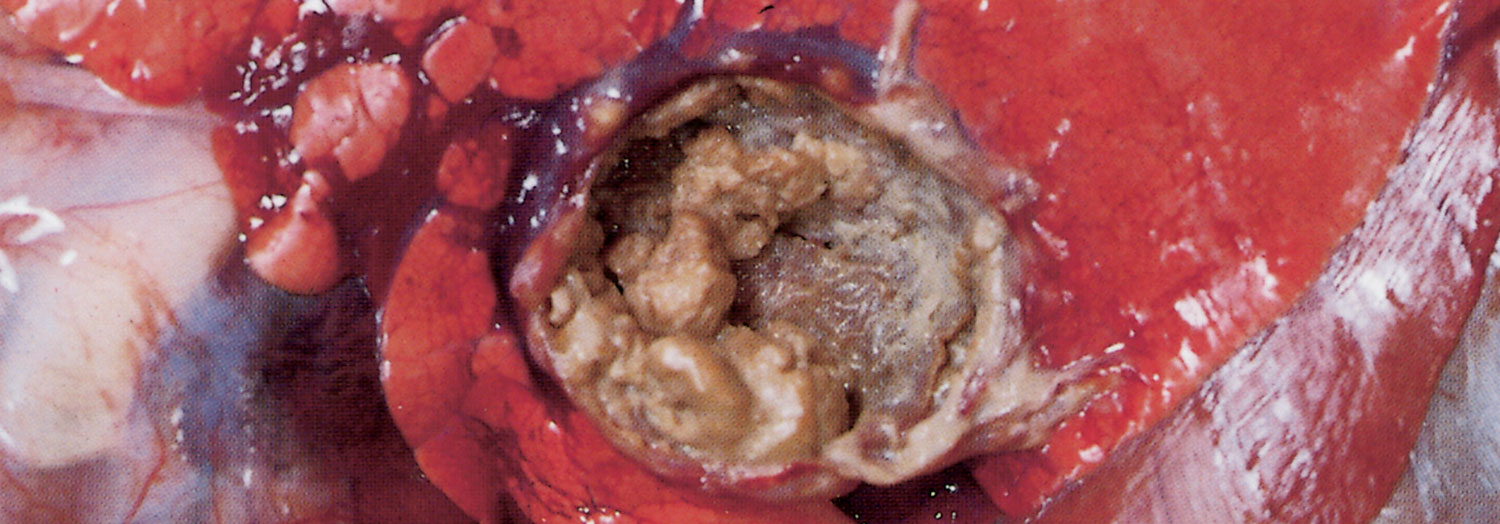
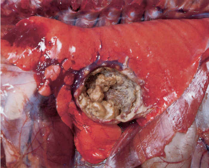
Trueperella pyogenes infection in cattle can cause chronic suppurative pneumonia173 and pulmonary abscesses,121 which may extend to involve the pleura, resulting in the formation of dense, adhesive, suppurative pleuritis or pyothorax with a foul-smelling exudate (Figure 1).26, 71 Bacterial emboli from a thrombus in the caudal vena cava may lead to embolic pneumonia or pulmonary abscesses. The organism can be an important complicating pathogen in the bovine respiratory disease complex (Mannheimia haemolytica, and/or Pasteurella multocida, or Histophilus somni infection), the most important illness of feedlot cattle,6 Mycoplasma bovis infections, or cases of infectious bovine rhinotracheitis.26
Adult cattle with chronic suppurative pneumonia are frequently afebrile but are tachypnoeic, demonstrate frequent and productive coughing, as well as halitosis and a purulent nasal discharge when the head is lowered.173 Lung lesions may be accompanied by the presence of metastatic abscesses elsewhere in the body.
In cattle, one or more liver abscesses can erode into the caudal vena cava or large hepatic vein and cause localized purulent thrombophlebitis. Septic emboli can then lead to right-sided mural or valvular endocarditis, or lodge in one or more pulmonary arteries resulting in embolic pulmonary abscesses or possibly the pulmonary embolic aneurysm (PEA or pulmonary arterial thromboembolism) syndrome. Similar septic emboli could also originate from traumatic reticulopericarditis. Rupture of a pulmonary aneurysm can then lead to pneumorrhagia, haemoptysis and epistaxis.130 Clinical signs of PEA range from an intermittent, blood-flecked, mucoid nasal discharge to severe, profuse, bilateral haemorrhaging from the nose, culminating in death. Pneumorrhagia may similarly be caused if a large blood vessel ruptures into a bronchus as a result of the erosion of its wall following pressure exerted on it by a primary lung abscess
Trueperella pyogenes pulmonary infections can also complicate bronchiolar-alveolar adenocarcinomas (jaagsiekte) in sheep, pneumonia due to M. haemolytica in sheep and goats, and atrophic rhinitis, mycoplasmal pneumonia or pleuropneumonia in pigs.71, 78
Cattle, and sometimes pigs, can develop chronic heart failure due to valvular, especially right atrioventricular, and/or mural endocarditis associated with T. pyogenes. The lesions on the valves or elsewhere on the endocardium tend to be large, vegetative, and firm but friable nodules. Such endocarditis may arise following embolic spread from hepatic abscesses, caudal vena cava thrombus, purulent metritis, mastitis or traumatic reticulopericarditis.160 Emboli originating from valvular endocarditis may be responsible for the development of septic infarcts and abscesses in the brain, lungs and kidneys, and osteomyelitis and septic arthritis.29, 160
Vegetative valvular endocarditis can lead to chronic passive congestion manifested by dependent oedema, transudation into body cavities, oedema of the abomasal wall, and cyanotic induration of the liver.160 A prominent jugular pulse may be noticeable. Despite extensive lesions in the heart, an electrocardiogram may be normal, and auscultation frequently does not reveal any evidence of cardiac murmurs.20
Gastrointestinal tract
The nozzles of dosing guns, particularly if burred, may penetrate the pharyngeal mucosa and cause injuries, which, if infected, may result in purulent inflammation and abscessation. The severity and extent of the lesions are greatly aggravated if the material that is administered is an irritant. The inflammatory process may extend to the facial and peripharyngeal tissues or down the neck in the loose peri-oesophageal and other connective tissues as far as the thoracic inlet. Affected animals have difficulty in swallowing or eating, and the infected tissues are often markedly swollen.
The awns of various grass seeds, including those of Themeda triandra (‘rooigras’), Stipagrostis and Aristida spp. (‘steekgras’), Heteropogon contortus (assegai grass) and Critesion spp. (wild barley or jackal tail), the seeds of Bidens pilosa (blackjack), the spines of prickly pears (Opuntia spp.) and other thorny plants (e.g. Acacia spp.), and fibrous weed material (e.g. Salvia reflexa)97, 162, 186, 192, 197 may injure the buccal mucous membranes or the skin. The abscesses and suppurative lesions46 that develop in the buccal and pharyngeal regions can be complicated by pneumonia,162 arteritis, polyarthritis, meningoencephalitis or uveitis.186 Lesions in the tissues of the lips and cheeks may interfere with mastication because of the pain and inhibition of movement of the parts due to intense fibroplasia and induration.
In an abattoir survey of the tonsils of grass-fed cattle that were clinically normal, a surprisingly high (29,3 per cent) prevalence of abscesses was found in which three out of four samples that were submitted for bacteriological examination were positive for T. pyogenes.45
In cases of carbohydrate overload or acidosis in ruminants, the predominant bacteria causing lesions in the forestomach are firstly, F. necrophorum, and secondly T. pyogenes, while fungi belonging to the Mucorales, including Mucor, Absidia and Rhizopus spp., can also be involved.107, 197
Colonization by T. pyogenes of the gastric mucosa of pigs with haemorrhagic gastritis or gastric ulceration has been shown to be surprisingly frequent.93
The migratory tracts of Oesophagostomum spp. larvae are often associated with miliary abscesses caused by T. pyogenes in the wall of the intestine.
Skin and adnexa
Abscesses in the skin and adnexa and in the regional lymph nodes are common in those parts of southern Africa where infestation with ticks possessing long mouth parts, such as Amblyomma spp., Hyalomma spp. and Rhipicephalus spp., are common. The percutaneous migration of helminth larvae, such as Strongyloides spp., as well as blowfly (Lucilia spp.) strike may result in purulent inflammation and abscessation.
The cutaneous and oral lesions due to lumpy skin disease71 and foot and mouth disease207 become secondarily infected with T. pyogenes.
Awns of certain plant seeds, such as blackjacks, ‘kakiebos’ (Tagetes minuta), and some grasses, such as Critesion, Aristida and Heteropogon spp., may penetrate the skin of woolled sheep in great numbers in those ventral aspects of the body that are most exposed to them. These are easily seen in the subcutis when the animal is skinned. The resulting lesions act as portals of entry for bacteria (often T. pyogenes),97 and the resulting subcutaneous abscesses, which are usually miliary, may cause severe discomfort, anorexia, loss of body weight and death. On histopathological examination, plant-awn material is frequently detected inside pyogranulomas or abscesses.162
Heavy adult sheep, particularly pregnant ewes continually exposed to excessive moisture when grazing on lush, wet pastures, may develop maceration of the interdigital skin. This predisposes to the penetration of bacteria and the development of interdigital dermatitis, which may progress and lead to ‘foot abscess’ and contagious footrot.122, 159 Lesions caused by tick bites and plant awns, or other sharp objects, may also predispose to these foot conditions.
It is possible that a degree of vascular stasis, which occurs in the digital cushion and interdigital dermis of the hind feet of pregnant ewes, may also predispose to foot infections.159 In cattle, necrobacillosis of the foot (also known as panaritium or foul-in-the-foot) is caused by a mixed infection of F. necrophorum and P. melaninogenica (see Fusobacterium necrophorum, Dichelobacter (Bacteroides)) nodosus and Bacteroides spp. infections).
Contagious footrot in small ruminants, on the other hand, is caused by Dichelobacter (Bacteroides) nodosus infection in association with F. necrophorum and other bacteria. Ovine ‘foot abscess’, which comprises ‘toe abscess’, in which the lamellae of the hoof are affected, and ‘heel abscess’ (or infective bulbar necrosis), in which the soft bulbar tissues of the digital cushion are involved,159 is caused by a combined infection involving F. necrophorum and T. pyogenes.209 (see Fusobacterium necrophorum, Dichelobacter (Bacteroides)) nodosus and Bacteroides spp. infections)
In sheep suffering from foot abscess, severe lameness is the outstanding sign and the affected foot is carried. Usually only one claw (particularly of a hind foot) is affected, but more than one may be involved.209 The hoof is usually not affected in the early stages, but it does become involved as the disease advances, and may become progressively deformed. Viscous, semi-liquid necrotic tissue may be expressed from the bulb of the heel through irregular openings in the interdigital skin, or the abaxial coronary band may bulge and rupture at focal areas to discharge greyish, blood-stained necrotic debris. The discharge eventually becomes more purulent.209 These areas may granulate and heal, only to fistulate again at the same site or elsewhere in the coronary skin. The interdigital infection spreads very easily to the coffin (distal interphalangeal) joint because the dorsal and palmar/plantar joint pouches extend up above the horn into the coronary area.208 The necrotizing infection in the distal interphalangeal joint may cause the axial collateral and interdigital ligaments to rupture, resulting in luxation of the distal phalanx and distal sesamoid bone.209 The pastern joint may also become involved. The course of foot abscess is about two months; slight lameness generally persists and, if ligaments are damaged, deformity of the digit may be permanent.159, 209
Neonatal piglets maintained on abrasive floors without bedding may develop lameness due to the development of foot abscesses mainly in the hind limbs, the medial claws in particular being infected. Bruising and erosion of the sole of the foot allows the penetration of polymicrobic infections that commonly include T. pyogenes, F. necrophorum and Bacteroides spp.57 In heavy sows, inflammation in the extremities causes oedema and pain and laminitis can develop rapidly, leading to swelling and heat in the coronary band, exudation of pus, and recumbency.87 Radiographic examination may reveal small abscesses, fractures of phalangeal bones, osteomyelitis and/or tendosynovitis.87
In a case-control study, T. pyogenes and E. coli have been isolated from cases of toe tip necrosis syndrome in Canadian feedlot cattle. The hypothesis is that this syndrome is caused by excessive wear along the white line that leads to separation and bacterial colonization of the third phalangeal bone and associated soft tissues.139
Skeletal system
Unhygienic environmental conditions may result in the infection of umbilical, docking and/or castration wounds in new-born animals with subsequent spread to joints, vertebral bodies and/or other sites in the body.
Trueperella pyogenes is one of the causes of purulent arthritis in calves, lambs, kids and pigs. Lameness is the outstanding clinical sign and, on palpation, affected joints are painful, hot and swollen. Only a few joints are generally involved. In chronic cases, the affected joints become thickened and may develop ankylosis, in which case their movements are severely restricted; and the joint capsule may even be partially mineralized.
Spinal epidural abscesses, tracking abscesses within the spinal cord, and purulent vertebral osteomyelitis followed by vertebral body abscessation and/or discospondylitis are quite common in calves, lambs, kids and piglets.24, 49, 80, 174, 179 These lesions may occur in the cervical, thoracic or lumbar regions and usually involve a single vertebral body or intervertebral disc. Adjacent vertebrae may also be affected.49 Since there are connections between the vertebral and the caval, portal and pulmonary venous systems, infection may arise from metastatic spread from pneumonic lesions, liver abscesses or omphalophlebitis.49, 179 Clinically, these animals develop progressive posterior paresis and paralysis that culminates in paraplegia or tetraparesis. Affected animals are normally bright and alert and, if only paraplegic, may drag themselves around using their fore legs. Staphylococcus spp. may also be the cause of vertebral body abscesses.49
Trueperella pyogenes infection is also associated with vertebral body osteomyelitis, in some cases with abscessation in adult cattle, pigs and, more rarely, sheep. The infection in most of these cases is assumed to be a result of haematogenous spread of bacteria from a primary site elsewhere in the body, the latter often not being established. In beef cattle in South Africa, particularly in bushveld (savannah) regions, it may at times become an economically significant problem in some herds. One unsubstantiated hypothesis as to its pathogenesis is three-pronged:196
• The primary site of the bacteria is infected skin lesions created by tick infestations, particularly when these are severe. Other infected skin wounds may also play a role, such as injuries by sharp projections in poorly constructed crushes or spray-races. These lead to periodic episodes of bacteraemia in which the bacteria may localize in distant sites, particularly if a preceding lesion is present (locus minores resistentiae).
• The second factor is the presence of nutritional osteomalacia, generally subclinical, due to a phosphorus deficiency, which, unless remedied, is common in parts of the southern Africa. This creates a situation in which microfractures (or even frank fractures) of bone are liable to develop. The localized bone trauma, often accompanied by a resultant haemorrhage, forms a ready site for the localization of bacteria from the blood stream.
• The third factor pertains to management procedures. Important among these is the driving of fractious animals into crushes or races during dipping, spraying, dosing or inoculation procedures. It frequently happens that cattle attempt to escape from a crush by turning or jumping, and this may lead to injuries that could include microfractures in a vertebra. In straining to turn, or in jumping, the lumbar region is often the most severely stressed part, which is the reason why—according to this hypothesis— one or more lumbar vertebral bodies are the most frequently affected. The suppurative osteomyelitis that develops may result in fracture with collapse or partial collapse of the vertebral body and pressure on the spinal cord, or the infection may extend to adjacent structures outside the bony tissue. Extension into the spinal canal may result in either cranial or caudal spread of the infection or its localization as an epidural abscess.24 In many cases, however, the exudate remains confined to the vertebra by the periosteum as an encapsulated abscess that protrudes into the spinal canal, with subsequent compression malacia of the spinal cord. Clinical signs in such an animal are those of progressively developing paresis and paralysis of the limbs. As many lesions are in the lumbar or thoracolumbar region, the hind limbs are most frequently involved, the final outcome being complete paraplegia. Affected animals assume a ‘dog-sitting’ position—hence the Afrikaans name ‘sitsiekte’—as they are still able to maintain an upright sitting position by the use of their fore limbs.196
Mandibular osteomyelitis caused by Actinomyces bovis (‘lumpy jaw’) (see Actinomyces bovis infections) may be complicated by other bacteria, such as T. pyogenes and F. necrophorum.33 In Brazil, a serious infectious periodontitis of calves, called ‘cara inchada’ (swollen face), is caused by P. melaninogenica and T. pyogenes and is associated with the altered ecological soil conditions that follow the clearing of forests and the establishment of cultivated pastures (Panicum maximum in particular).17, 39 Nutritional secondary hyperparathyroidism also plays a role.17 The bacteria produce enzymes and endotoxins that damage periodontal tissues, leading to a purulent periodontitis and secondary ossifying alveolar periostitis.39 Also, antibiotics produced by soil actinomycetes, such as streptomycin and actinomycin, have been shown to increase the adherence of P. melaninogenica to epithelial cells of the gums.64, 101
In young piglets, complications of tail-biting include embolic pneumonia,198 spinal epidural abscessation and vertebral osteomyelitis,49, 141 cerebral abscesses199 and septic embolic lesions elsewhere.24 In older pigs, T. pyogenes causes purulent arthritis, chronic purulent osteomyelitis of the costochondral junctions, aseptic necrosis of cartilage and bone, separation of epiphyses (especially of the femur), pathological fractures, massive disfiguring abscesses in periarticular muscle, associated phlebitis, cervical lymphadenitis and sepsis.87, 88, 191 Affected joints can be extremely swollen, deformed, hot and painful and yellow-green pus may drain from one or more fistulae.87 Organisms isolated from abscesses and other lesions in pigs include T. pyogenes, Staphylococcus spp., haemolytic Streptococcus spp., Erysipelothrix rhusiopathiae, coliforms, as well as anaerobic and facultatively anaerobic bacteria, in particular Bacteroides spp.9, 49, 198, 199
Central nervous system
A highly fatal neurological disorder known as basilar empyema or pituitary abscess syndrome is sporadically encountered in cattle, goats, sheep and pigs.8, 69, 100, 142 In the majority of cases it is characterized by the development of suppurative meningitis of the ventral or basilar aspect of the brain and abscessation with or without involvement of the pituitary gland itself and adjacent brain stem. In some, however, abscessation in the pituitary gland itself is the primary cause of the syndrome. Trueperella pyogenes, either alone or as part of a mixed infection with other organisms such as Streptococcus spp., Pasteurella spp., F. necrophorum, Pseudomonas aeruginosa, Staphylococcus aureus, Haemophilus spp. and others, is the most common bacterium isolated from the lesions.7, 69, 100, 100, 142 While the pathogenesis of the syndrome remains to be elucidated, it seems probable that the most common routes that organisms use to reach the parts concerned are venous, arterial or lymphogenous, the infection being secondary to a chronic suppurative process elsewhere in the body.142 A complex mesh of arterioles, known as the rete mirabile, surrounds the pituitary gland of cattle, goats, sheep and pigs. The rete mirabile and the pituitary are bathed in a venous bed, the cavernous sinus, which is part of the sinuses of the dura mater. By means of diploic veins that traverse the bones of the skull, these sinuses are connected to the subcutaneous veins of the head.8, 142 As they are valveless, blood in them can flow in both directions. It is thought that the complex vascular dynamics make this part susceptible to the embolic deposition of bacteria. In addition, bacteria might invade via lymphatics,142 as the existence of a communication between the cerebrospinal fluid and the lymphatic system has been documented, particularly in the area of the nasal mucosa and the cribriform plate. Another route of entry for bacteria is by direct extension of an otitis interna.83
In South Africa, the pituitary abscess syndrome is most frequently encountered in Boer goats, particularly horned animals, in which it seems to be associated with the attachment of ticks, such as Rhipicephalus evertsi, to the skin of the head, especially that of the caudal aspects at the base of the horns. It is thought that the tick bites provide a portal of entry for pyogenic bacteria that are responsible for the development of a suppurative inflammatory process in the skin and subcutis, with subsequent venous distribution of the bacteria to the cavernous sinus. It is possible that some cases of the syndrome in cattle may also be associated with the attachment of ticks, such as R. evertsi, to the skin of the head and ears. Other cases in cattle have been associated with septic rhinitis that developed after nose-ringing100, 142 or broken-off plant sticks in the nasal cavity where they had presumably tried to alleviate allergic nasal pruritus due to, for example, nasal granuloma. In rams, infection following fighting injuries or sinusitis may lead to the condition.69, 100 Other primary sites of bacterial infection thought to play a possible role include polyarthritis, suppurative nephritis, mastitis, pneumonia, tooth abscess, and parotid, mediastinal and mandibular lymph node abscessation.142
The clinical signs of pituitary abscessation resemble those of heartwater to some extent, and for this reason many cases in southern Africa are possibly misdiagnosed. A wide variety of signs are manifested by affected animals; this precludes the description of a ‘typical’ case. Signs include several of the following: obtundation, blindness, pupil-diameter abnormalities, absent pupillary light reflex, ventrolateral strabismus, nystagmus, dropped jaw, decreased tongue tone, facial paralysis, head-tilt, dysphagia, head-pressing, circling, ataxia, lateral recumbency, opisthotonus and paddling movements of the legs.112 There is usually no fever or evidence of pituitary endocrine disturbances. Recovery of these animals is unusual; most die within days. Treatment is usually of no avail, and for this reason is generally not advocated. It should be borne in mind that by the time the clinical signs are manifested or death has occurred, ticks that played a pathogenetic role may already have detached.
Cerebral abscesses due to T. pyogenes may arise following severe Oestrus ovis infestation or otitis in sheep,24, 80 or septic embolism from endocarditis24 or as a complication of dehorning in cattle.24 Animals suffering from epidural or subdural abscesses of the spinal cord, which will probably have arisen from septic embolism, may present with similar signs to those associated with vertebral body abscesses.24
Outbreaks of uni- or bilateral otitis media in calves have been described in Germany and Israel.150, 211 Clinical signs include apathy, anorexia, fever, head tilting, drooping of the affected ear(s) and purulent discharge from the external ear canal. Otitis media may follow outbreaks of enzootic pneumonia.150 Organisms isolated included Pasteurella spp., T. pyogenes and Candida spp.211
Diagnosis
The clinical signs of T. pyogenes infections in animals depend on the size, severity and location of the lesions. Cattle with liver abscesses generally do not show clinical signs unless one or more superficial ones rupture into the peritoneal cavity or perforate the caudal vena cava.130 Liver abscesses are normally only detected at slaughter. Although there are limitations, especially cost, ultrasonography may be useful in detecting abscesses in research contexts.130 A diagnosis should be confirmed by the isolation of the bacterium from specimens of affected tissues or organs. An array of laboratory tests exists in addition to bacterial isolation that can be used to confirm the diagnosis (See Aetiology).
Recording and monitoring lung sounds combined with ultrasound has been successfully used to accurately diagnose chronic suppurative pneumonia in adult cattle.173
In summer mastitis, the putrid smell of the milk, abscess development and systemic illness are characteristic.31 Mastitic milk should be cultured aerobically and anaerobically,182 as T. pyogenes is seldom the sole pathogen present.
To confirm a diagnosis of pituitary abscessation at necropsy it is often necessary to expose the pituitary gland by making an incision through the diaphragma sellae of the dura mater that covers the gland.
If T. pyogenes is the cause of an abortion, it is readily isolated from foetal lung, abomasal content, placenta or vaginal mucus.76, 172, 184 Foetal lung is a useful specimen for bacteriology and histopathology in the confirmation of the cause of abortion, as T. pyogenes usually causes foetal bronchopneumonia.113
Differential diagnosis
The differential diagnosis depends on the particular presenting syndrome. Purulent processes and abscesses are caused by many other species of pyogenic bacteria, the most common being Staphylococcus spp., Streptococcus spp. and C. pseudotuberculosis, which is the cause of caseous lymphadenitis. Other organisms that can cause lymphadenitis and abscesses in small ruminants include Actinomyces hyovaginalis and Staphylococcus aureus subsp. anaerobius.35 Lesions from which T. pyogenes may be isolated in pigs at slaughter may resemble tuberculosis.25
Purulent polyarthritis due to T. pyogenes in sheep may mimic polyarthritis due to E. rhusiopathiae, Streptococcus spp., S. aureus and C. pseudotuberculosis. The clinical signs in an animal suffering from the basilar empyema may resemble those of heartwater, cerebral babesiosis and theileriosis, rabies, listeriosis,100, 112 cerebrocortical necrosis, lead poisoning, cerebral and other forms of abscessation within the cranium, and thrombotic meningoencephalitis (Histophilus somni infection) in cattle; and of focal symmetrical encephalomalacia and heartwater in sheep and goats. Other encephalitides and space-occupying lesions (e.g. Coenuris cerebralis) should also be considered.142
Orchitis or epididymitis caused by T. pyogenes in rams should be differentiated from the lesions caused by Brucella ovis, Histophilus ovis, Actinobacillus seminis, C. pseudotuberculosis and Chlamydophila spp. and from non-infectious spermatic granulomas and varicoceles.
Foot abscess should be differentiated from footrot (Dichelobacter nodosus infection), ovine interdigital dermatitis (foot scald), strawberry footrot (Dermatophilus congolensis infection), laminitis, and from foot lesions caused by bluetongue, injuries and joint-ill.
In sheep, abortion due to T. pyogenes should be differentiated from that caused by certain viruses, such as Rift Valley fever and Akabane and related Simbu-group viruses, Salmonella serovars, Campylobacter spp., Listeria monocytogenes, Coxiella burnetii, Toxoplasma gondii, and Chlamydophila abortus. Bacteria other than T. pyogenes that cause uterine infections in cows include E. coli and other coliforms, Pasteurella spp., Streptococcus spp.18, 67, 188 and Rhodococcus equi.32 Uterine infections or abortion caused by T. pyogenes in mares should be differentiated from bacterial, rickettsial, protozoal and viral infections that cause reproductive failure.
Although T. pyogenes is a frequent cause of vertebral osteomyelitis in cattle, other bacteria, such as F. necrophorum, Staphylococcus spp., Streptococcus spp. and coliforms may also be implicated.49 Differential diagnoses of this syndrome also include fracture of a vertebral body, nutritional muscular dystrophy, ‘dumb’ rabies, Histophilus somni infection, Hypoderma bovis infestation, meningitis of any cause, spinal lymphosarcoma, the downer cow syndrome, and certain hereditary conditions, such as progressive ataxia in Charolais, degenerative myeloencephalopathy in Brown Swiss cattle, and congenital spinal dysraphism.49, 179
It is important to note that Mycoplasma bovis infection can also cause otitis media, chronic pneumonia, polyarthritis, mastitis, abortion, and vesicular adenitis in bulls;123 T. pyogenes may also be isolated from such cases. Differential diagnoses for paraplegia caused by vertebral body abscesses in pigs include trauma and organic arsenic or organic mercurial poisonings.49
In sheep, conditions which may be confused clinically with vertebral osteomyelitis and abscessation include swayback (which is a copper deficiency), white muscle disease, tick paralysis caused by Rhipicephalus evertsi or Ixodes rubicundus, botulism, hypocalcaemia, trauma and Coenuris cerebralis (particularly cysts of Taenia multiceps located in the spinal column).
Control
Tick control is of major importance in the control of subcutaneous abscesses, pituitary abscessation and abscesses in the skin of the udder of domestic ruminants. Sites of tick attachment, such as behind the horns and the escutcheon, should be sprayed regularly with an acaricide.8 Dehorning goats, or the breeding of polled goats, can be considered where pituitary abscessation is a problem.8 Acaricide footbaths or pour-ons should be used where ticks on the feet have caused abscesses. Appropriate anthelmintics will control Oesophagostomum infestation.
Strict hygiene must be applied when docking or castrating, particularly when elastic rings are used for castration. Omphalophlebitis and subsequent pyaemia may be prevented by the disinfection of the umbilical stump with povidone iodine at birth and again four hours later.174 Pregnant animals should not be permitted to give birth on infected premises.
It may be necessary to avoid certain pastures or veld types at certain times of the year in order to lower the exposure of animals, particularly woolled sheep, to plant awns and seeds. The point of the nozzle of dosing guns must be free of burrs and, in order to prevent injury to the pharyngeal region while dosing, the nozzle must not be forced to the back of the mouth—it must be borne in mind that many animals jump forward during the dosing process.
Abscesses which are accessible to surgical intervention should preferably be excised intact.59 Alternatively, they may be lanced and drained (following local or general anaesthesia), and the abscess cavity packed with iodine gauze or flushed with povidone iodine, hydrogen peroxide or chlorhexidine.59 Success with antimicrobial treatment alone is unlikely to be successful.121
In sheep with foot abscesses, surgical drainage and irrigation of the lesion with chlorhexidine antiseptic solution can be attempted; the foot should be bandaged to prevent further contamination and to reduce splaying of the affected digit.159, 209 Systemic antibiotic treatment can be instituted where lesions are in the acute stage. Formalin (3 to 5 per cent) footbaths can be utilized on a weekly basis to reduce the incidence of foot infections.
Purulent arthritis may be treated surgically by external drainage and repeated flushing of the joint with antimicrobials, but the prognosis is guarded. Prolonged systemic treatment of such cases with a long-acting oxytetracycline may sterilize the infection, but affected joints rarely recover completely.
Mild cases of T. pyogenes endometritis in cows can be treated successfully with intrauterine infusions of gentamicin; however, the problem of tissue and milk residues should be considered.5, 67 In pyometra with placental retention, the uterine exudate should be siphoned off using a 10-mm-diameter tube. Parenteral antibacterial drugs are beneficial when systemic signs are present.51 Antimicrobial instillation in these cases is of questionable value, as many antimicrobials are rapidly inactivated by lochial fluids. Although cloprostenol (prostaglandin F2α analog) treatment can give excellent results when a corpus luteum is present,51 the efficacy of treatment with intrauterine antimicrobials and PGF2α to obtain a clinical cure, and positive reproductive outcome, is controversial.200 Prevention of postparturient endometritis involves the improvement of hygiene and the rotation of calving paddocks.51
Since obligate anaerobic bacteria, such as P. melaninogenica and F. necrophorum, are frequently present in cases of T. pyogenes infection,47, 163 metronidazole treatment can be considered when the expense is justified. Use of metronidazole eliminates the anaerobes and, in so doing, reduces their inhibitory effect on neutrophil phagocytosis, thus allowing the host’s phagocytic defences to operate against T. pyogenes.82
The costs of antibiotic treatment and detrimental effects on reproduction, loss of milk production and survivability have been estimated at nearly US$400 per cow with metritis.138
Cows suffering from pyogenic or summer mastitis or discharging abscesses should be isolated.31, 70 In those parts of the world where they are a problem, flies should be controlled using insecticide sprays, insecticide-impregnated ear tags, or pour-ons.31 Parenteral treatment with oxytetracyclines can be attempted during the peracute stage, but the response to intensive treatment is usually disappointing.31 Failure of treatment may be due to the extent of the suppuration and not antimicrobial resistance per se.31 Amputation of the teat to facilitate drainage can be considered but even intensive treatment fails in 80 per cent of quarters.31 Affected quarters are probably best treated by permanently drying off by the infusion of 0,5 per cent iodine after complete milk out and the administration of 1 mg/kg flunixin meglumine intravenously.31 Intramammary prophylaxis using semi-synthetic penicillins (such as cloxacillin and ampicillin) at regular intervals can be successful.31, 193 Polymicrobial sensitivity data show susceptibility to penicillin G, amoxicillin, amoxicillin-clavulanate, cefoxitin, clindamycin and chloramphenicol.94 The likelihood of recovery of affected quarters is inversely proportional to the severity of onset.84 There is evidence that cows that recover from pathogen specific clinical mastitis are unlikely to be protected against recurrence.27 First-parity cows with T. pyogenes mastitis have a high risk for having to be culled.28
With respect to the bovine respiratory disease complex, multi-drug resistance appears to be an increasing trend regarding important pathogens, such as T. pyogenes.6 With the advent of multi-drug resistant strains and the ability of T. pyogenes to form biofilms, recent work has shown that bacteriophages may be effective in preventing such biofilm formation. Bacteriophages are non-toxic, effective and economically viable alternatives to antibiotics.42 In one case study, daily treatment with procaine penicillin for six weeks was successful in treating chronic suppurative pneumonia in cattle.173
From an economics perspective, liver abscesses are second only to respiratory diseases as causes of losses in feedlot cattle.92 In the USA, sub-therapeutic antibiotic feed additives (tylosin, chlor- and oxytetracycline, bacitracin and virginiamycin) are approved for the control of liver abscessation in feedlot cattle. The mode of action of these growth-promoting antibiotics is probably via the inhibition of F. necrophorum. Tylosin is the most successful (and bacitracin the least) and reduces abscess incidence by 40 to 70 per cent and increases weight gain and feed conversion92, 129 due to the effect against F. necrophorum in the rumen.130 Of interest, though, is that the incidence of liver abscesses containing T. pyogenes increases in cattle fed tylosin.128 However, many isolates have demonstrated resistance to antimicrobials,15, 41, 92, 195 so development of vaccines is now more urgent than ever.91 For the prevention of liver abscesses, the most important consideration is sound nutritional management, particularly increased roughage and decreased easily fermentable carbohydrates.130
Antimicrobial resistance
Antimicrobial resistance is common in T. pyogenes.195 In a study of 94 T. pyogenes isolates from 83 cattle with respiratory disease, every isolate was resistant to at least two of the classes of antimicrobials tested and 80 per cent of isolates were resistant to four or more classes of antimicrobials. Extreme multi-drug resistance was present in 36 per cent of isolates.6 Trueperella pyogenes isolates had especially high levels of tetracycline resistance (>90 per cent) and macrolide resistance (>75 per cent). It is disturbing that the antimicrobials to which little resistance was observed (enrofloxacin 0 per cent and ceftiofur 1,1 per cent) are regarded as drugs of great importance in human medicine and thus veterinary use is discouraged.6 Similarly, 69 of 72 uterine isolates (96 per cent) of T. pyogenes exhibited resistance to at least two antimicrobials tested (amoxicillin, ampicillin, chloramphenicol, florfenicol, oxytetracycline, penicillin and tetracycline), and multi-drug resistance was observed in 64 (89 per cent) of isolates.167 In this study over 50 per cent of isolates were resistant to oxytetracycline.167 In a study out of China, more than 60 per cent of T. pyogenes isolates were resistant to tetracycline and doxycycline, and 3,1 per cent showed resistance to all tetracyclines.214 Another recent study found that florfenicol, cefoperozone, cephalexin and ceftiofur were more than 90 per cent effective against T. pyogenes.154 The use of ceftiofur and ampicillin against E. coli and T. pyogenes in acute puerperal metritis in dairy cows has shown that increasing minimum inhibitory concentrations of these antimicrobials could indicate the emergence of resistance, particularly regarding E. coli.145
Resistance to tetracyclines is usually mediated by active efflux of tetracycline from bacteria, or ribosomal protection from tetracycline effects. TetW is a ribosomal protection tetracycline resistance protein encoded by the tetW gene, usually carried on a mobile genetic element capable of transfer among strains. Its presence has been reported in T. pyogenes.15 Other tetracycline resistance genes (tetK, tetL, tetM, tetO, tetZ, tet33 and otrA) as well as genes conferring resistance to macrolide-lincosamide-streptogramin B, aminoglycosides, trimethoprim, phenicols and beta-lactams have been identified in T. pyogenes.48, 214 Furthermore, specific ribosomal RNA mutations are associated with macrolide-lincosamide resistance in T. pyogenes.48
In general, it appears that ceftiofur and ampicillin constitute the best choices for antibacterial treatment of T. pyogenes infections. Antimicrobials should be used with appropriate restraint to limit the development of antimicrobial resistance.
Vaccination
Many years ago, attempts to immunize animals effectively by employing A. pyogenes cells or toxoid met with variable results.21, 147 Although multiple doses of such a bacteria-toxoid vaccine of high potency induced a substantial antibody response,22 protection against infection was generally poor38, 81, 187 and use of the vaccine was thought to suppress cell-mediated immunity.81
In the USA, two vaccines, one containing a F. necrophorum bacterin and a second containing a leukotoxoid of F. necrophorum combined with the T. pyogenes PLO bacterin have been used commercially in feedlots.4, 130 The latter vaccine, however, is no longer available.4 There was no additive effect in the reduction of liver abscesses when feedlot cattle received tylosin plus vaccination.90, 130
Experiments to prevent puerperal metritis in dairy cows has shown that subcutaneous, as opposed to intravaginal, vaccination with inactivated bacterial components and/or protein subunits of E. coli, F. necrophorum and T. pyogenes caused a significant increase in serum IgG titres against all antigens and led to improved reproduction.116 On the other hand, an experimental herd-specific multivalent vaccine containing inactivated whole bacterial cells of T. pyogenes, E. coli, S. uberis, Bacteroides spp., and Peptostreptoccus spp., administered subcutaneously, had no effect on reducing the risk of uterine disease or reproduction or overall production in dairy cows.54
An autogenous T. pyogenes vaccine administered to pregnant sows enhanced the immune potential of colostrum and reduced economic losses in suckling piglets and weaners.102
Promising work has also begun on the efficacy of a DNA vaccine based on the PLO gene against T. pyogenes infections in mice.79
References
- ABDULMAWJOOD, A., WICKHORST, J., HASHIM, O., SAMMRA, O., HASSAN, A. A., ALSSAHEN, M., LÄMMLER, C., PRENGER-BERNINGHOFF, E. & KLEIN, G., 2016. Application of a loop-mediated isothermal amplification (LAMP) assay for molecular identification of Trueperella pyogenes isolated from various origins. Molecular and Cellular Probes, 30, 205-210.
- ADAMU, S. A. & SPERRY, J. F., 1981. Polymorphonuclear neutrophil chemotaxis induced and inhibited by Bacteroides spp. Infection and Immunity, 33, 806-810.
- ADDO, P. B. & DENNIS, S. M., 1979. Experimental production of Corynebacterium pyogenes abortion in sheep. Cornell Veterinarian, 69, 20-32.
- AMACHAWADI, R. G. & NAGARAJA, T. G., 2016. Liver abscesses in cattle: A review of incidence in Holsteins and of bacteriology and vaccine approaches to control in feedlot cattle. Journal of Animal Science, 94, 1620-1632.
- ANETZHOFER, J. V., 1989. Die behandlung der Actinomyces pyogenes-endometritis durch intrauterine Gentaseptin(R)-applikation. Schweizer Archiv für Tierheilkunde, 139, 495-498.
- ANHOLT, R. M., KLIMA, C., ALLAN, N., MATHESON-BIRD, H., SCHATZ, C., AJITKUMAR, P., OTTO, S. J., PETERS, D., SCHMID, K., OLSON, M., MCALLISTER, T. & RALSTON, B., 2017. Antimicrobial susceptibility of bacteria that cause bovine respiratory disease complex in Alberta, Canada. Frontiers in Veterinary Science, 4, 207.
- BATH, G. F., 1989. Department of Theriogenology, Faculty of Veterinary Science, Onderstepoort. Unpublished data.
- BATH, G. F., 1994. Peripituitary abscessation of goats in Southern Africa. Kenya Veterinarian, 16, 41-42.
- BENNO, Y., T., M. & SHIRASAKA, S., 1982. Anaerobic bacteria isolated from abscesses in pigs. Japanese Journal of Veterinary Science, 44, 309-315.
- BEVERLEY, J. K. A. & WATSON, W. A., 1962. Further studies on toxoplasmosis and ovine abortion in Yorkshire. Veterinary Record, 74, 548-552.
- BICALHO, M. L. S., LIMA, F. S., MACHADO, V. S., MEIRA, E. B., JR., GANDA, E. K., FODITSCH, C., BICALHO, R. C. & GILBERT, R. O., 2016. Associations among Trueperella pyogenes, endometritis diagnosis, and pregnancy outcomes in dairy cows. Theriogenology, 85, 267-274.
- BICALHO, M. L. S., MACHADO, V. S., OIKONOMOU, G., GILBERT, R. O. & BICALHO, R. C., 2012. Association between virulence factors of Escherichia coli, Fusobacterium necrophorum, and Arcanobacterium pyogenes and uterine diseases of dairy cows. Veterinary Microbiology, 157, 125-131.
- BICALHO, M. L. S., SANTIN, T., RODRIGUES, M. X., MARQUES, C. E., LIMA, S. F. & BICALHO, R. C., 2017. Dynamics of the microbiota found in the vaginas of dairy cows during the transition period: Associations with uterine diseases and reproductive outcome. Journal of Dairy Science, 100, 3043-3058.
- BILLINGTON, S. J., H., J. B., A., C. W., R., B. K. & SONGER, J. G., 1997. The Arcanobacterium (Actinomyces) pyogenes hemolysin, pyolysin, is a novel member of the thiol-activated cytolysin family. Journal of Bacteriology, 179, 6100-6106.
- BILLINGTON, S. J. & JOST, B. H., 2006. Multiple genetic elements carry the tetracycline resistance gene tet(W) in the animal pathogen Arcanobacterium pyogenes. Antimicrobial Agents and Chemotherapy, 50, 3580-3587.
- BILLINGTON, S. J., POST, K. W. & JOST, B. H., 2002. Isolation of Arcanobacterium (Actinomyces) pyogenes from cases of feline otitis externa and canine cystitis. Journal of Veterinary Diagnostic Investigation, 14, 159-162.
- BLOBEL, H., J., D., F., L. F. G. & ROSA, I. V., 1984. Bacterial isolations from ‘cara inchada’-lesions of cattle. Pesquisa Veterinária Brasileira, 4, 73-77.
- BONNETT, B. N., W., M. S., J., G. V. P., B., M. R. & ETHERINGTON, W. G., 1991. Endometrial biopsy in Holstein-Friesian dairy cows III. Bacteriological analysis and correlations with histological findings. Canadian Journal of Veterinary Research, 55, 168-173.
- BRITO, F., ROSA, G. J., PINEDO, P., SANTOS, J. E., SCHUENEMANN, G. M., BICALHO, R. C., CHEBEL, R. C., GALVAO, K., GILBERT, R. O., RODRIGEZ-ZAS, S. L., SEABURY, C. M., FETROW, J. & THATCHER, W., 2018. Genetic and functional relationships among reproductive traits in US Holstein cows. Abstracts of the 2018 American Dairy Science Association® Annual Meeting June 24–27, 2018 Knoxville, Tennessee. Journal of Dairy Science, 243.
- CABANA, E. M., R., K. W., W., D. R. C. & O’BOYLE, D. O., 1990. A case of bovine valvular endocarditis caused by Corynebacterium pseudodiphtheriticum. Veterinary Record, 126, 41-42.
- CAMERON, C. M., 1966. Corynebacterium pyogenes: The problem of vaccination against infection. Journal of the South African Veterinary Medical Association, 37, 11-15.
- CAMERON, C. M., F., B. W. & SMIT, B. H. J., 1976. Antibody response to and immunity induced by Corynebacterium pyogenes vaccine. Onderstepoort Journal of Veterinary Research, 43, 97-104.
- CAMERON, H. S. & BRITTON, J. W., 1943. Chronic ovine laryngitis. Cornell Veterinarian, 33, 265-268.
- CANTILE, C. & YOUSSEF, S., 2016. Nervous system. In: MAXIE, M. G., (ed.). Jubb, Kennedy, and Palmer's Pathology of Domestic Animals. 6th edn. St. Louis, Missouri, USA: Elsevier.
- CARDOSO-TOSET, F., GÓMEZ-LAGUNA, J., AMARILLA, S. P., VELA, A. I., CARRASCO, L., FERNÁNDEZ-GARAYZÁBAL, J. F., ASTORGA, R. J. & LUQUE, I., 2015. Multi-etiological nature of tuberculosis-like lesions in condemned pigs at the slaughterhouse. PLoS One, 10.
- CASWELL, J. L. & WILLIAMS, K. J., 2016. Respiratory system. In: MAXIE, M. G., (ed.). Jubb, Kennedy, and Palmer's Pathology of Domestic Animals.6th edn. St. Louis, Missouri, USA: Elsevier.
- CHA, E., HERTL, J., SCHUKKEN, Y., TAUER, L., WELCOME, F. & GRÖHN, Y., 2016. Evidence of no protection for a recurrent case of pathogen specific clinical mastitis from a previous case. Journal of Dairy Research, 83, 72-80.
- CHA, E., HERTL, J. A., SCHUKKEN, Y. H., TAUER, L. W., WELCOME, F. L. & GRÖHN, Y. T., 2013. The effect of repeated episodes of bacteria-specific clinical mastitis on mortality and culling in Holstein dairy cows. Journal of Dairy Science, 96, 4993-5007.
- CIANCIOLO, R. E. & MOHR, F. C., 2016. Urinary system. In: MAXIE, M. G., (ed.). Jubb, Kennedy, and Palmer's Pathology of Domestic Animals. 6th edn. St. Louis, Missouri, USA: Elsevier.
- COLLINS, M. D. & JONES, D., 1982. Reclassification of Corynebacterium pyogenes (Glage) in the genus Actinomyces, as Actinomyces pyogenes comb. nov. Journal of General Microbiology, 128, 901-903.
- CONSTABLE, P. D., HINCHCLIFF, K. W., DONE, S. H. & GRÜNBERG, W., 2017. Veterinary Medicine A Textbook of the Diseases of Cattle, Horses, Sheep, Pigs, and Goats, St. Louis, Missouri, Elsevier.
- CRAIG, J. F. & DAVIES, G. O., 1940. Corynebacterium equi in bovine pyometra. Veterinary Journal, 96, 417-419.
- CRAIG, L. E., DITTMER, K. E. & THOMPSON, K. G., 2016. Bones and joints. In: MAXIE, M. G., (ed.).Jubb, Kennedy, and Palmer's Pathology of Domestic Animals. 6th edn. St. Louis, Missouri, USA: Elsevier.
- CULLEN, J. M. & STALKER, M. J., 2016. Liver and biliary system. In: MAXIE, M. G., (ed.). Jubb, Kennedy, and Palmer's Pathology of Domestic Animals.6th edn. St. Louis, Missouri, USA: Elsevier.
- DE LA FUENTE, R., DE LAS HERAS, M., TORRIJOS, C., DIEZ DE TEJADA, P., PÉREZ-SANCHO, M., CARRION, F. J., ORDEN, J. A. & DOMINGUEZ-BERNAL, G., 2017. Short communication: Isolation frequency of bacteria causing lymphadenitis and abscesses in small ruminants in central Spain. Small Ruminant Research, 154, 5-8.
- DEL VECCHIO, R. P., MATSAS, D. J., FORTIN, S., SPONENBERG, D. P. & LEWIS, G. S., 1994. Spontaneous uterine infections are associated with elevated prostaglandin F alpha metabolite concentrations in postpartum dairy cows. Theriogenology, 41, 413–421.
- DENNIS, S. M. & BAMFORD, V. W., 1966. The role of corynebacteria in perinatal lamb mortality. Veterinary Record, 79(4), 105-108.
- DERBYSHIRE, J. B. & MATTHEWS, P. R. J., 1963. Immunological studies with Corynebacterium pyogenes in mice. Research in Veterinary Science, 4, 537-542.
- DÖBEREINER, J., 1990. Zur ätiologie der ‘cara inchada’ einer parodontalen erkrankung der jungrinder in Brasilien. Deutsche Tierärztliche Wochenschrift, 97, 482-490.
- DOHMEN, M. J. W., JOOP, K., STURK, A., BOLS, P. E. J. & LOHUIS, J. A. C. M., 2000. Relationship between intra-uterine bacterial contamination, endotoxin levels and the development of endometritis in postpartum cows with dystocia or retained placenta. Theriogenology, 54, 1019-1032.
- DONG, W. L., KONG, L. C., WANG, Y., GOU, C. L., XU, B., MA, H. X. & GAO, Y. H., 2017. Aminoglycoside resistance of Trueperella pyogenes isolated from pigs in China. Journal of Veterinary Medical Science, 79, 1836-1839.
- DUARTE, V. D. S., DIAS, R. S., KROPINSKI, A. M., XAVIER, A. D. S., FERRO, C. G., VIDIGAL, P. M. P., DA SILVA, C. C. & DE PAULA, S. O., 2018. A T4virus prevents biofilm formation by Trueperella pyogenes. Veterinary Microbiology, 218, 45-51.
- DUARTE, V. D. S., TREU, L., CAMPANARO, S., DIAS, R. S., SILVA, C. C. D., GIACOMINI, A., CORICH, V. & DE PAULA, S. O., 2017. The complete genome sequence of Trueperella pyogenes UFV1 reveals a processing system involved in the quorum-sensing signal response. Genome Announcements, 5.
- DUBUC, J., DUFFIELD, T. F., LESLIE, K. E., WALTON, J. S. & LEBLANC, S. J., 2010. Risk factors for postpartum uterine diseases in dairy cows. Journal of Dairy Science, 93, 5764-5771.
- DUNCANSON, G. R. & SLATER, C. A., 1986. Post mortem health inspection of bovine tonsils. Veterinary Record, 118, 459.
- EDGAR, G., 1935. Mandibular abscess of sheep. Australian Veterinary Journal, 11, 19-22.
- FARIN, P. W., BALL, L., OLSON, J. D., MORTIMER, R. G., JONES, R. L., ADNEY, W. S. & MCCHESNEY, A. E., 1989. Effect of Actinomyces pyogenes and Gram-negative anaerobic bacteria on the development of bovine pyometra. Theriogenology, 31, 979-989.
- FESSLER, A. T. & SCHWARZ, S., 2017. Antimicrobial resistance in Corynebacterium spp., Arcanobacterium spp., and Trueperella pyogenes. Microbiology Spectrum, 5.
- FINLEY, G. G., 1975. A survey of vertebral abscesses in domestic animals in Ontario. Canadian Veterinary Journal, 16, 114-117.
- FIVAZ, B. H., 1981. Current aspects of the post-partum period of the dairy cow. Zimbabwe Veterinary Journal, 12, 22-26.
- FIVAZ, B. H. & SWANEPOEL, R., 1978. Bovine post-partum metritis and the reconception period. Rhodesian Veterinary Journal, 9, 17-23.
- FÖLDI, J., KULCSÁR, M., PÉCSI, A., HUYGHE, B., DE SA, C., LOHUIS, J. A. C. M., COX, P. & HUSZENICZA, G., 2006. Bacterial complications of postpartum uterine involution in cattle. Animal Reproduction Science, 96, 265-281.
- FOSTER, R. A., 2016. Male genital system. In: MAXIE, M. G., (ed.). Jubb, Kennedy, and Palmer's Pathology of Domestic Animals. 6th edn. St. Louis, Missouri, USA Elsevier.
- FREICK, M., KUNZE, A., PASSARGE, O., WEBER, J. & GEIDEL, S., 2017. Metritis vaccination in Holstein dairy heifers using a herd-specific multivalent vaccine - Effects on uterine health and fertility in first lactation. Animal Reproduction Science, 184, 160-171.
- FUNK, P. G., J., S. J., M., H., G., N. T. & CHENGAPPA, M. M., 1996. Identification and partial characterization of an Actinomyces pyogenes hemolysin. Veterinary Microbiology, 50, 129-142.
- GAHRN-HANSEN, B. & FREDERIKSEN, W., 1992. Human infections with Actinomyces pyogenes (Corynebacterium pyogenes). Diagnostic Microbiology and Infectious Disease, 15, 349-354.
- GARDNER, I. A., W., H. D., M., S. N. & PIERCE, R. J., 1990. Clinical, pathologic, and microbiologic findings of foot abscess in neonatal pigs. Journal of the American Veterinary Medical Association, 196, 1791-1794.
- GEY, A., WERCKENTHIN, C., POPPERT, S. & STRAUBINGER, R. K., 2013. Identification of pathogens in mastitis milk samples with fluorescent in situ hybridization. Journal of Veterinary Diagnostic Investigation, 25, 386-394.
- GEZON, H. M., BITHER, H. D., HANSON, L. A. & THOMPSON, J. K., 1991. Epizootic of external and internal abscesses in a large goat herd over a 16-year period. Journal of the American Veterinary Medical Association, 198, 257–263.
- GEZON, H. M., D., B. H., A., H. L. & THOMPSON, J. K., 1991. Epizootic of external and internal abscesses in a large goat herd over a 16-year period. Journal of the American Veterinary Medical Association, 198, 257-263.
- GILBERT, R. O., 2016. Management of reproductive disease in dairy cows. Veterinary Clinics of North America Food Animal Practice, 32, 387-410.
- GILBERT, R. O. & SANTOS, N. R., 2016. Dynamics of postpartum endometrial cytology and bacteriology and their relationship to fertility in dairy cows. Theriogenology, 85, 1367-1374.
- GOLDSTONE, R. J., AMOS, M., TALBOT, R., SCHUBERTH, H. J., SANDRA, O., SHELDON, I. M. & SMITH, D. G. E., 2014. Draft genome sequence of Trueperella pyogenes, isolated from the infected uterus of a postpartum cow with metritis. Genome Announcements, 2.
- GRASSMANN, B., J., D., S., D. I., A., K. P. & BLOBEL, H., 1997. Adherence and experimental infection of bacteria associated with periodontal infections of young cattle in Brazil (‘cara inchada’). Pesquisa Veterinaria Brasileira, 17, 123-125.
- GRIFFIN, J. F. T., J., H. P. & NUNN, W. R., 1974. Non-specific uterine infection and bovine fertility. I. Infection patterns and endometritis during the first seven weeks postpartum. Theriogenology, 1, 91-106.
- GUNTER, J. J., J., C. W., J., O., M., S. A., W., S. J. & ALFORD, J. A., 1955. A survey of the bacteria in the reproductive tract of dairy animals and their relationship to infertility. American Journal of Veterinary Research, 16, 282-285.
- HADDAD, N. S., M., P. W., L., C. R. & RAVIS, W. R., 1986. Concentrations of gentamicin in serum, milk, urine, endometrium, and skeletal muscle of cows after repeated intrauterine injections. American Journal of Veterinary Research, 47, 1597-1601.
- HANCOCK, J. L. & KELLY, W. R., 1948. Corynebacterium pyogenes in bull semen. Veterinary Record, 60, 669-670.
- HARTLEY, W. J. & KATER, J. C., 1962. Observations on diseases of the central nervous system of sheep in New Zealand. New Zealand Veterinary Journal, 10, 128-142.
- HEIDRICH, H. J. & RENK, W., 1967. Diseases of the Mammary Glands of Domestic Animals. Philadelphia, London: W.B. Saunders Company.
- HENNING, M. W., 1956. Animal Diseases in South Africa. 3rd edn. Pretoria, South Africa: Central News Agency Ltd.
- HERATH, S., LILLY, S. T., FISCHER, D. P., WILLIAMS, E. J., DOBSON, H., BRYANT, C. E. & SHELDON, I. M., 2009. Bacterial lipopolysaccharide induces an endocrine switch from prostaglandin F2α to prostaglandin E2 in bovine endometrium. Endocrinology, 150, 1912-1920.
- HIJAZIN, M., ÜLBEGI-MOHYLA, H., ALBER, J., LÄMMLER, C., HASSAN, A. A., ABDULMAWJOOD, A., PRENGER-BERNINGHOFF, E., WEISS, R. & ZSCHÖCK, M., 2011. Molecular identification and further characterization of Arcanobacterium pyogenes isolated from bovine mastitis and from various other origins. Journal of Dairy Science, 94, 1813-1819.
- HILLERTON, J. E. & BRAMLEY, A. J., 1989. Infection following challenge of the lactating and dry udder of dairy cows with Actinomyces pyogenes and Peptostreptococcus indolicus. British Veterinary Journal, 145, 148-158.
- HILLERTON, J. E., BRAMLEY, A. J. & THOMAS, G., 1990. The role of Hydrotaea irritans in the transmission of summer mastitis. British Veterinary Journal, 146, 147-156.
- HINTON, M., 1972. Bovine abortion associated with Corynebacterium pyogenes. Veterinary Bulletin, 42, 753-756.
- HINTON, M., 1974. Corynebacterium pyogenes and bovine abortion. Journal of Hygiene, 72, 365-368.
- HOIE, S., K., F. & LIUM, B. M., 1991. An abattoir survey of pneumonia and pleuritis in slaughter weight swine from nine selected herds. IV. Bacteriological findings in chronic pneumonic lesions. Acta Veterinaria Scandinavica, 32, 395-402.
- HUANG, T., ZHAO, K., ZHANG, Z., TANG, C., ZHANG, X. & YUE, B., 2016. DNA vaccination based on pyolysin co-immunized with IL-1 beta enhances host antibacterial immunity against Trueperella pyogenes infection. Vaccine, 34, 3469-3477.
- HULL, F. E. & TAYLOR, E. L., 1941. Abscesses affecting the central nervous system of sheep. American Journal of Veterinary Research, 2, 356-357.
- HUNTER, P., J., V. D. L. J. & GOUWS, J. J., 1990. Failure of an Actinomyces pyogenes vaccine to protect sheep against an intravenous challenge. Onderstepoort Journal of Veterinary Research, 57, 239-241.
- INGHAM, H. R., R., S. P., D., T., B., S. J. & CODD, A. A., 1977. Inhibition of phagocytosis, in vitro by obligate anaerobes. Lancet, 2, 1252-1254.
- INNES, J. R. M. & SAUNDERS, L. Z., 1962. Comparative Neuropathology. New York: Academic Press.
- ISHIYAMA, D., MIZOMOTO, T., UEDA, C., TAKAGI, N., SHIMIZU, N., MATSUURA, Y., MAKUUCHI, Y., WATANABE, A., SHINOZUKA, Y. & KAWAI, K., 2017. Factors affecting the incidence and outcome of Trueperella pyogenes mastitis in cows. Journal of Veterinary Medical Science, 79, 626-631.
- IWATA, W. & TSUJI, M., 2016. Field survey on cervical abscess in pigs at meat hygiene inspection office. Journal of Veterinary Medicine, Japan, 69, 675-677.
- JANSEN, B. C., 1983. The epidemiology of bacterial infection of the genitalia in rams. Onderstepoort Journal of Veterinary Research, 50, 275-282.
- JAROSZ, L. S., GRĄDZKI, Z. & KALINOWSKI, M., 2014. Trueperella pyogenes infections in swine: clinical course and pathology. Polish Journal of Veterinary Sciences, 17, 395-404.
- JENSEN, H. E., B., J., J., A. & AALBAEK, B., 1999. Chronic purulent osteomyelitis complicated by chondritis at the costochondral junction of slaughter pigs. European Journal of Veterinary Pathology, 5, 3-7.
- JEON, S. J. & GALVAO, K. N., 2018. An advanced understanding of uterine microbial ecology associated with metritis in dairy cows. Genomics & Informatics, 16, e21.
- JONES, G., JAYAPPA, H., HUNSAKER, B., SWEENEY, D., RAPP-GABRIELSON, V., WASMOEN, T., NAGARAJA, T. G., SWINGLE, S. & BRANINE, M., 2004. Efficacy of an Arcanobacterium pyogenes-Fusobacterium necrophorum bacterin-toxoid as an aid in the prevention of liver abscesses in feedlot cattle. Bovine Practitioner, 38, 36-44.
- JOST, B. H. & BILLINGTON, S. J., 2005. Arcanobacterium pyogenes: molecular pathogenesis of an animal opportunist. Antonie van Leeuwenhoek, 88, 87-102.
- JOST, B. H., FIELD, A. C., TRINH, H. T., SONGER, J. G. & BILLINGTON, S. J., 2003. Tylosin resistance in Arcanobacterium pyogenes is encoded by an Erm X determinant. Antimicrobial Agents and Chemotherapy, 47, 3519-3524.
- JOST, B. H., POST, K. W., SONGER, J. G. & BILLINGTON, S. J., 2002. Isolation of Arcanobacterium pyogenes from the porcine gastric mucosa. Veterinary Research Communications, 26, 419-425.
- JOUSIMIES-SOMER, H., S., P. & KANERVO, A., 1996. Susceptibilities of bovine summer mastitis bacteria to antimicrobial agents. Antimicrobial Agents and Chemotherapy, 40, 157-160.
- KANOE, M., 1990. Fusobacterium necrophorum haemolysin in bovine hepatic abscess. Journal of Veterinary Medicine, Series B, 37, 770-773.
- KARSTRUP, C. C., KLITGAARD, K., JENSEN, T. K., AGERHOLM, J. S. & PEDERSEN, H. G., 2017. Presence of bacteria in the endometrium and placentomes of pregnant cows. Theriogenology, 99, 41-47.
- KELLERMAN, T. S., W., C. J. A. & NAUDÉ, T. W., 1988. Plant Poisonings and Mycotoxicoses of Livestock in Southern Africa. Cape Town: Oxford University Press Southern Africa.
- KOLAR, J., 1963. Corynebacterium pyogenes à l’origine de l’avortement chez les bovins. Bulletin de L’Office International des Épizooties, 60, 341-353.
- KONIG, C. D. W., 1977. Mond- en klauwzeervaccinatie en Corynebacterium pyogenes. Tijdschrift voor Diergeneeskunde, 102, 698-699.
- KONRADT, G., BASSUINO, D. M., PRATES, K. S., BIANCHI, M. V., SNEL, G. G. M., SONNE, L., DRIEMEIER, D. & PAVARINI, S. P., 2017. Suppurative infectious diseases of the central nervous system in domestic ruminants. Pesquisa Veterinaria Brasileira, 37, 820-828.
- KOPP, P. A., S., D. I., J., D., M., S., B., G. & BLOBEL, H., 1996. Estreptomicina aumenta a aderencia em celulas epiteliais de Bacteroides melaninogenicus associado as lesoes peridentarias da ‘cara inchada’ dos bovinos. Pesquisa Veterinaria Brasileira, 16, 53-57.
- KOSTRO, K., LISIECKA, U., ZMUDA, A., NIEMCZUK, K., STOJECKI, K. & TASZKUN, I., 2014. Selected parameters of immune response in pregnant sows vaccinated against Trueperella pyogenes. Bulletin of the Veterinary Institute in Pulawy, 58, 17-21.
- KUNE, T., TAINAKA, M., SAITO, M., HIRUMA, M., NISHIO, S., KASHIWAZAKI, M., MITANI, K. & NAKAJIMA, Y., 1983. Research on experimental Corynebacterium pyogenes infections in pigs. Kitasato Archives of Experimental Medicine, 56, 119-135.
- LÄMMLER, C., 1990. Untersuchungen zu möglichen pathogenitätsfaktoren von Actinomyces pyogenes Übersichtsreferat. Berliner und Münchener Tierärztliche Wochenschrift, 103, 121-125.
- LEAN, I. J., J., E. A., G., S. & VILLANUEVA, M., 1987. Corynebacterium pyogenes mastitis outbreak in unbred heifers in a California dairy. Cornell Veterinarian, 77, 367-373.
- LEBLANC, S. J., 2014. Reproductive tract inflammatory disease in postpartum dairy cows. Animal, 8, 54-63.
- LECHTENBERG, K. F., NAGARAJA, T. G., LEIPOLD, H. W. & CHENGAPPA, M. M., 1988. Bacteriologic and histologic studies of hepatic abscesses in cattle. American Journal of Veterinary Research, 49, 58-62.
- LEDGARD, A. M., SMOLENSKI, G. A., HENDERSON, H. & LEE, R. S. F., 2015. Influence of pathogenic bacteria species present in the postpartum bovine uterus on proteome profiles. Reproduction, Fertility and Development, 27, 395-406.
- LEIFSSON, P. S., BASSE, A., JENSEN, H. E., BLOCH, B. & AALBAEK, B., 1995. Pulmonary intravascular macrophages in the pathogenesis of bovine pulmonary lesions caused by Actinomyces pyogenes. Journal of Comparative Pathology, 112, 197-206.
- LEVY, C. E., PEDRO, R. J., VON NOWAKONSKI, A., HOLANDA, L. M., BROCCHI, M. & RAMOS, M. C., 2009. Arcanobacterium pyogenes sepsis in farmer, Brazil. Emerging Infectious Diseases, 15, 1131-1132.
- LIN, C. C., CHEN, T. H., SHYU, C. L., SU, N. Y. & CHAN, J. P. W., 2010. Disseminated abscessation complicated with bone marrow abscess caused by Arcanobacterium pyogenes in a goat. Journal of Veterinary Medical Science, 72, 1089-1092.
- LOPES CÂMARA, A. C., DO VALE, A. M., BATISTA, J. S., FEIJÓ, F. M. C. & SOTO-BLANCO, B., 2014. Suppurative intracranial processes in 15 domestic ruminants. Pesquisa Veterinaria Brasileira, 34, 421-426.
- LOPEZ, A., A., P. & ANGULO, G., 1989. Distribution of lesions in the lungs of aborted bovine fetuses. Canadian Veterinary Journal, 30, 519-521.
- LOVELL, R., 1937. Studies on Corynebacterium pyogenes with special reference to toxin production. Journal of Pathology and Bacteriology, 45, 339-355.
- LOVELL, R., 1944. Further studies on the toxin of Corynebacterium pyogenes. Journal of Pathology and Bacteriology, 56, 525-529.
- MACHADO, V. S. & BICALHO, R. C., 2014. Complete genome sequence of Trueperella pyogenes, an important opportunistic pathogen of livestock. Genome Announcements, 2.
- MACHADO, V. S., OIKONOMOU, G., BICALHO, M. L., KNAUER, W. A., GILBERT, R. & BICALHO, R. C., 2012. Investigation of postpartum dairy cows' uterine microbial diversity using metagenomic pyrosequencing of the 16S rRNA gene. Veterinary Microbiology, 159, 460-469.
- MADSEN, M., H., S. G. & AALBAEK, B., 1990. Summer mastitis in heifers: A bacteriological examination of secretions from clinical cases of summer mastitis in Denmark. Veterinary Microbiology, 22, 319-328.
- MAHAJAN, A. K. & KATOCH, R. C., 1997. Aerobic microbial flora associated with endometritis in sheep and goats. Indian Journal of Animal Sciences, 67, 290-291.
- MANNS, J. G., NKUUHE, J. R. & BRISTOL, F., 1985. Prostaglandin concentrations in uterine fluid of cows with pyometra. Canadian Journal of Comparative Medicine, 49, 436-438.
- MATES, C. I., PALL, E., SPINU, M. & NICULAE, M., 2017. Trueperella pyogenes associated lung abscesses in calves - a case report. Lucrari Stiintifice - Universitatea de Stiinte Agricole a Banatului Timisoara, Medicina Veterinara, 50, 76-80.
- MAULDIN, E. A. & PETERS-KENNEDY, J., 2016. Integumentary system. In: MAXIE, M. G., (ed.). Jubb, Kennedy, and Palmer's Pathology of Domestic Animals.6th edn. St. Louis, Missouri, USA: Elsevier.
- MAUNSELL, F. P., WOOLUMS, A. R., FRANCOZ, D., ROSENBUSCH, R. F., STEP, D. L., WILSON, D. J. & JANZEN, E. D., 2011. Mycoplasma bovis infections in cattle. Journal of Veterinary Internal Medicine, 25, 772-783.
- MAXWELL, H., 1943. The occurrence in cattle of abortion, macerated fetuses, and acute suppurative mastitis, associated with Corynebacterium pyogenes. Cornell Veterinarian, 33, 315-318.
- MOORE, S. G., ERICSSON, A. C., POOCK, S. E., MELENDEZ, P. & LUCY, M. C., 2017. Hot topic: 16S rRNA gene sequencing reveals the microbiome of the virgin and pregnant bovine uterus. Journal of Dairy Science, 100, 4953-4960.
- MORENO, L. Z., MATAJIRA, C. E. C., DA COSTA, B. L. P., FERREIRA, T. S. P., SILVA, G. F. R., DUTRA, M. C., GOMES, V. T. M., SILVA, A. P. S., CHRIST, A. P. G., SATO, M. I. Z. & MORENO, A. M., 2017. Characterization of porcine Trueperella pyogenes by matrix-assisted laser desorption ionization-time of flight mass spectrometry (MALDI-TOF MS), molecular typing and antimicrobial susceptibility profiling in Sao Paulo State. Comparative Immunology Microbiology and Infectious Diseases, 51, 49-53.
- MOTTA, R. G., MARTINS, L. S. A., MOTTA, I. G., GUERRA, S. T., DE PAULA, C. L., DAZA BOLANOS, C. A., DA SILVA, R. C. & RIBEIRO, M. G., 2017. Multidrug resistant bacteria isolated from septic arthritis in horses. Pesquisa Veterinaria Brasileira, 37, 325-330.
- NAGARAJA, T. G., BEHARKA, A. B., CHENGAPPA, M. M., CARROLL, L. H., RAUN, A. P., LAUDERT, S. B. & PARROTT, J. C., 1999. Bacterial flora of liver abscesses in feedlot cattle fed tylosin or no tylosin. Journal of Animal Science, 77, 973-978.
- NAGARAJA, T. G. & CHENGAPPA, M. M., 1998. Liver abscesses in feedlot cattle: a review. Journal of Animal Science, 76, 287-298.
- NAGARAJA, T. G. & LECHTENBERG, K. F., 2007. Liver abscesses in feedlot cattle. Veterinary Clinics of North America: Food Animal Practice, 23, 351-369.
- NAGIB, S., RAU, J., SAMMRA, O., LÄMMLER, C., SCHLEZ, K., ZSCHÖCK, M., PRENGER-BERNINGHOFF, E., KLEIN, G. & ABDULMAWJOOD, A., 2014. Identification of Trueperella pyogenes isolated from bovine mastitis by Fourier transform infrared spectroscopy. PLoS One, 9.
- NARAYANAN, S., NAGARAJA, T. G., STAATS, J., CHENGAPPA, M. M. & OBERST, R. D., 1998a. Biochemical and biological characterizations and ribotyping of Actinomyces pyogenes and Actinomyces pyogenes-like organisms from liver abscesses in cattle. Veterinary Microbiology, 61, 289-303.
- NARAYANAN, S., NAGARAJA, T. G., WALLACE, N., STAATS, J., CHENGAPPA, M. M. & OBERST, R. D., 1998b. Biochemical and ribotypic comparison of Actinomyces pyogenes and A. pyogenes-like organisms from liver abscesses, ruminal wall, and ruminal contents of cattle. American Journal of Veterinary Research, 59, 271-276.
- NATTERMANN, H. & HORSCH, F., 1977. Die Corynebacterium pyogenes- infektion des rindes. Archiv für Experimentelle Veterinärmedizin, 31, 405-413.
- NOAKES, D. E., M., W. L. & SMITH, G. R., 1990. Pyometra in a Friesian heifer: Bacteriological and endometrial changes. Veterinary Record, 126, 509.
- OCHI, Y. & ZAIZEN, K., 1936. Studies on Corynebacterium pyogenes, I. Journal of the Japanese Society of Veterinary Science, 15, 13-14.
- OIKONOMOU, G., MACHADO, V. S., SANTISTEBAN, C., SCHUKKEN, Y. H. & BICALHO, R. C., 2012. Microbial diversity of bovine mastitic milk as described by pyrosequencing of metagenomic 16s rDNA. PLoS One, 7(10), e47671.
- OVERTON, M. & FETROW, J., 2008. Economics of postpartum uterine health. In: Proceedings of Dairy Cattle Reproduction Council Convention. Omaha, Nebraska. Dairy Cattle Reproductive Council, 39-43.
- PAETSCH, C., FENTON, K., PERRETT, T., JANZEN, E., CLARK, T., SHEARER, J. & JELINSKI, M., 2017. Prospective case-control study of toe tip necrosis syndrome (TTNS) in western Canadian feedlot cattle. Canadian Veterinary Journal-Revue Veterinaire Canadienne, 58, 247-254.
- PANCIERA, R. J., J., P. L., A., B. C., J., M. R. & SWANSON, J. E., 1989. Observations on calf diphtheria in the commercial feedlot. Agri-practice, 10, 12-17.
- PEELEN, J. P. J., J., V. D. B., U., N., M., N. J. F., D., O. B. & VERDIJK, A. T. M., 1981. Afwijkingen bij slachtdieren. I. Wervelfracturen bij zeugen. Tijdschrift voor Diergeneeskunde, 106, 72-74.
- PERDRIZET, J. A. & DINSMORE, P., 1986. Pituitary abscess syndrome. Compendium on Continuing Education for the Practicing Veterinarian, 8, 5311-5318.
- PETZER, I. M., KARZIS, J., DONKIN, E. F., WEBB, E. C. & ETTER, E. M. C., 2017. Validity of somatic cell count as indicator of pathogen-specific intramammary infections. Journal of the South African Veterinary Association, 88.
- PLAMONDON, M., MARTINEZ, G., RAYNAL, L., TOUCHETTE, M. & VALIQUETTE, L., 2007. A fatal case of Arcanobacterium pyogenes endocarditis in a man with no identified animal contact: case report and review of the literature. European Journal of Clinical Microbiology & Infectious Diseases, 26, 663-666.
- POHL, A., LÜBKE-BECKER, A. & HEUWIESER, W., 2018. Minimum inhibitory concentrations of frequently used antibiotics against Escherichia coli and Trueperella pyogenes isolated from uteri of postpartum dairy cows. Journal of Dairy Science, 101, 1355-1364.
- POLAT, B., CENGIZ, M., CANNAZIK, O., COLAK, A., ORUC, E., ALTUN, S., SALAR, S. & BASTAN, A., 2015. Endometrial echotexture variables in postpartum cows with subclinical endometritis. Animal Reproduction Science, 155, 50-55.
- PRESCOTT, J. F. & MUCKLE, C. A., 1986. Corynebacterium. In: GYLES, C. L. & THOEN, C. O., (eds.). Pathogenesis of bacterial infections in animals. Ames: Iowa State University Press.
- QUEEN, C., WARD, A. C. S. & HUNTER, D. L., 1994. Bacteria isolated from nasal and tonsillar samples of clinically healthy Rocky Mountain bighorn and domestic sheep. Journal of Wildlife Diseases, 30, 1-7.
- QUINN, A. K., VERMUNT, J. J. & TWISS, D. P., 2002. Arcanobacterium pyogenes mastitis in an 18-month-old heifer. New Zealand Veterinary Journal, 50, 167-168.
- RADEMACHER, G., H., S. & DIRKSEN, G., 1991. Otitis-Enzootie in einem Kalberbestand. Tierärztliche Praxis, 19, 253-257.
- RAMOS, C. P., G., F. & COLLINS, M. D., 1997. Phylogenetic analysis of the genus Actinomyces based on 16S rRNA gene sequences: Description of Arcanobacterium phocae sp. nov., Arcanobacterium bernardiae comb. nov., and Arcanobacterium pyogenes comb. nov. International Journal of Systematic Bacteriology, 47, 46-53.
- RAMPACCI, E., PASSAMONTI, F., BOTTINELLI, M., STEFANETTI, V., CERCONE, M., NANNARONE, S., GIALLETTI, R., BECCATI, F., COLETTI, M. & PEPE, M., 2017. Umbilical infections in foals: microbiological investigation and management. Veterinary Record, 180, 543.
- REDDY, C. A., P., C. C. & FRAGA, A. M., 1980. Chemically defined growth medium for Corynebacterium pyogenes. American Journal of Veterinary Research, 41, 843-845.
- RIBEIRO, M. G., RISSETI, R. M., BOLAÑOS, C. A. D., CAFFARO, K. A., DE MORAIS, A. C. B., LARA, G. H. B., ZAMPROGNA, T. O., PAES, A. C., LISTONI, F. J. P. & FRANCO, M. M. J., 2015. Trueperella pyogenes multispecies infections in domestic animals: a retrospective study of 144 cases (2002 to 2012). Veterinary Quarterly, 35, 82-87.
- RICCI, A., BONIZZI, G., SARASSO, G., GALLO, S., DONDO, A., ZOPPI, S. & VINCENTI, L., 2017. Subclinical endometritis in beef cattle in early and late postpartum: Cytology, bacteriology, haptoglobin and test strip efficiency to evaluate the evolution of the disease. Theriogenology, 94, 86-93.
- RISSETI, R. M., ZASTEMPOWSKA, E., TWARUŻEK, M., LASSA, H., PANTOJA, J. C. F., DE VARGAS, A. P. C., GUERRA, S. T., BOLAÑOS, C. A. D., DE PAULA, C. L., ALVES, A. C., COLHADO, B. S., PORTILHO, F. V. R., TASCA, C., LARA, G. H. B. & RIBEIRO, M. G., 2017. Virulence markers associated with Trueperella pyogenes infections in livestock and companion animals. Letters in Applied Microbiology, 65, 125-132.
- ROBERTS, D. S., 1967a. The pathogenic synergy of Fusiformis necrophorus and Corynebacterium pyogenes. Part I. Influence of the leukocidal exotoxin of F. necrophorus. British Journal of Experimental Pathology, 48, 665-673.
- ROBERTS, D. S., 1967b. The pathogenic synergy of Fusiformis necrophorus and Corynebacterium pyogenes. Part II. The response of F. necrophorus to a filterable product of C. pyogenes. British Journal of Experimental Pathology, 48, 674-679.
- ROBERTS, D. S., GRAHAM, N. P. H., EGERTON, J. R. & PARSONSON, I. M., 1968. Infective bulbar necrosis (heel-abscess) of sheep, a mixed infection with Fusiformis necrophorus and Corynebacterium pyogenes. Journal of Comparative Pathology, 78, 1-8.
- ROBINSON, W. F. & ROBINSON, N. A., 2016. Cardiovascular system. In: MAXIE, M. G., (ed.). Jubb, Kennedy, and Palmer's Pathology of Domestic Animals. 6th edn. St. Louis, Missouri, USA: Elsevier.
- ROGOVSKYY, A. S., LAWHON, S., KUCZMANSKI, K., GILLIS, D. C., WU, J., HURLEY, H., ROGOVSKA, Y. V., KONGANTI, K., YANG, C. Y. & DUNCAN, K., 2018. Phenotypic and genotypic characteristics of Trueperella pyogenes isolated from ruminants. Journal of Veterinary Diagnostic Investigation, 30, 348-353.
- ROSSITER, D. L., A., R. R., P., O. L. T. & HANSON, J. O., 1984. Lingual abscesses in suckling and weaned lambs. American Journal of Veterinary Research, 185, 1552.
- RUDER, C. A., SASSER, R. G., WILLIAMS, R. J., ELY, J. K., BULL, R. C. & BUTLER, J. E., 1981. Uterine infections in the postpartum cow. II. Possible synergistic effect of Fusobacterium necrophorum and Corynebacterium pyogenes. Theriogenology, 15, 573-580.
- RYFF, J. F. & BROWNE, J., 1954. Corynebacterium pyogenes—a cultural and serological study. American Journal of Veterinary Research, 15, 617-621.
- SAINI, S. S., SHARMA, J. K. & KWATRA, M. S., 1992. Actinomyces pyogenes mastitis among lactating cows following foot-and-mouth disease. Veterinary Record, 131, 152.
- SANTOS, T. M. & BICALHO, R. C., 2012. Diversity and succession of bacterial communities in the uterine fluid of postpartum metritic, endometritic and healthy dairy cows. PLoS One, 7, e53048.
- SANTOS, T. M. A., CAIXETA, L. S., MACHADO, V. S., RAUF, A. K., GILBERT, R. O. & BICALHO, R. C., 2010. Antimicrobial resistance and presence of virulence factor genes in Arcanobacterium pyogenes isolated from the uterus of postpartum dairy cows. Veterinary Microbiology, 145, 84-89.
- SANTOS, T. M. A., GILBERT, R. O. & BICALHO, R. C., 2011. Metagenomic analysis of the uterine bacterial microbiota in healthy and metritic postpartum dairy cows. Journal of Dairy Science, 94, 291-302.
- SARASHINA, T., H., Y. & SENO, N., 1991. Summer mastitis in Hokkaido: Clinical, bacteriological and pathological findings in spontaneous cases. [Japanese] Research Bulletin of Obihiro University, I, 17, 109-115.
- SCHAAL, K. P., 1986. Genus Actinomyces. In: SNEATH, P. H. A., MAIR, N. S., SHARPE, M. E. & HOLT, J. G., (eds.). Bergey’s Manual of Systematic Bacteriology. Baltimore, London: Williams & Wilkins, 2.
- SCHIEFER, B., PANTEKOEK, J. F. C. A. & MOFFATT, R. E., 1974. The pathology of bovine abortion due to Corynebacterium pyogenes. Canadian Veterinary Journal, 15, 322-326.
- SCHLAFER, D. H. & FOSTER, R. A., 2016. Female genital system. In: MAXIE, M. G., (ed.). Jubb, Kennedy, and Palmer's Pathology of Domestic Animals. 6th edn. St. Louis, Missouri, USA: Elsevier.
- SCOTT, P. R., 2013. Clinical presentation, auscultation recordings, ultrasonographic findings and treatment response of 12 adult cattle with chronic suppurative pneumonia: case study. Irish Veterinary Journal, 66.
- SCOTT, P. R., D., P. C. & MURRAY, L. D., 1991. A field study of eight ovine vertebral body abscess cases. New Zealand Veterinary Journal, 39, 105-107.
- SEINHORST, J. W., SOL, J. & VECHT, U., 1991. Effect of damage to the teat end on the experimental induction of mastitis in dry cows with Corynebacterium pyogenes. Veterinary Record, 128, 54-56.
- SEMAMBO, D. K. N., R., A. T., S., B. J. & TAYLOR, D. J., 1991. Early abortion in cattle induced by experimental intrauterine infusion with pure cultures of Actinomyces pyogenes. Veterinary Record, 129, 12-16.
- SHELDON, I. M., LEWIS, G. S., LEBLANC, S. & GILBERT, R. O., 2006. Defining postpartum uterine disease in cattle. Theriogenology, 65, 1516-1530.
- SHELDON, I. M., RYCROFT, A. N., DOGAN, B., CRAVEN, M., BROMFIELD, J. J., CHANDLER, A., ROBERTS, M. H., PRICE, S. B., GILBERT, R. O. & SIMPSON, K. W., 2010. Specific strains of Escherichia coli are pathogenic for the endometrium of cattle and cause pelvic inflammatory disease in cattle and mice. PLoS One, 5, e9192.
- SHERMAN, D. M. & AMES, T. R., 1986. Vertebral body abscesses in cattle: a review of five cases. Journal of the American Veterinary Medical Association, 188, 608-611.
- SHONE, D. K., R., P. J., M., R. R. & CHRISTIE, G. J., 1958. Some aetiological agents of bovine abortions in Southern Rhodesia. Journal of the South African Veterinary Medical Association, 29, 55-62.
- SILVA, E., GAIVÃO, M., LEITÃO, S., JOST, B. H., CARNEIRO, C., VILELA, C. L., DA COSTA, L. L. & MATEUS, L., 2008. Genomic characterization of Arcanobacterium pyogenes isolates recovered from the uterus of dairy cows with normal puerperium or clinical metritis. Veterinary Microbiology, 132, 111-118.
- SLEE, K. J. & MCORIST, S., 1985. Mastitis due to a group of pyogenic bacteria. Australian Veterinary Journal, 62, 63-65.
- SMITH, R. E., M., R. I., W., C. G. & MILBURY, J. A., 1971. Fetoplacental effects of Corynebacterium pyogenes in sheep. Cornell Veterinarian, 61, 573-590.
- SMITH, R. E., REYNOLDS, I. M., CLARK, G. W. & MILBURY, J. A., 1971. Fetoplacental effects of Corynebacterium pyogenes in sheep. Cornell Veterinarian, 61, 573-590.
- SMITH, T. J. A., 1970. Abortion due to C. pyogenes. Veterinary Record, 87, 519-520.
- SORDEN, S. D. & RADOSTITS, O. M., 1996. Lingual arteritis, multifocal meningoencephalitis, and uveitis induced by barley spikelet clusters in a two-year-old heifer. Canadian Veterinary Journal, 37, 227-229.
- STUART, P., D., B. & LANGRIDGE, R. G., 1951. Bacteriological examination of secretions from cases of ‘summer mastitis’ and experimental infection of non-lactating bovine udders. Veterinary Record, 63, 451-453.
- STUDER, E. & MORROW, D. A., 1978. Postpartum evaluation of bovine reproductive potential: Comparison of findings from genital tract examination per rectum, uterine culture, and endometrial biopsy. Journal of the American Veterinary Medical Association, 172, 489-494.
- SWANEPOEL, R., K., B. N., P., L. K., B., V. D. & LEWIS, A. R., 1975. An investigation of infectious infertility and abortion of cattle. Rhodesian Veterinary Journal, 6, 42-55.
- TAKÁCS, T., I., G., Z., M. & BAJMÓCY, E., 1990. Bacterial contamination of the uterus after parturition and its effect on the reproductive performance of cows on large-scale dairy farms. Theriogenology, 33, 851-865.
- TAYLOR, D. J., 1995. Pig Diseases. 6th edn. Published by the author, 31 North Birbiston Rd, Lennoxtown, Glasgow, G65 7LZ, UK.
- THOMAS, A. D., 1931. Actinobacillosis and other complications in sheep which may arise from the feeding of prickly pear (Opuntia sp.). In: SERVICES, T. R. O. T. D. O. V., (ed.). South Africa.
- TIMONEY, J. F., GILLESPIE, J. H., SCOTT, F. W. & BARLOUGH, J. E., 1988. Hagan and Bruner’s Microbiology and Infectious Diseases of Domestic Animals, 8th edn. Ithaca and London: Comstock Publishing Associates.
- TOLLE, A., V., F. & REICHMUTH, J., 1983. Zur C. pyogenes-mastitis bakteriologische aspekte. Deutsche Tierärztliche Wochenschrift, 90, 256-260.
- TRINH, H. T., BILLINGTON, S. J., FIELD, A. C., SONGER, J. G. & JOST, B. H., 2002. Susceptibility of Arcanobacterium pyogenes from different sources to tetracycline, macrolide and lincosamide antimicrobial agents. Veterinary Microbiology, 85, 353-359.
- TUSTIN, R. C., 1992. Faculty of Veterinary Science, University of Pretoria. Unpublished data.
- UZAL, F. A., PLATTNER, B. L. & HOSTETTER, J. M., 2016. Alimentary system. In: MAXIE, M. G., (ed.). Jubb, Kennedy, and Palmer's Pathology of Domestic Animals. 6th edn. St. Louis, Missouri, USA: Elsevier.
- VAN DEN BERG, J., U., N., M., N. J. F., D., O. B., J., P. J. P. & SOETHOUT, A. E. E., 1981. Afwijkingen bij slachtdieren. II. Ontstoken staart met een embolische pneumonie bij varkens. Tijdschrift voor Diergeneeskunde, 106, 407-410.
- VAN REES, A. L. A., 1964. Osteomyelitiden bij jonge en oude varkens. Tijdschrift voor Diergeneeskunde, 89, 136-145.
- WAGENER, K., GABLER, C. & DRILLICH, M., 2017. A review of the ongoing discussion about definition, diagnosis and pathomechanism of subclinical endometritis in dairy cows. Theriogenology, 94, 21-30.
- WAGENER, K., GRUNERT, T., PRUNNER, I., LING-SCHULZ, M. E. & DRILLICH, M., 2014. Dynamics of uterine infections with Escherichia coli, Streptococcus uberis and Trueperella pyogenes in post-partum dairy cows and their association with clinical endometritis. Veterinary Journal, 202, 527-532.
- WAGENER, K., PRUNNER, I., POTHMANN, H., DRILLICH, M. & EHLING-SCHULZ, M., 2015. Diversity and health status specific fluctuations of intrauterine microbial communities in postpartum dairy cows. Veterinary Microbiology, 175, 286-293.
- WARETH, G., EL-DIASTY, M., MELZER, F., MURUGAIYAN, J., ABDULMAWJOOD, A., SPRAGUE, L. D. & NEUBAUER, H., 2018. Trueperella pyogenes and Brucella abortus coinfection in a dog and a cat on a dairy farm in Egypt with recurrent cases of mastitis and abortion. Veterinary Medicine International.
- WATSON, E. D., 1989a. In vitro function of bovine neutrophils against Actinomyces pyogenes. American Journal of Veterinary Research, 50, 455-458.
- WATSON, E. D., 1989b. Specific antibody in milk whey and phagocytosis of Actinomyces pyogenes by neutrophils in vitro. Research in Veterinary Science, 47, 253-256.
- WATSON, E. D., DIEHL, N. K. & EVANS, J. F., 1990. Antibody response in the bovine genital tract to intrauterine infusion of Actinomyces pyogenes. Research in Veterinary Science, 48, 70-75.
- WEINBERG, M., FORGEOT, P. & RICHART, A., 1938. Complications pseudo-gangréneuses de la fièvre aphteuse causées par le bacille de poëls, seul on associé. Bulletin de L’Académie Vétérinaire de France, 9, 217-218.
- WEST, D. M., 1983a. Anatomical considerations of the distal interphalangeal joint in sheep. New Zealand Veterinary Journal, 31, 58-60.
- WEST, D. M., 1983b. A study of naturally occurring cases of ovine foot abscess in New Zealand. New Zealand Veterinary Journal, 31, 152-156.
- YASSIN, A. F., HUPFER, H., SIERING, C. & SCHUMANN, P., 2011. Comparative chemotaxonomic and phylogenetic studies on the genus Arcanobacterium Collins et al. 1982 emend. Lehnen et al. 2006: proposal for Trueperella gen. nov and emended description of the genus Arcanobacterium. International Journal of Systematic and Evolutionary Microbiology, 61, 1265-1274.
- YERUHAM, I., D., E. & LIBERBOIM, M., 1999. Clinical and microbiological study of an otitis media outbreak in calves in a dairy herd. Journal of Veterinary Medicine, Series B, 46, 145-150.
- YONEMICHI, H. & SARASHINA, T., 1991. Summer mastitis in Hokkaido: Clinical and pathological findings in heifers experimentally infected with Actinomyces pyogenes. [Japanese] Research Bulletin of Obihiro University, I, 17, 117-122.
- ZASTEMPOWSKA, E. & LASSA, H., 2012. Genotypic characterization and evaluation of an antibiotic resistance of Trueperella pyogenes (Arcanobacterium pyogenes) isolated from milk of dairy cows with clinical mastitis. Veterinary Microbiology, 161, 153-158.
- ZHANG, D., ZHAO, J., WANG, Q., LIU, Y., TIAN, C., ZHAO, Y., YU, L. & LIU, M., 2017. Trueperella pyogenes isolated from dairy cows with endometritis in Inner Mongolia, China: Tetracycline susceptibility and tetracycline-resistance gene distribution. Microbial Pathogenesis, 105, 51-56.
- ZHANG, W., MENG, X. & WANG, J., 2013. Sensitive and rapid detection of Trueperella pyogenes using loop-mediated isothermal amplification method. Journal of Microbiological Methods, 93, 124-126.
- ZHAO, K., TIAN, Y., YUE, B., WANG, H. & ZHANG, X., 2013. Virulence determinants and biofilm production among Trueperella pyogenes recovered from abscesses of captive forest musk deer. Archives of Microbiology, 195, 203-209.


