- Infectious Diseases of Livestock
- Part 3
- Tyzzer's disease
- GENERAL INTRODUCTION: SPIROCHAETES
- Swine dysentery
- Borrelia theileri infection
- Borrelia suilla infection
- Lyme disease in livestock
- Leptospirosis
- GENERAL INTRODUCTION: AEROBIC ⁄ MICRO-AEROPHILIC, MOTILE, HELICAL ⁄ VIBROID GRAM-NEGATIVE BACTERIA
- Genital campylobacteriosis in cattle
- Proliferative enteropathies of pigs
- Campylobacter jejuni infection
- GENERAL INTRODUCTION: GRAM-NEGATIVE AEROBIC OR CAPNOPHILIC RODS AND COCCI
- Moraxella spp. infections
- Bordetella bronchiseptica infections
- Pseudomonas spp. infections
- Glanders
- Melioidosis
- Brucella spp. infections
- Bovine brucellosis
- Brucella ovis infection
- Brucella melitensis infection
- Brucella suis infection
- Brucella infections in terrestrial wildlife
- GENERAL INTRODUCTION: FACULTATIVELY ANAEROBIC GRAM NEGATIVE RODS
- Klebsiella spp. infections
- Escherichia coli infections
- Salmonella spp. infections
- Bovine salmonellosis
- Ovine and caprine salmonellosis
- Porcine salmonellosis
- Equine salmonellosis
- Yersinia spp. infections
- Haemophilus and Histophilus spp. infections
- Haemophilus parasuis infection
- Histophilus somni disease complex in cattle
- Actinobacillus spp. infections
- Actinobacillus equuli infections
- Gram-negative pleomorphic infections: Actinobacillus seminis, Histophilus ovis and Histophilus somni
- Porcine pleuropneumonia
- Actinobacillus suis infections
- Pasteurella and Mannheimia spp. infections
- Pneumonic mannheimiosis and pasteurellosis of cattle
- Haemorrhagic septicaemia
- Pasteurellosis in sheep and goats
- Porcine pasteurellosis
- Progressive atrophic rhinitis
- GENERAL INTRODUCTION: ANAEROBIC GRAM-NEGATIVE, IRREGULAR RODS
- Fusobacterium necrophorum, Dichelobacter (Bacteroides) nodosus and Bacteroides spp. infections
- GENERAL INTRODUCTION: GRAM-POSITIVE COCCI
- Staphylococcus spp. infections
- Staphylococcus aureus infections
- Exudative epidermitis
- Other Staphylococcus spp. infections
- Streptococcus spp. infections
- Strangles
- Streptococcus suis infections
- Streptococcus porcinus infections
- Other Streptococcus spp. infections
- GENERAL INTRODUCTION: ENDOSPORE-FORMING GRAM-POSITIVE RODS AND COCCI
- Anthrax
- Clostridium perfringens group infections
- Clostridium perfringens type A infections
- Clostridium perfringens type B infections
- Clostridium perfringens type C infections
- Clostridium perfringens type D infections
- Malignant oedema⁄gas gangrene group of Clostridium spp.
- Clostridium chauvoei infections
- Clostridium novyi infections
- Clostridium septicum infections
- Other clostridial infections
- Tetanus
- Botulism
- GENERAL INTRODUCTION: REGULAR, NON-SPORING, GRAM-POSITIVE RODS
- Listeriosis
- Erysipelothrix rhusiopathiae infections
- GENERAL INTRODUCTION: IRREGULAR, NON-SPORING, GRAM-POSITIVE RODS
- Corynebacterium pseudotuberculosis infections
- Corynebacterium renale group infections
- Bolo disease
- Actinomyces bovis infections
- Trueperella pyogenes infections
- Actinobaculum suis infections
- Actinomyces hyovaginalis infections
- GENERAL INTRODUCTION: MYCOBACTERIA
- Tuberculosis
- Paratuberculosis
- GENERAL INTRODUCTION: ACTINOMYCETES
- Nocardiosis
- Rhodococcus equi infections
- Dermatophilosis
- GENERAL INTRODUCTION: MOLLICUTES
- Contagious bovine pleuropneumonia
- Contagious caprine pleuropneumonia
- Mycoplasmal pneumonia of pigs
- Mycoplasmal polyserositis and arthritis of pigs
- Mycoplasmal arthritis of pigs
- Bovine genital mycoplasmosis
- Neurotoxin-producing group of Clostridium spp.
- Contagious equine metritis
- Tyzzer's disease
- MYCOTIC AND ALGAL DISEASES: Mycoses
- MYCOTIC AND ALGAL DISEASES: Pneumocystosis
- MYCOTIC AND ALGAL DISEASES: Protothecosis and other algal diseases
- DISEASE COMPLEXES / UNKNOWN AETIOLOGY: Epivag
- DISEASE COMPLEXES / UNKNOWN AETIOLOGY: Ulcerative balanoposthitis and vulvovaginitis of sheep
- DISEASE COMPLEXES / UNKNOWN AETIOLOGY: Ill thrift
- Eperythrozoonosis
- Bovine haemobartonellosis
Tyzzer's disease
This content is distributed under the following licence: Attribution-NonCommercial CC BY-NC  View Creative Commons Licence details here
Tyzzer’s disease
J J VAN DER LUGT
Introduction
Tyzzer’s disease was first described in 1977 by Tyzzer 26 in laboratory mice. Since then, naturally occurring disease has been reported in a variety of laboratory and wild rodents, rhesus monkeys, dogs, cats,6, 8, 14 and several species of wildlife.13, 31 In horses, Tyzzer’s disease was first reported in 1973 21 and has subsequently been diagnosed in foals in many countries,2, 10, 22, 23, 25, 30 including South Africa.27 It has also been encountered in two calves.15, 29 The disease in foals and calves is an acute, fatal infection characterized by a multifocal, necrotizing hepatitis. It is caused by the bacterium Clostridium piliforme.
In laboratory animals losses may be extensive,8 but in horses the disease occurs only sporadically in individual foals.
Aetiology
The causative organism was previously classified as Bacillus piliformis although it did not belong to the genus Bacillus, whose members are Gram-positive, spore-forming rods.3, 12, 17 Bacillus piliformis was not listed in Bergey’s Manual of Systemic Bacteriology of 1984.16 Using16S rRNA sequence analysis, the phylogenetic relatedness of the Tyzzer’s bacillus to the genera Clostridium was demonstrated and the causative agent was subsequently reclassified as Clostridium piliforme.5 In their review of Tyzzer’s disease, Ganaway et al.8 described C. piliforme as a Gram negative, spore-forming, pleomorphic rod, 0,5 μm wide and 8 to 10 μm long. It may occasionally be up to 40 μm in length. The bacterium is an obligate, intracellular parasite and is motile, propulsion being by means of peritrichous flagella. In tissue sections, it sometimes appears beaded or banded. This may represent transitional stages in the development of spores.8
The vegetative form of C. piliforme is very labile and is rapidly destroyed in tissues undergoing autolysis. Organisms are also destroyed by freezing, storage at low temperature, heating and centrifugation.8
Spores, on the other hand, survive for as long as a year in soiled bedding of mice,26 and resist repeated cycles of freezing and thawing, heat and certain disinfectants, such as 0,3 per cent sodium hypochlorite solution.7, 8
Epidemiology
Little is known about the epidemiology of Tyzzer’s disease in horses. The disease occurs sporadically in foals between 7 and 42 days of age.25 Seldom is more than one foal from a single farm affected at any one time.22 There appears to be no sex or breed predilection, and predisposing factors incriminated in rodents, such as cortisone administration, poor diet, poor sanitation and sulfonamide treatment,8 have not been identified in foals.22, 25, 30 Horses may be susceptible to at least two distinct strains.12 Although Tyzzer’s disease was diagnosed in two Arabian foals suffering from the combined immunodeficiency (CID) syndrome, the prevalence of the disease in foals with CID is not statistically different from that in foals without CID.25
The isolated and sporadic nature of the disease suggests that it is not highly contagious. Because the vegetative form of C. piliforme is labile and rapidly loses infectivity outside host cells, it is assumed that natural transmission occurs through the ingestion of spores from an environment contaminated by the faeces of infected animals. Many adult horses may be asymptomatic carriers of C. piliforme, and foals can become infected by organisms that are shed in the faeces of such horses.20 Nurse mares may be of particular importance in spreadingthe infection between different farms.20 As foals are naturally coprophagous during the first few days after birth, the most likely route of infection would seem to be per os.2 Rabbits, mice, rats, muskrats and cats, which are all susceptible to natural infections, may also act as reservoirs for environmental contamination.10 Interspecies transmission of Tyzzer’s disease under natural conditions has not been reported.31 Transplacental transmission of C. piliforme has been demonstrated in mice 31 but not in horses.
Pathogenesis
The pathogenesis of Tyzzer’s disease has not been elucidated, but is believed to involve a primary intestinal infection with spread of the organism to the liver and heart.31 Organisms invade hepatocytes, resulting in their death and the formation of necrotic foci. Occasionally, both acute and chronic liver lesions are present in foals, indicating successive cycles of hepatocellular necrosis after initial infection of the liver.27, 30 Foals may suffer from subclinical liver necrosis for some time before death ensues or clinical signs develop.30
Clinical signs
Foals are often found dead having shown no premonitory signs or prior history of illness.20, 25, 30 If clinical signs do occur they usually are of sudden onset and may include fever, listlessness, depression, anorexia, dehydration, and icterus.2, 10, 22, 25, 27 Tachypnoea, tachycardia and diarrhoea are less commonly manifested.4, 19, 24 Terminally affected animals may show convulsions or become comatose before death, which invariably ensues within 48 hours after the first clinical signs.20, 22, 25, 30
Pathology
Few records of clinical pathological data have been reported, presumably because the disease has such a short clinical course and is of such sporadic occurrence. There are increased serum concentrations of sorbitol dehydrogenase, alkaline phosphatase, aspartate transaminase, and gammaglutamyl transferase accompanied by bilirubinaemia and hypoglycaemia, all of which are indicative of hepatocellular necrosis and intrahepatic cholestasis.1, 19 Foals may show neutropenia or neutrophilia, dependingon the stage of the disease.1, 19, 30
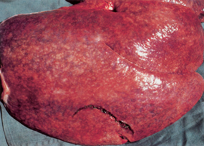
At necropsy, the liver of affected foals is moderately to markedly enlarged and mottled, and contains numerous yellowish-grey foci of necrosis, 1 to 3mm in diameter, throughout the parenchyma 22, 25, 30 (Figure 208.1). The hepatic, mesenteric and other abdominal lymph nodes may be enlarged and oedematous or haemorrhagic.9, 20, 30 In some there is a catarrhal enterocolitis.10, 20 There maybe icterus, and petechiae or ecchymoses in various organs.19, 22, 25
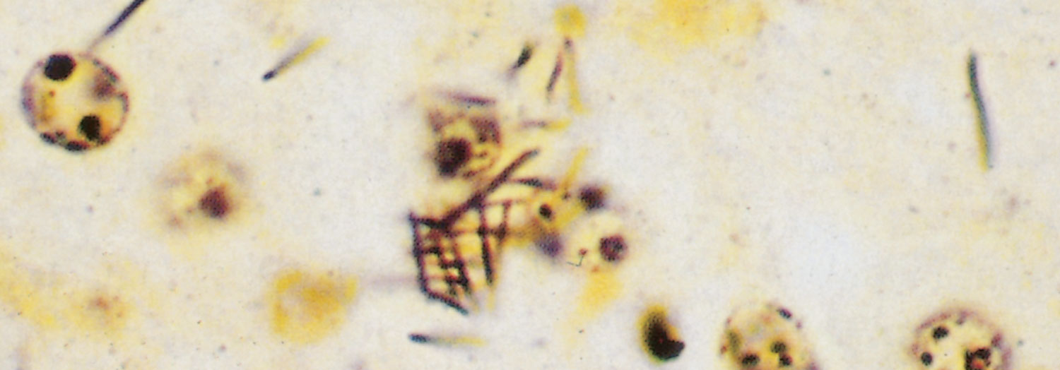
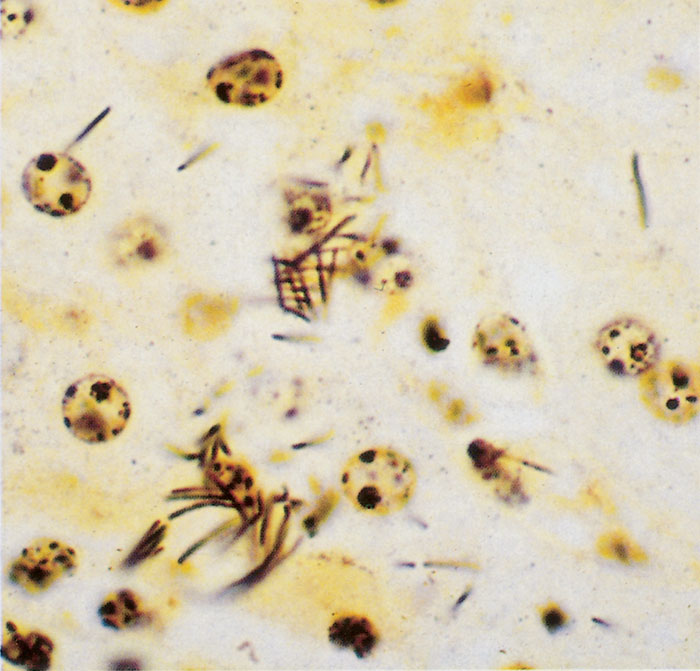
Microscopically there are multiple, often coalescingfoci of coagulative necrosis randomly distributed throughout the liver. Some foci contain abundant fibrin and cellular debris, while an inflammatory exudate consisting primarily of neutrophils and a few macrophages and lymphocytes surrounds many of the necrotic areas.10, 11, 20, 22, 25 In some foals the portal triads are infiltrated by varying numbers of neutrophils and mononuclear cells. Fibrosis of portal and centrilobular areas may also be evident.10, 27, 30 Numerous C. piliforme organisms arranged in parallel or haphazard criss-cross patterns occur in apparently intact hepatocytes at the periphery of the necrotic foci.10, 11, 18, 20, 22, 25 The staining characteristics of the bacteria vary somewhat;2, 23, 25, 27 they may or may not stain faintly basophilic with haematoxylin and eosin; are usually weakly positive with the periodic acid-Schiff reaction; and stain weakly Gram-negative (or are sometimes Gram-variable). They are best demonstrated with Giemsa and, in particular, with silver impregnation techniques such as that of Warthin-Starry 8 (Figure 208.2).
Lesions in other organs are less common, and include necrosis of lymphocytes in lymphoid follicles in the spleen and lymph nodes;10, 22, 30 necrosis of crypt epithelium in the intestines; acute inflammation and haemorrhage in the small and large intestines, accompanied by the presence of bacilli in intestinal glands;2, 30 and foci of myocardial necrosis and myocarditis with bacteria within these foci and adjacent myocardial fibres.2, 30
Diagnosis
In foals the disease occurs between one and six weeks of age, has a rapid onset, and a short course of illness. Clostridium piliforme cannot be cultured in cell-free media,6, 8 and the few reports of its successful cultivation remain unconfirmed. 31 The organism can be cultivated in the yolk sac of embryonated chicken eggs,8 but this method is seldom used in routine diagnostic bacteriology. The diagnosis of Tyzzers’s disease is therefore mostly based upon the histological demonstration of typical organisms in the cytoplasm of hepatocytes bordering on necrotic foci,8 especially by silver impregnation techniques. Immunofluorescent techniques to demonstrate the causative agent and tests to detect serum antibodies against C. piliforme have been developed to diagnose the disease in laboratory animals.28, 31 Recently, a PCR technique usingthe 196-bp DNA fragment specific to 16S rRNA of C. piliforme were used to confirm Tyzzer’s disease in paraffin-embedded tissue sections of a calf.15
Differential diagnosis
Other bacterial infections such as those caused by Salmonella serovars, Actinobacillus equuli and Rhodococcus equi may cause death with occasional liver involvement in foals one to six weeks old. Icterus may be pronounced in isoimmune haemolytic anaemia, congenital babesiosis and equid herpesvirus 1 infection, although in the latter, foals usually die within the first week of life.
Control
The results of treatment of foals with confirmed Tyzzer’s disease have been unrewarding, although affected foals may respond temporarily to treatment with antimicrobial drugs and fluids containingg lucose.1, 20 Based on trials in rodents, it has been claimed that penicillin, streptomycin, erythromycin and tetracycline have some therapeutic effect, but that sulfonamides and chloramphericol do not.6, 8
References
- brown, c.m., ainsworth, d.m., personett, l.a. & derksen, f.j., 1983. Serum biochemical and haematological findings in two foals with focal bacterial hepatitis (Tyzzer’s disease). Equine Veterinary Journal, 15, 375–376.
- carrigan, m.j., pedrana, r.g. & mckibbin, a.w., 1984. Tyzzer’s disease in foals. Australian Veterinary Journal, 61, 199–200.
- carter, g.r., 1986. Essentials of Veterinary Bacteriology and Mycology. 3rd edn. Philadelphia: Lea & Febiger.
- copland, m.d., robartson, c.w., pry, j. & wilson, g., 1984. Tyzzer’s disease in a foal. Australian Veterinary Journal, 61, 302–304.
- duncan, a.j., carman, r.j., olsen, g.j. & wilson, k.h., 1993. Assignment of the agent of Tyzzer’s disease to Clostridium piliforme comb. Nov. on the basis of 16S rRNA sequence analysis. International Journal of Systematic Bacteriology. 43, 314–318.
- fujiwara, k., 1978. Tyzzer’s disease. Japanese Journal of Experimental Medicine, 48, 467–480.
- ganaway, j.r., 1980. Effect of heat and selected chemical disinfectants upon infectivity of spores of Bacillus piliformis. Laboratory Animal Science, 30, 192–196.
- ganaway, j.r., allen, a.m. & moore, t.d., 1971. Tyzzer’s disease. American Journal of Pathology, 64, 717–732.
- hall, w.c. & van kruiningen, h.j., 1974. Tyzzer’s disease in a horse. Journal of the American Veterinary Medical Association, 164, 1187–1189.
- harrington, d.d., 1975. Naturally-occurringTyzzer’s disease (Bacillus piliformis infection) in horse foals. The Veterinary Record, 96, 59–63.
- harrington, d.d., 1976. Bacillus piliformis infection (Tyzzer’s disease) in two foals. Journal of the American Veterinary Medical Association, 168, 58-60.
- hook, r.r., riley, l.k., franklin, c.l. & besch-williford, c.l., 1995. Seroanalysis of Tyzzer’s disease in horses: Implications that multiple strains can infect Equidae. Equine Veterinary Journal, 27, 8–12.
- hum, s. & best, f.g., 1988. Tyzzer’s disease in a wombat. Australian Veterinary Journal, 65, 89-91.
- ikegami, t., shirota, k., goto, k., takakura, a., itoh, t., kawamuru, s. une, y., nomura, y. & fujiwara, k., 1999. Enterocolitis associated with dual infection by Clostridium piliforme and feline panleukopaenia virus in three kittens. Veterinary Pathology, 36, 613–615.
- ikegami, t., shirota, k., une, y., nomura, y., wada, y., goto, k., takakura, a., itoh, t. & fujiwara, k., 1999. Naturally occurringTyzzer’s disease in a calf. Veterinary Pathology, 36, 253–255.
- krieg, n.r. & holt, j.g., 1984. Bergey’s Manual of Systematic Bacteriology, Vol 1. Baltimore: Williams and Wilkins.
- logan, n.a., 1988. Bacillus species of medical and veterinary importance. Journal of Medical Microbiology, 25, 157–165.
- pulley, l.t. & shively, j.n., 1974. Tyzzer’s disease in a foal. Veterinary Pathology, 11, 203–211.
- scarratt, w.k., saunders, g.k., welker, f.h., halpern, n.e., cordes, d.o. & camp, g.m., 1985. Bacillus piliformis infection (Tyzzer’s disease) in two Virginia foals. Journal of Equine Veterinary Science, 5, 135–138.
- swerczek, t.w., 1977. Multi-focal hepatic necrosis and hepatitis in foals caused by Bacillus piliformis (Tyzzer’s disease). Veterinary Annual, 17, 130–132.
- swerczek, t.w., crowe, m.w., prickett, m.e. & bryans, j.t., 1973. Focal bacterial hepatitis in foals: Preliminary report. Modern Veterinary Practice, 54, 66–67.
- taylor, r.f. & mullaney, t.p., 1983. Tyzzer’s disease in Michigan foals: A review. Twenty-sixth Annual Proceedings of the American Association of Veterinary Laboratory Diagnosticians.
- thomson, g.w., wilson, r.w., hall, e.a. & physick-sheard, p., 1977. Tyzzer’s disease in the foal: Case reports and review. Canadian Veterinary Journal, 18, 41–43.
- trent, a.m. & walsh, k.m., 1983. Tyzzer’s disease in a foal. Equine Practice, 5, 8–17.
- turk, m.a.m., gallina, a.m. & perryman, l.e., 1981. Bacillus piliformis infection (Tyzzer’s disease) in foals in northwestern United States: A retrospective study of 21 cases. Journal of the American Veterinary Medical Association, 178, 279–281.
- tyzzer, e.e., 1917. A fatal disease of the Japanese Waltzingmouse caused by a spore-bearingbacillus (Bacillus piliformis, n. sp.). Journal of Medical Research, 37, 307–338.
- van der lugt, j.j., coetzer, j.a.w., jordaan, p. & marlow, c.h.b., 1985. Suspected Tyzzer’s disease in two foals. Journal of the South African Veterinary Association, 56, 107–108.
- waggie, k.s., spencer, t.h. & ganaway, j.r., 1987. An enzyme-linked immunosorbent assay for detection of anti-Bacillus piliformis serum antibody in rabbits. Laboratory Animal Science, 37, 176–179.
- webb, d.m., harrington, d.d. & boehm, p.n., 1987. Bacillus piliformis infection (Tyzzer’s disease) in a calf. Journal of the American Veterinary Medical Association, 191, 431–434.
- whitwell, k.e., 1976. Four cases of Tyzzer’s disease in foals in England. Equine Veterinary Journal, 8, 118–122.
- wobeser, g., 1981. Tyzzer’s disease. In: davis, j.w., karstad, l.h. & trainer, d.o., (eds). Infectious Diseases of Wild Mammals. 2nd edn. Iowa: Iowa State University Press.


