- Infectious Diseases of Livestock
- Part 2
- Crimean-Congo haemorrhagic fever
- GENERAL INTRODUCTION: PARAMYXOVIRIDAE AND PNEUMOVIRIDAE
- Rinderpest
- Peste des petits ruminants
- Parainfluenza type 3 infection
- Bovine respiratory syncytial virus infection
- Hendra virus infection
- Paramyxovirus-induced reproductive failure and congenital defects in pigs
- Nipah virus disease
- GENERAL INTRODUCTION: CALICIVIRIDAE AND ASTROVIRIDAE
- Vesicular exanthema
- Enteric caliciviruses of pigs and cattle
- GENERAL INTRODUCTION: RETROVIRIDAE
- Enzootic bovine leukosis
- Jaagsiekte
- Visna-maedi
- Caprine arthritis-encephalitis
- Equine infectious anaemia
- GENERAL INTRODUCTION: PAPILLOMAVIRIDAE
- Papillomavirus infection of ruminants
- Papillomavirus infection of equids
- GENERAL INTRODUCTION: ORTHOMYXOVIRIDAE
- Equine influenza
- Swine influenza
- GENERAL INTRODUCTION: CORONAVIRIDAE
- Porcine transmissible gastroenteritis
- Porcine respiratory coronavirus infection
- Porcine epidemic diarrhoea
- Porcine haemagglutinating encephalomyelitis virus infection
- Porcine deltacoronavirus infection
- Bovine coronavirus infection
- Ovine coronavirus infection
- Equine coronavirus infection
- GENERAL INTRODUCTION: PARVOVIRIDAE
- Porcine parvovirus infection
- Bovine parvovirus infection
- GENERAL INTRODUCTION: ADENOVIRIDAE
- Adenovirus infections
- GENERAL INTRODUCTION: HERPESVIRIDAE
- Equid herpesvirus 1 and equid herpesvirus 4 infections
- Equid gammaherpesvirus 2 and equid gammaherpesvirus 5 infections
- Equine coital exanthema
- Infectious bovine rhinotracheitis/infectious pustular vulvovaginitis and infectious pustular balanoposthitis
- Bovine alphaherpesvirus 2 infections
- Malignant catarrhal fever
- Pseudorabies
- Suid herpesvirus 2 infection
- GENERAL INTRODUCTION: ARTERIVIRIDAE
- Equine viral arteritis
- Porcine reproductive and respiratory syndrome
- GENERAL INTRODUCTION: FLAVIVIRIDAE
- Bovine viral diarrhoea and mucosal disease
- Border disease
- Hog cholera
- Wesselsbron disease
- Louping ill
- West nile virus infection
- GENERAL INTRODUCTION: TOGAVIRIDAE
- Equine encephalitides caused by alphaviruses in the Western Hemisphere
- Old World alphavirus infections in animals
- Getah virus infection
- GENERAL INTRODUCTION: BUNYAVIRIDAE
- Diseases caused by Akabane and related Simbu-group viruses
- Rift Valley fever
- Nairobi sheep disease
- Crimean-Congo haemorrhagic fever
- GENERAL INTRODUCTION: ASFARVIRIDAE
- African swine fever
- GENERAL INTRODUCTION: RHABDOVIRIDAE
- Rabies
- Bovine ephemeral fever
- Vesicular stomatitis and other vesiculovirus infections
- GENERAL INTRODUCTION: REOVIRIDAE
- Bluetongue
- Ibaraki disease in cattle
- Epizootic haemorrhagic disease
- African horse sickness
- Equine encephalosis
- Palyam serogroup orbivirus infections
- Rotavirus infections
- GENERAL INTRODUCTION: POXVIRIDAE
- Lumpy skin disease
- Sheeppox and goatpox
- Orf
- Ulcerative dermatosis
- Bovine papular stomatitis
- Pseudocowpox
- Swinepox
- Cowpox
- Horsepox
- Camelpox
- Buffalopox
- GENERAL INTRODUCTION: PICORNAVIRIDAE
- Teschen, Talfan and reproductive diseases caused by porcine enteroviruses
- Encephalomyocarditis virus infection
- Swine vesicular disease
- Equine picornavirus infection
- Bovine rhinovirus infection
- Foot-and-mouth disease
- GENERAL INTRODUCTION: BORNAVIRIDAE
- Borna disease
- GENERAL INTRODUCTION: CIRCOVIRIDAE AND ANELLOVIRIDAE
- Post-weaning multi-systemic wasting syndrome in swine
- GENERAL INTRODUCTION: PRION DISEASES
- Scrapie
- Bovine spongiform encephalopathy
- Transmissible spongiform encephalopathies related to bovine spongiform encephalopathy in other domestic and captive wild species
Crimean-Congo haemorrhagic fever
This content is distributed under the following licence: Attribution-NonCommercial CC BY-NC  View Creative Commons Licence details here
Crimean-Congo haemorrhagic fever
Previous authors: R Swanepoel and FJ Burt
Current authors:
F J BURT - Professor, Medical Scientist, PhD, Division of Virology, Francois Retief Building, DF Malherbe Street, Bloemfontein, 9300, South Africa
D GOEDHALS - Division of Virology, Francois Retief Building, DF Malherbe Street, Bloemfontein, 9300, South Africa
R SWANEPOEL - Extraordinary Lecturer, BVSc, DTVM, PhD, Department of Veterinary Tropical Diseases, Faculty of Veterinary Science, University of Pretoria, Private Bag X04, Onderstepoort, Pretoria, Gauteng, 0110, South Africa
Introduction
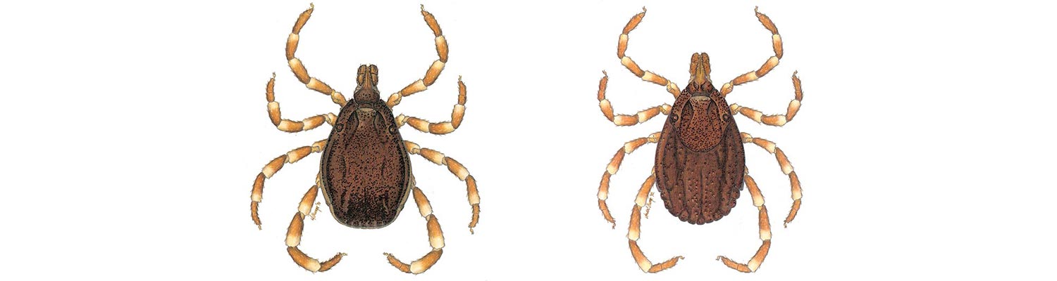
Crimean-Congo haemorrhagic fever (CCHF) is a tick-borne disease found in Africa, Asia, eastern Europe and the Balkans. The geographic distribution of the CCHF virus (CCHFV) correlates with that of ticks belonging to the genus Hyalomma, which are considered to be the principal vectors. The virus causes benign infection with viraemia in cattle, sheep and small mammals such as hares. Humans become infected by contact with infected blood or other tissues of livestock or human patients, or from tick bite. The human disease is usually characterized by a febrile illness with a petechial rash, often followed by a haemorrhagic state and necrotic hepatitis.
A disease given the name Crimean haemorrhagic fever was first described in people bitten by ticks while harvesting crops on the Crimean Peninsula in 1944. It was demonstrated in 1945, through the inoculation of filtered tick-suspensions and blood from patients into human subjects, that the disease was caused by a tick-transmitted virus, but the virus was only isolated in laboratory hosts, namely mice, in 1967.22 In 1969 it was shown that the agent of Crimean haemorrhagic fever was identical to a virus named Congo which had been isolated in 1956 from the blood of a febrile child in Stanleyville (now Kisangani) in what was then the Belgian Congo (now Democratic Republic of Congo, DRC), and since that time the two names have been used in combination.20, 22, 23, 93
Crimean-Congo haemorrhagic fever virus, or antibody to it, has been found in many countries of eastern Europe, Asia and Africa, mainly in the course of surveys. In some countries of eastern Europe and Asia, however, the presence of the virus first became evident in nosocomial outbreaks, or in epidemics that arose in circumstances where humans were exposed to ticks and livestock on a large scale, such as in major land reclamation or resettlement schemes in Bulgaria and parts of the former USSR.45, 51, 52, 80, 81, 94, 102
In February 1981, the first case of CCHF to be recognized in South Africa occurred in a child bitten by a Hyalomma tick in the North West Province. From 1981 to 2019 just over 200 cases have been confirmed in southern Africa with a fatality rate of approximately 30 per cent.43, 59, 90, 95, 96, 98, 99, 100, 101, 105 The virus is likely present throughout Africa, and causes disease that is no less severe than that which occurs in eastern Europe and Asia.
In southern Africa, antibody to CCHF was found to be widely distributed in the sera of livestock and wild vertebrates in South Africa, Zimbabwe and Namibia, including sera that had been in frozen storage since 1964.16, 89, 91, 95, 99, 101 This implies that the virus must have been in southern Africa long before its presence was recognized, and it is believed that the regular diagnosis of cases of CCHF in the subcontinent in recent years probably stems from the increased awareness among medical clinicians that resulted from wide publicity given to the disease. Hoogstraal51, 52 pointed out that mechanisms for the dissemination of ticks (and hence virus), which include the movement of birds that migrate annually on a north-south axis,53, 54 must have operated in Eurasia and Africa for millennia. In addition, ticks can be dispersed between continents by movement of livestock. Although there is evidence of CCHF outbreaks in the United Arab Emirates resulting from trade of livestock from Africa, long established endemicity of CCHF in the region cannot be excluded.
The emergence and re-emergence of CCHFV in Balkan countries and south western regions of the Russian Federation, and more recently in southern regions of Europe, highlights the potential for this pathogen to spread to non-endemic regions where ticks belonging to the genus Hyalomma are present, although there is also the possibility of late recognition of the infection in areas where the virus has long been present.
Aetiology
Crimean-Congo haemorrhagic fever virus is classified as a member of the genus Orthonairovirus, order Bunyavirales, family Nairoviridae, (formerly the genus Nairovirus of the family Bunyaviridae).21, 26 Species within the genus were originally grouped on the basis of antigenic affinities and recently revised using morphological and genetic relatedness.1, 18 Twelve orthonairovirus species are currently recognized: CCHF, Dera Ghazi Khan, Dugbe, Hughes, Qalyub, Sakhalin, Thiafora, Burana, Hazara, Kasokera, Keterah and Nairobi sheep disease.1 Crimean-Congo haemorrhagic fever virus is regarded as the only member of an eponymous group.
The orthonairoviruses are spherical, 90-120 nm in diameter, and have a host-cell-derived bilipid envelope incorporating virus-coded glycoproteins that form indistinct surface projections.29, 67 The single-stranded, negative-sense RNA genome has three segments with a total molecular weight of 6.2-7.5 x 10⁶, and each of the three RNA segments, L (large), M (medium) and S (small), is contained in a separate nucleocapsid within the virion.24 Complementary non-coding regions are present at the 5’ and 3’ termini of each segment flanking a single transcriptional unit. The complementarity results in base pairing of the regions and formation of panhandles with circular conformation. The virions contain three major structural proteins: two envelope glycoproteins, GN and GC, and a nucleocapsid protein (N) plus minor quantities of L (large) protein.24, 41 The S segment has ambisense coding: a long open reading frame that encodes the N protein, plus negative sense coding of a non-structural protein NSs. The N protein of CCHFV is the most antigenic viral protein and appears to be the most conserved. Expression of the N protein has been used to prepare safe recombinant antigen for diagnostic tests.34 The M segment encodes a non-structural protein, NSm, and a glycoprotein precursor (GPC). Maturation of the GPC yields two major structural glycoproteins, GN and GC, plus secreted non-structural glycoproteins GP160, GP85 and GP38.73, 82
The L segment contains a viral RNA-dependent RNA polymerase (L protein) domain and ovarian tumour domain with deubiquitinating and deISGylating activities. The RNA segments are encapsidated in the viral encoded NP to form ribonucleoprotein particles within the virion. By analogy with other related genera it can be assumed that glycoproteins are responsible for recognition of receptor sites on susceptible cells and consequently cell tropism and pathogenicity of the virus in humans, for the induction of protective immune response, and probably play a role in tick host selection.83 Genetic studies have shown that despite a high degree of variability between CCHF isolates from geographically distinct regions, the isolates appear to comprise a single virus species.
Little information is available on the stability of CCHFV, but being enveloped, it is sensitive to lipid solvents,60 and it is known that infectivity is destroyed by low concentrations of formalin and beta-propriolactone. The virus is labile in infected human tissues after death,51 but the examination of specimens from human patients appears to show that infectivity is preserved for at least a few days at ambient temperature in separated serum. Infectivity is destroyed by boiling or autoclaving, but the virus is stable at temperatures below -60°C.
Crimean-Congo haemorrhagic fever virus replicates in a wide variety of primary- and line-cell cultures, including Vero, CER and BHK21 cells, but not usually to high titres. The virus is poorly cytopathic, so that infectivity is generally titrated by immunofluoresence staining of infected cells using virus-specific antisera.24, 51, 107 In the past the virus was usually isolated and titrated by intracerebral inoculation of suckling mice.51
Because of its propensity for human-to-human transmission, its ability to cause laboratory infections, and the severity of the human disease, CCHF is now placed in biohazard class 4 in countries that have biosafety guidelines, and this dictates that culture of the virus is only permitted in maximum-security laboratories, although molecular and serologic diagnostic procedures may be performed under less stringent conditions where the virus is endemic.108
Molecular diversity and evolution
Crimean-Congo haemorrhagic fever virus shows a relatively high rate of genetic diversity in comparison to other arboviruses, where evolution is often constrained by the need to maintain competence in both arthropod vectors and mammalian hosts. The highest level of diversity is seen in the M segment, with 31 per cent variation at the nucleotide level, while the S and L segments are relatively more conserved (20 per cent and 22 per cent respectively).28 Based on phylogenetic analysis of complete or partial genome sequences, CCHFV isolates are separated into six or seven lineages. The lineages or clades reflect the geographic origins of viral isolates, with clades I to III originating in Africa, clade IV (which is sometimes divided into two clades) originating in Asia, and clades V and VI originating in Europe.6, 20, 28
Genetic diversity is driven by three processes, namely point mutations or genetic drift, reassortment, and recombination. Genetic drift results from the accumulation over time of point mutations due to the error-prone viral RNA-dependent RNA-polymerase. This process is the main determinant of the genetic diversity of CCHFV isolates.6 Due to its segmented genome, genetic reassortment also occurs whereby exchange of an entire segment can occur during dual infection. Reassortment of the M segment has been most commonly described and is thought to occur due to co-infection of the tick vectors.28, 50 This phenomenon is well described amongst South African CCHFV isolates where M segments cluster with Asian isolates (clade IV), while the S and L segments cluster with African isolates (clade III).14, 44 Recombination is best described in the S segment of CCHFV and appears to occur less frequently than reassortment.64
Epidemiology
The distribution of the disease coincides with that of the principal vectors of the virus, ticks of the genus Hyalomma. Cases of naturally acquired human infection (based on virus isolation or detection of viral RNA) have been documented in the following Asian and eastern European countries: Afghanistan, Albania, Bulgaria, China, Georgia, Greece, Turkey, Iran, Iraq, Kazakhstan, Kosovo (formerly Yugoslavia), Oman, Pakistan, Russia, Saudi Arabia, Tajikistan, Turkey, United Arab Emirates, Uzbekistan, and the following African countries: Burkina Faso, Central African Republic, Democratic Republic of Congo (formerly Zaire), Egypt, Kenya, Mauritania, Namibia, Nigeria, Senegal, South Africa, Tanzania, Uganda.3, 9, 10, 32, 34, 35, 51, 61, 63, 70, 74, 85, 107, 109 More recently, emergence of the disease has been identified in India and Spain.69, 71 Evidence of virus circulation based on isolation from ticks or non-human mammals, or serological evidence, has been reported from Afghanistan, Algeria, Benin, Cameroon, Equatorial Guinea, Ethiopia, Ghana, Guinea, Hungary, Madagascar, Mali, Morocco, Tunisia and Zimbabwe.107 Limited serological observations have been reported from Portugal, France, and Kuwait.106 Excluding Turkey where more than 10 000 cases have been described since 2002, most countries report sporadic outbreaks.
In South Africa, the prevalence of antibody to CCHFV in cattle sera was found to be lowest in a strip extending along the southern coast from Cape Town to the environs of East London, with many herds lacking evidence of infection in this area where H. truncatum is the sole representative of the three species of Hyalomma known to occur in the country.58, 99 In the interior of the country the prevalence of antibody was found to be high, with up to 96 per cent of the cattle sera tested in some herds being positive.16, 99 The prevalence of antibody in cattle sera was also found to be high in Zimbabwe, but was somewhat lower in Namibia, where comparatively few sera have been tested. On farms where sheep were tested, they generally had a lower prevalence of antibody than cattle, which probably reflects the preference of adult Hyalomma ticks for larger hosts.100
The prevalence of antibody to CCHF in the sera of wild vertebrates in South Africa and Zimbabwe is generally low, but is highest in large herbivores with a mass similar to or greater than kudu antelope, such as the zebra, eland, buffalo, rhinoceros and giraffe, which are the preferred hosts of adult Hyalomma ticks.16, 91 Antibody has also been found in the sera of farmed ostriches, but not in wild passerines or water birds.89 Immature Hyalomma ticks feed on small mammals and ground-frequenting birds, and, among these, antibody is most prevalent in hares, but also in a low proportion of myomorph rodents (rats and mice) and guinea fowl.91, 95 It has been shown that the prevalence of antibody to CCHFV in wild vertebrates was lower in the Kruger National Park along the north-eastern border of South Africa than elsewhere in the country and in Zimbabwe.16, 95 As on the southern Cape coast, H. truncatum is the only Hyalomma present in the Kruger National Park, and the implication is that this tick may be a less efficient vector of CCHFV than H. rufipes and H. glabrum, although this has still to be confirmed.55, 56, 57, 58, 59, 93
Crimean-Congo haemorrhagic fever virus has been isolated from at least 30 species of ticks, but for most species there is no evidence that they are capable of serving as vectors, and in some instances the virus recovered from engorged ticks may merely have been present in the blood meal imbibed from a viraemic host.51, 101, 107 Members of three genera of ixodid ticks, Hyalomma, Dermacentor and Rhipicephalus, have been shown to be capable of transmitting the virus transstadially after feeding on viraemic hosts, but the coincidence in distribution of CCHFV and Hyalomma ticks strongly suggests that members of this genus are the most important vectors of the virus.51, 99, 107 A few species of all three of the above genera have been shown to be capable of transmitting the infection transovarially, but it has been suggested that this probably does not occur with sufficient frequency to ensure indefinite perpetuation of the virus in the absence of amplification of infection in vertebrate hosts.51, 107 In particular, it is believed that the infection of small vertebrates constitutes an important amplifying mechanism that facilitates transstadial transmission of infection by adult ticks to large vertebrates.
The occurrence of viraemia has been demonstrated in various small mammals of Eurasia and Africa, such as little susliks, hedgehogs, hares and certain myomorph rodents, and in some instances it has been shown that these hosts are able to infect ticks.22, 48, 52, 65 Domestic ruminants also develop demonstrable viraemia and are capable of infecting ticks.22, 52, 66 In all of the above species, viraemia lasts up to a week and is of low to moderate intensity, with maximum titres of infectivity ranging from about 102,7 to 104,2 mouse intracerebral 50 per cent lethal doses/ml. 22, 48, 52, 65 Viraemia is slightly more intense and of longer duration in humans than in other animals.
Serological surveillance remains a highly useful tool for identifying areas in which the virus is circulating.92 Identification of immune responses in wild vertebrates can provide information on the possible role of various species in maintenance of the virus in nature. In limited observations, passerine birds, domestic chickens and a few species of wild birds in West Africa have been found to be refractory to the virus, while experimentally infected guinea-fowls developed transient viraemia of very low intensity and an antibody response that remained demonstrable for a few weeks only, so it is unlikely that birds are able to infect ticks directly.51, 89, 107, 110 Nevertheless, infection can be transferred between infected and non-infected ticks through the so-called phenomenon of ’non-viraemic transmission ‘, and migratory birds carrying immature ticks could serve to disseminate virus that has been transmitted transovarially in the ticks.53, 54 The relatively high prevalence and titres of antibody found in ostriches, which are hosts to adult Hyalomma ticks, suggested that they may be more susceptible to infection than other birds. An outbreak of CCHF among workers at an ostrich abattoir in Oudtshoorn, South Africa in 1996 in which 17 workers were infected and one fatality was recorded, prompted an investigation of the nature of the infection in ostriches in order to determine if there was a potential risk for consumers of ostrich meat and to devise a protective strategy for abattoir workers.13, 97 Following experimental infection ostriches showed no clinical signs, but developed viraemia that was demonstrable for up to four days, and a strong antibody response.97 In addition, virus was detectable in visceral organs such as spleen, liver and kidney. It was concluded that human infections could be prevented if ostriches were kept free of ticks for 14 days prior to slaughter.19, 97
Apart from nosocomial infections, the disease has never been recorded in a town dweller in South Africa without a history of exposure to ticks or fresh blood and tissues of livestock in a rural setting or abattoir. There has been no indication that CCHFV constitutes a public health hazard in meat processed and matured according to normal health regulations.51, 107 During the maturation of meat in abattoirs there is a sharp drop in pH, and it is possible that this destroys the virus infectivity, thus rendering the meat safe.
Crimean-Congo haemorrhagic fever virus causes only inapparent infection or mild fever in livestock.98, 99, 100 Calves and lambs acquire antibody from colostrum, but it has not been determined whether or not this is protective, and many animals seroconvert after the occurrence of natural infection early in life.40, 95, 99 Thus humans commonly become infected when they come into contact with viraemic blood of young animals in the course of performing procedures such as castrations, vaccinations, inserting ear tags or slaughtering the animals. 51, 98, 99, 100 The available evidence suggests that the infection in humans is acquired through contact of viraemic blood with broken skin, and this accords with the fact that nosocomial infection in medical personnel usually results from accidental sticking with needles contaminated with the blood of patients, or similar mishaps.51, 90, 105, 107
Many of the human infections diagnosed in southern Africa resulted directly from tick bites, and most of the patients concerned were able to verify that they had been bitten by ticks that fit the description of Hyalomma spp.99 It has been observed in both South Africa and the former USSR that humans can also become infected from merely squashing the ticks between the fingers.51, 99
A few southern African patients were unable to recall contact with blood or other tissues of livestock, or having been bitten by ticks, but lived in or visited a rural environment where such exposure to infection was possible.99 The majority of patients in southern Africa have been adult males engaged in the livestock industry, such as farmers, labourers, slaughtermen and veterinarians, and most cases of the disease have occurred in North West, western Free State and Northern and Western Cape Provinces in South Africa, where the relatively arid climate suits the xerophilic Hyalomma ticks.96, 99 A few patients were town dwellers who had contact with animal tissues or were bitten by ticks while on hunting or hiking trips. Human disease has been diagnosed in most months of the year, but there has been a slight preponderance of cases in February-March and October-November, when adult Hyalomma spp. tend to manifest peak questing activity.76 The pattern of human infection differs markedly in regions such as the Balkans and the Near East where women and children participate to a greater extent in the livestock industry.
In view of the serological evidence that infection of livestock occurs on a wide scale, it is surprising that so few human infections are diagnosed. This raises the possibility that many human infections are asymptomatic or mild and pass unnoticed, but the low prevalence of antibody detected in surveys and the sparse evidence of infection encountered among cohorts of cases of the disease suggest that a high proportion of CCHFV infections do, in fact, come to medical attention.40, 98, 99, 100, 106 Possible explanations for the low incidence of human infection include the facts that viraemia in livestock is short-lived and of low intensity compared to that in other zoonotic diseases such as Rift Valley fever, which is more readily acquired from contact with infected tissues, plus the facts that immature Hyalomma ticks feed only on small animals and humans are not the preferred hosts of the adult ticks.
Pathogenesis
The pathogenesis of the disease is not completely understood but by analogy with other arthropod-borne virus infections it can be surmised that CCHFV may undergo some replication at the site of inoculation, and that there is haematogenous and lymph-borne spread of infection to target organs such as the liver, which are the major sites of replication.17, 88, 99 Immunohistochemical staining of tissues to demonstrate localization of the virus has shown that mononuclear phagocytes and endothelial cells are major targets of CCHFV infection.5 A similar tropism is exhibited by many lethal haemorrhagic fever viruses. Involvement of the mononuclear phagocyte system may represent a mechanism for viral clearance in some patients, or in others replication of the virus in these cells may enhance viraemia by shedding of virus back into the circulation. Activation of mononuclear phagocytes and infection of endothelial cells may play a role in the pathogenesis of CCHF through secretion of physiologically active substances, including cytokines and other inflammatory mediators.
The hepatocytes, a main target of the virus, can be infected by passive transfer of the virus or release of replicating virus from the Kupffer cells. Minimal inflammatory infiltrates suggest that hepatocellular necrosis may be mediated by a direct viral cytopathic effect. Hepatocellular necrosis may lead to release of tumour necrosis factor and other procoagulants into the circulation, and ultimately to impairment of the synthesis of coagulation factors to replace those that are consumed. Disseminated intravascular coagulation results in further depletion of clotting factors and platelets, thus contributing to the bleeding diathesis.96 Hepatocellular damage and monocyte activation also result in proinflammatory cytokine release, which contributes to disease severity.37, 72 Widespread infection of endothelium, with degenerative change rather than necrosis, could signify capillary dysfunction, which contributes to the occurrence of a haemorrhagic diathesis and the generation of a petechial rash. The endothelial dysfunction may result from both direct infection as well as immunologically mediated effects.25, 62
Clinical signs
The incubation period commonly ranges from 1-3 days after a person has been infected by tick bite, but occasionally extends to 7 days, and it is usually 5-6 days in people exposed to infected blood or other tissues of livestock or human patients, but may occasionally extend to 9 days or more.96, 99
Onset of the disease is usually very sudden, with severe headache often being the first symptom. This is frequently accompanied by dizziness, neck pain and stiffness, sore eyes, photophobia, fever, rigor and chills. Patients rapidly develop general myalgia and malaise, with intense backache or leg pains. Nausea, sore throat and vomiting commonly occur early in the illness and patients may experience non-localized abdominal pain and diarrhoea at this stage. Fever is often intermittent and patients may undergo sharp changes of mood over the next two days with feelings of confusion and aggression. By the second to fourth day of illness patients may exhibit lassitude, depression and somnolence, and may have a flushed appearance with injected conjunctivae or chemosis. By this time, tenderness may be localized in the right upper quadrant of the abdomen, and hepatomegaly may be discernible. Tachycardia is common and patients may be slightly hypotensive. There may be lymphadenopathy and exanthema and petechiae of the throat, tonsils and buccal mucosa.99
A petechial rash appears on the trunk and limbs by the third to sixth day of illness, and this may be followed rapidly by the appearance of large bruises and ecchymoses, especially in the antecubital fossae, upper arms, axillae and groin. Epistaxis, haematemesis, haematuria, melaena, gingival bleeding and bleeding from the vagina or other orifices may commence on the fourth or fifth day of illness, or even earlier. Sometimes a haemorrhagic tendency is evident only from the oozing of blood from injection or venipuncture sites. There may be internal bleeding, including retroperitoneal and intracranial haemorrhage. Severely ill patients may enter a state of hepatorenal and pulmonary failure from about the fifth day onwards, and progressively become drowsy, stuporous and comatose. Jaundice may become apparent during the second week of illness.99
The mortality rate varies from 5 to 30 per cent or more, and deaths generally occur on the fifth to fourteenth day of illness.37, 46, 51, 99 Strains of low virulence circulate in Greece and Turkey.36, 75 Patients who recover usually begin to improve on day nine or ten, but asthenia, conjunctivitis, slight confusion and amnesia may continue for a month or longer. 96, 99 With follow up at 12 months, symptoms of post-traumatic stress disorder (PTSD) have been reported in approximately half of patients who recover, with PTSD diagnosed in nearly 20 per cent according to DSM-V-TR criteria. Patients with bleeding and those who require blood transfusions and admission to intensive care units have an increased risk of PTSD, probably related to the severity of disease.47
Crimean-Congo haemorrhagic fever in pregnancy results in a high rate of maternal and foetal/neonatal deaths (34 per cent and 59 per cent respectively), regardless of the duration of pregnancy at the time of infection. Maternal haemorrhage is associated with an increased risk of maternal and foetal/neonatal mortality. A high rate of nosocomial transmissions linked to maternal cases have been reported; however, transmission by means of breast feeding has not been documented.33
Crimean-Congo haemorrhagic fever in children presents similarly to that in adults, but with tonsillopharyngitis, abdominal pain, diarrhoea, myalgia and cardiac involvement more commonly reported.33, 104 It has been suggested that disease may be milder in children, but reported case fatality rates have varied from 1.65 – 26.5 per cent.87, 104 The variation may represent differences in viral strains, host factors, access to and quality of health care services and co-morbidities.104
Crimean-Congo haemorrhagic fever virus causes only inapparent infection or mild fever in livestock and wild animals.98, 99, 100
Pathology
Changes in the cellular and chemical composition of blood recorded during the first few days of illness in human patients include leukocytosis or leukopenia and elevated aspartate transaminase, alanine transaminase, gamma-glutamyltransferase, lactic dehydrogenase, alkaline phosphatase and creatine kinase levels, while bilirubin, creatinine and urea levels increase and serum protein levels decline during the second week.30, 58, 60 Thrombocytopenia, elevation of the prothrombin ratio, activated thromboplastin time, thrombin time and fibrin degradation products, as well as depression of fibrinogen and haemoglobin values are evident during the first few days of illness. While a number of laboratory parameters have been evaluated as severity criteria in acute infection, findings have been variable, with viral load identified as the strongest predictor of mortality.2, 49, 79
Complete autopsies are seldom performed on patients that die of CCHF, and examination of tissues is often confined to liver samples taken with biopsy needles. Lesions in the liver vary from disseminated foci of necrosis, mainly mid-zonal in distribution, to massive necrosis, involving over 75 per cent of hepatocytes, and a variable degree of haemorrhage.4, 17, 59, 99 Necrotic areas are frequently marked by haemorrhage and cell loss and associated with eosinophilic change of hepatocytes with prominent Councilman-type bodies. Inflammatory mononuclear cell infiltrates in necrotic areas are absent or mild and unrelated to the extent of hepatocellular damage. Common findings include fatty change, hyperplastic and hypertrophic Kupffer cells containing phagocytosed debris, and portal inflammatory infiltrates. Limited observations of splenic tissue show lymphoid depletion, focal necrosis, and scattered lymphoblasts in periarterial sheaths. In addition, diffuse alveolar damage, intra-alveolar haemorrhage, hyaline membrane formation, and mononuclear interstitial pneumonitis have been observed in the lung, and congestion and slight interstitial oedema have been noted in the heart. Lesions in other organs include congestion, haemorrhage and focal necrosis in the central nervous system, kidneys and adrenals, and general depletion of lymphoid tissues. None of the histopathologic features are pathognomonic and similar features can be seen in other viral, rickettsial and bacterial infections as in toxic exposures.
Diagnosis
A diagnosis of CCHF should be suspected when severe febrile illness with sudden onset and short incubation period, usually less than one week, occurs in persons exposed to tick bites or fresh blood and other tissues of livestock or human patients. The disease is easier to recognize once a rash appears and there are haemorrhagic signs such as epistaxis, haematemesis, and melaena.
Aetiologic investigation of suspected CCHF infection is generally performed in a biosafety level (BSL) 4 laboratory, but in countries with a high incidence of disease it has been found that molecular and serologic diagnostic procedures can be performed safely in BSL2 laboratories.108 Laboratory diagnosis of the infection in live patients during the acute phase of illness consists of detection of viral nucleic acid or demonstration of viral antigen by enzyme-linked immunoassay (ELISA) in serum or plasma samples, or by isolation of the virus.11, 12, 13, 88, 90, 96, 99 In samples collected later during the illness, diagnosis is confirmed by demonstration of an immune response. The reverse-transcription polymerase chain reaction (RT-PCR) using conventional thermocycling or real-time PCR constitutes a rapid and sensitive technique for diagnosing CCHF infection during the early stages of illness before an antibody response is demonstrable, or in fatal cases where an antibody response is frequently not demonstrable.13, 31, 77, 86 Detection of viral RNA using molecular techniques has several distinct advantages compared to isolation of virus. A result can be obtained in less than 24 hours compared with virus isolation which requires, on average, three to six days. Amplification of viral RNA is more sensitive than virus isolation or antigen detection and non-infectious viral nucleic acid can be detected after infectious virus is no longer present.
Various nucleic acid amplification platforms have been developed. The nucleoprotein gene is frequently targeted for diagnostic assays as it is the most conserved among genetically and geographically distinct strains. Complete and partial nucleotide sequence data for viral genes available on public data bases from isolates worldwide have facilitated development of assays with global application accommodating genetic diversity. In addition to conventional RT-PCR methods, various real time RT-PCR platforms have been developed and commercial assays are available using primers and probe technology. For low resource countries, assays have been described that can be performed without the need for sophisticated thermal cyclers and software. Assays that can be performed at one temperature such as loop-mediated isothermal amplification and recombinase polymerase assay could have application in a low resource setting. Laboratories with maximum containment facilities can attempt virus isolation. Virus may be isolated in cell cultures, commonly Vero cells, or by intracerebral inoculation of day-old mice. The virus is detected and identified in cell cultures by performing an immunofluorescence test. Isolation of the virus in cell cultures can be achieved in one to five days, compared to five to eight days in mice, but mouse inoculation is more sensitive for isolating virus present in low concentration.
Antibodies, both IgG and IgM, become demonstrable by indirect immunofluorescence from about day four of illness onwards, and are present in the sera of all survivors of the disease by day nine at the latest. The IgM antibody activity declines to undetectable levels often by the fourth month after infection but has been detected up to 12 months after illness, and although IgG titres may begin to decline gradually at this stage, IgG remains demonstrable for at least ten years and likely for life. Recent or current infection is confirmed by demonstrating seroconversion, a four-fold or greater increase in antibody activity in paired serum samples, or IgM activity in a single specimen. A limited number of commercial immunofluorescent assays and ELISA are available.
Patients who succumb rarely develop a demonstrable antibody response, and the diagnosis is confirmed by isolation of virus or detection of viral nucleic acid in serum samples or liver samples taken after death, or by demonstration of CCHF antigen using immunohistochemical techniques on paraffin-embedded liver sections. Virus antigen may sometimes be demonstrated in liver impression smears by immunofluorescence staining, or in serum or liver homogenate by ELISA. Observation of necrotic lesions compatible with CCHF in sections of liver provides presumptive evidence in support of the diagnosis.
Differential diagnosis
The disease should be distinguished from other tick-borne virus infections and particularly from rickettsial infection (Rickettsia conorii infection, commonly known as tick-bite fever), which is highly prevalent in southern Africa, and in which there is often a characteristic necrotic lesion, or eschar, at the site of the tick bite.15 Tick-bite fever has an incubation period of seven to ten days, and a more insidious onset than CCHF. Tick-bite fever is associated with a petechial rash and is capable of causing fatal disease in humans, with haemorrhagic manifestations similar to CCHF, but is amenable to treatment with broad-spectrum antibiotics. Rift Valley fever can also be acquired from contact with the tissues of livestock in Africa, but this usually occurs in the context of massive epidemics involving abortion and death of sheep and cattle at irregular intervals of years when heavy rains favour the breeding of the mosquito vectors of the virus. Less than one per cent of Rift Valley fever infections in humans manifest as fatal haemorrhagic disease.
Many other infectious conditions of humans may present as haemorrhagic disease, including bacterial septicaemias, such as meningococcaemia, salmonellosis, brucellosis, leptospirosis, and borreliosis but also leishmaniasis, ehrlichiosis, Q fever (Coxiella burnetii) and malaria.37, 68 Viral causes include hepatitis A-E and herpes simplex virus,8, 37 but of special interest are the other viral haemorrhagic fevers of Africa that are most likely to be encountered in patients who reside in or visit tropical areas of the continent. In brief, they include Marburg disease and Ebola fever, caused by members of the family Filoviridae, and Lassa fever and Lujo haemorrhagic fever, caused by rodent-borne viruses of the family Arenaviridae. A further group of rodent-associated viruses belonging to the family Hantaviridae can also cause haemorrhagic manifestations, but are found in Europe, Asia and America. Although there is some evidence of infection of humans and rodents with viruses of this family in Africa, the significance of the findings remains uncertain. Yellow fever and dengue are mosquito-borne flaviviruses that are known to be present in West and East Africa, and are capable of causing fatal haemorrhagic disease in humans in the context of large urban outbreaks. However, dengue infection is usually benign in Africa, and outbreaks associated with haemorrhagic disease tend to occur on Indian Ocean islands, Asia and elsewhere. Non-infectious causes should also be considered, including haematological diseases, malignancies, drugs, collagen tissue and auto-immune disorders, vitamin B12 deficiency, febrile neutropaenia, and HELLP syndrome (haemolytic anaemia, elevated liver enzymes, low platelet count) in pregnancy.37, 38, 103, 105
Distinguishing between the possible causes of suspected cases of viral haemorrhagic fever is a specialized task, normally undertaken in laboratories dedicated to the purpose.
Control
The control of CCHF through the application of acaricides to livestock is impractical, particularly under the extensive farming conditions that prevail in the arid areas where Hyalomma ticks are most prevalent. Pyrethroid preparations are available that can be used to kill ticks that come into contact with human clothing. Following the outbreak of CCHF in an ostrich abattoir in South Africa in 1996, it was decided that ostriches should be treated for ticks and kept in a tick-free environment for two weeks prior to slaughter in order to reduce the risk of exposing abattoir workers to infection.19, 97 Veterinarians, slaughtermen and others involved with livestock should be aware of the disease and take practical steps, such as wearing of gloves, to limit or avoid exposure of naked skin to fresh blood and other tissues of animals. Inactivated vaccines prepared from infected mouse brain were used for the protection of humans in eastern Europe and the former USSR, but currently remain available only in Bulgaria.51
Treatment of the disease involves the use of barrier-nursing techniques for the protection of medical staff, and consists essentially of supportive and replacement therapy with blood products. Immune-plasma has been used but the efficacy of the treatment is not clear. Anti-viral agents such as ribavirin and T-705 (favipiravir) show some efficacy in animal and in vitro studies. Promising results were obtained in a trial of ribavirin39, 95 particularly when administered before day five after onset of illness. However, there are conflicting reports on the efficacy of ribavirin, emphasizing the need for development of novel vaccines and anti-viral drugs.
Vaccine development
The inactivated mouse brain vaccine used in Bulgaria requires boosters to induce effective immune responses, and safety concerns with regard to auto immune and allergic responses render it unlikely that the vaccine will gain approval for use in other countries.
Vaccine development has been hampered by the lack of a suitable animal model for the disease. However, there are now several animal models available, including interferon deficient mice and a non-human primate model.5, 7, 42, 48 STAT-1 knockout mice that have defective type I, II and III interferon signaling and type 1 interferon receptor knockout mice exhibit 100 per cent mortality when inoculated with challenge virus. In contrast, disease produced in the non-human primate model more closely resembles disease in humans, with severity of illness ranging from mild disease to severe and fatal. The primate model presents ethical and cost implications that render knockout mice more suitable for initial studies.
Candidate immunogens including subunit vaccine, transcriptionally competent virus-like particles, DNA vaccines, viral vectors carrying CCHF segments and antigens produced in genetically modified plants have been investigated.27, 30, 78, 84, 111, 112 The challenge remains that the immune correlates of protection are not well defined. Notably, the induction of neutralizing antibody does not appear to confer complete protection, suggesting a role for both humoral and cellular immunity. In conclusion, it appears likely that the evolving understanding of the immune mechanisms involved will lead to development of effective vaccines and therapeutics.
References
- ADAMS, M. J., LEFKOWITZ, E. J., KING, A. M. Q., HARRACH, B., HARRISON, R. L., KNOWLES, N. J., KROPINSKI, A. M., KRUPOVIC, M., KUHN, J. H., MUSHEGIAN, A. R., NIBERT, M., SABANADZOVIC, S., SANFAÇON, H., SIDDEL, S. G., SIMMONDS, P., VARSANI, A., ZERBINI, F. M., GORBALENYA, A. E. & DAVISON, A. J., 2017. Changes to taxonomy and the International Code of Virus Classifications and Nomenclature ratified by the International Committee on Taxonomy of Viruses. Archives of Virology, 162, 2505-2539.
- AKINCI, E., BODUR, H., SUNBUL, M. & LEBLEBICIOGLU, H., 2016. Prognostic factors, pathophysiology and novel biomarkers in Crimean-Cong hemorrhagic fever. Antiviral Research, 132, 233-243.
- ARADAIB, I. E., ERICKSON, B. R., MUSTAFA, M. E., KRISTOVA, M. L., SAEED, N. S., ELAGEB, R. M. & NICHOL, S. T., 2010. Nosocomial outbreak of Crimean-Congo hemorrhagic fever, Sudan. Emerging Infectious Diseases, 16, 837-839.
- BASKERVILLE, A., SATTI, A., MURPHY, F. A. & SIMPSON, D. I., 1981. Congo-Crimean haemorrhagic fever in Dubai: histopathological studies. Journal of Clinical Pathology, 34, 871-874.
- BENTE, D. A., ALIMONTI, J. B., SHIEH, W. J., CAMUS, G., STRÖHER, U., ZAKI, S. & JONES, S. M., 2010. Pathogenesis and immune response of Crimean-Congo hemorrhagic fever virus in a STAT-1 knockout mouse model. Journal Virology, 84, 11089-11100.
- BENTE, D. A., FORRESTER, N. L., WATTS, D. M., MCAULEY, A. J., WHITEHOUSE, C. A. & BRAY, M., 2013. Crimean-Congo hemorrhagic fever: history, epidemiology, pathogenesis, clinical syndrome and genetic diversity. Antiviral Research, 100, 159-189.
- BERECZKY, S., LINDEGREN, G., KARLBERG, H., AKERSTRÖM, S., KLINGSTRÖM, J. & MIRAZIMI, A., 2010. Crimean-Congo hemorrhagic fever virus infection is lethal for adult type I interferon receptor-knockout mice. Journal of General Virology, 91, 1473-1477.
- BONNEY, J. H., OSEI-KWASI, M., ADIKU, T. K., BARNOR, J. S., AMESIYA, R., KUBIO, C., AHADZIE, L., OLSCHLÄGER, S., LELKE, M., BECKER-ZIAJA, B., PAHLMANN, M. & GÜNTHER, S., 2013. Hospital-based surveillance for viral hemorrhagic fevers and hepatitides in Ghana. PLoS Neglected Tropical Diseases, 7, e2435.
- BUKBUK, D. N., DOWALL, S. D., LEWANDOWSKI, K., BOSWORTH, A., BABA, S. S., VARGHESE, A., WATSON, R. J., BELL, A., ATKINSON, B. & HEWSON, R., 2016. Serological and virological evidence of Crimean-Congo haemorrhagic fever virus circulation in the human population of Borno State, Northeastern Nigeria. PLoS Neglected Tropical Diseases, 10, e0005126.
- BURNEY, M. I., GHAFOOR, A., SALEEN, M., WEBB, P. A. & CASALS, J., 1980. Nosocomial outbreak of viral hemorrhagic fever caused by Crimean hemorrhagic fever-Congo virus in Pakistan, January 1976. The American Journal of Tropical Medicine and Hygiene, 29, 941-947.
- BURT, F. J., 2011. Laboratory diagnosis of Crimean-Congo hemorrhagic fever virus infections. Future Virology, 6, 831-841.
- BURT, F. J., LEMAN, P. A., ABBOTT, J. C. & SWANEPOEL, R., 1994. Serodiagnosis of Crimean-Congo haemorrhagic fever. Epidemiology and Infection, 113, 551-562.
- BURT, F. J., LEMAN, P. A., SMITH, J. F. & SWANEPOEL, R., 1998. The use of a reverse transcription-polymerase chain reaction for the detection of viral nucleic acid in the diagnosis of Crimean-Congo haemorrhagic fever. Journal of Virological Methods, 70, 129-137.
- BURT, F. J., PAWESKA, J. T., ASHKETTLE, B. & SWANEPOEL, R., 2009. Genetic relationship in southern African Crimean-Congo haemorrhagic fever virus isolates: evidence for occurrence of reassortment. Epidemiology and Infection, 137, 1302-1308.
- BURT, F. J., SPENCER, D. C., LEMAN, P. A., PATTERSON, B. & SWANEPOEL, R., 1996. Investigation of tick-borne viruses as pathogens of humans in South Africa and evidence of Dugbe virus infection in a patient with prolonged thrombocytopenia. Epidemiology and Infection, 116, 353-361.
- BURT, F. J., SWANEPOEL, R. & BRAACK, L. E. O., 1993. Enzyme-linked immunosorbent assays for the detection of antibody to Crimean-Congo haemorrhagic fever virus in the sera of livestock and wild vertebrates. Epidemiology and Infection, 111, 547-557.
- BURT, F. J., SWANEPOEL, R., SHIEH, W. J., SMITH, J. F., LEMAN, P. A., GREER, P. W., COFFIELD, L. M., ROLLIN, P. E., KSIAZEK, T. G., PETERS, C. J. & ZAKI, S. R., 1997. Immunohistochemical and in situ localization of Crimean-Congo hemorrhagic fever (CCHF) virus in human tissues and implications for CCHF pathogenesis. Archives of Pathology and Laboratory Medicine, 121, 839-846.
- CALISHER, C. H. & KARABATSOS, N., 1989. Arbovirus serogroups: Definition and geographic distribution. In: MONATH, T. P., (ed.). The Arboviruses: Epidemiology and Ecology. Boca Raton, Florida:CRC Press, Inc, 1.
- CAPUA, I., 1998. Crimean-Congo haemorrhagic fever in ostriches: a public health risk for countries of the European Union. Avian Pathology, 27, 117-120.
- CARROLL, S. A., BIRD, B. H., ROLLIN, P. E. & NICHOL, S. T., 2010. Ancient common ancestry of Crimean-Congo hemorrhagic fever virus. Molecular Phylogenetics and Evolution, 55, 1103-1110.
- CASALS, J. & TIGNOR, G. H., 1980. The Nairovirus genus: Serological relationships. Intervirology, 14, 144-147.
- CHUMAKOV, M. P., 1974. Contribution to 30 years of investigation of Crimean-Congo haemorrhagic fever. In: CHUMAKOV, M. P., (ed.). Medical Virology, 22, 5-18. Trudy Inst Polio Virus Enstef Akad Med Nauk SSSR (In Russian. English translation: NAMRU3-T950.
- CHUMAKOV, M. P., SMIRNOVA, S. E. & TKACHENKO, E. A., 1970. Relationship between strains of Crimean haemorrhagic fever and Congo viruses. Acta Virologica, 14, 82-85.
- CLERX, J. P., CASALS, J. & BISHOP, D. H., 1981. Structural characteristics of nairoviruses (genus Nairovirus, Bunyaviridae). Journal of General Virology, 55, 165-178.
- CONNOLLY-ANDERSEN, A. M., MOLL, G., AKERSTRŐM, S., KARLBERG, H., DOUAGI, I. & MIRAZIMI, A., 2011. Crimean-Congo hemorrhagic fever virus activates endothelial cells. Journal of Virology, 85, 7766-7774.
- DAVIES, F. G., CASALS, J., JESSET, D. M. & OCHIENG, P., 1978. The serological relationships of Nairobi sheep disease virus. Journal of Comprehensive Pathology, 88, 519-523.
- DEVIGNOT, S., BERGERON, E., NICHOL, S., MIRAZIMI, A. & WEBER, F., 2015. A virus-like particle system identifies the endonuclease domain of Crimean-Congo hemorrhagic fever virus. Journal of Virology, 89, 5957-67.
- DEYDE, V. M., KHRISTOVA, M. L., ROLLIN, P. E., KSIAZEK, T. G. & NICHOL, S. T., 2006. Crimean-Congo hemorrhagic fever virus genomics and global diversity. Journal of Virology, 80, 8834-8842.
- DONETS, M. A., CHUMAKOV, M. P., KOROLEV, M. B. & RUBIN, S. G., 1977. Physicochemical characteristics, morphology and morphogenesis of virions of the causative agent of Crimean hemorrhagic fever. Intervirology, 8, 294-308.
- DOWALL, S. D., CARROLL, M. W. & HEWSON, R., 2017. Development of vaccines against Crimean-Congo haemorrhagic fever virus. Vaccine, 35, 6015-6023.
- DROSTEN, C., GOTTIG, S., SCHILLING, S., ASPER, M., PANNING, M., SCHMITZ, H. & GUNTHER, S., 2002. Rapid detection and quantification of RNA of Ebola and Marburg Viruses, Lassa Virus, dengue Virus, and yellow fever Virus by real-time reverse transcription-PCR. Journal of Clinical Microbiology, 40, 2323-2330.
- DROSTEN, C., MINNAK, D., EMMERICH, P., SCHMITZ, H. & REINECKE, T., 2002. Crimean-Congo hemorrhagic fever in Kosovo. Journal of Clinical Microbiology, 40, 1122-1123.
- EBRAY, A., CEVIK, M. A., ONGURU, P., GÖZEL, G., AKINCI, E., KUBAR, A. & BODUR, H., 2008. Breastfeeding in Crimean-Congo haemorrhagic fever. Scandinavian Journal of Infectious Diseases, 40, 186-188.
- EL-BAHNASAWY, M. M., SABAH, A. A., SALEH, H. A. & MORSY, T. A., 2012. The tick-borne Crimean-Congo hemorrhagic fever in Africa, Asia, Europe, and America: what about Egypt? Journal of the Egyptian Society of Parasitology, 42, 373-384.
- EL AZAZY, O. M. & SCRIMGEOUR, E. M., 1997. Crimean-Congo haemorrhagic fever virus infection in the western province of Saudi Arabia. Transactions of the Royal Society of Tropical Medicine and Hygiene, 91, 275-278.
- ELEVLI, M., OZKUL, A. A., CIVILIBAL, M., MIDILLI, K., GARGILI, A. & DURU, N. S., 2010. A newly identified Crimean-Congo hemorrhagic fever virus strain in Turkey. International Journal of Infectious Diseases, 14S, e213-e216.
- ERGŐNŰL, O., 2006. Crimean-Congo haemorrhagic fever. Lancet Infectious Diseases, 6, 203-214.
- ERGŐNŰL, O., CELIKBAS, A., YILDRIM, U., ZENCIROGLU, A., ERDOGAN, D., ZIRAMAN, I., SARACOGLU, F., DEMIREL, N., CAKMAK, O. & DOKUZOGUZ, B., 2010. Pregnancy and Crimean-Congo haemorrhagic fever. Clinical Microbiology and Infection,16, 647-650.
- FISHER-HOCH, S. P., KHAN, J. A., REHMAN, S., MIRZA, S., KHURSHID, M. & MCCORMICK, J. B., 1995. Crimean Congo-haemorrhagic fever treated with oral ribavirin. Lancet, 346, 472-475.
- FISHER-HOCH, S. P., MCCORMICK, J. B., SWANEPOEL, R., VAN MIDDLEKOOP, A., HARVEY, S. & KUSTNER, H. G., 1992. Risk of human infections with Crimean-Congo hemorrhagic fever virus in a South African rural community. American Journal of Tropical Medicine and Hygiene, 47, 337-345.
- FOULKE, R. S., ROSATO, R. R. & FRENCH, G. R., 1981. Structural polypeptides of Hazara virus. Journal of General Virology, 53, 169-172.
- GARRISON, A. R., SHOEMAKER, C. J., GOLDEN, J. W., FITZPATRICK, C. J., SUSCHAK, J. J., RICHARDS, M. J., BADGER, C. V., SIX, C. M., MARTIN, J. D., HANNAMAN, D., ZIVCEC, M., BERGERON, E., KOEHLER, J. W. & SCHMALJOHN, C. S., 2017. A DNA vaccine for Crimean-Congo hemorrhagic fever protects against disease and death in two lethal mouse models. PLoS Neglected Tropical Diseases, 11, e0005908.
- GEAR, J. H., THOMSON, P. D., HOPP, M., ANDRONIKOU, S., COHN, R. J., LEDGER, J. & BERKOWITZ, F. E., 1982. Congo-Crimean haemorrhagic fever in South Africa. Report of a fatal case in the Transvaal. South African Medical Journal, 62, 576-580.
- GOEDHALS, D., BESTER, P. A., PAWESKA, J. T., SWANEPOEL, R. & BURT, F. J., 2014. Next-generation sequencing of southern African Crimean-Congo haemorrhagic fever virus isolates reveals a high frequency of M segment reassortment. Epidemiology and Infection, 142, 1952-1962.
- GONZALEZ, J. P., LEGUENNO, B., GUILLAUD, M. & WILSON, M. L., 1990. A fatal case of Crimean-Congo haemorrhagic fever in Mauritania: virological and serological evidence suggesting epidemic transmission. Transactions of the Royal Society of Tropical Medicine and Hygiene, 84, 573-576.
- GOZALAN, A., ESEN, B., FITZNER, J., TAPAR, F. S., OZKN, A. P., GEORGES-COURBOT, M. C., UZUN, R., GUMUSLU, F., AKIN, L. & ZELLER., H., 2007. Crimean-Congo haemorrhagic fever cases in Turkey. Scandanavian Journal of Infectious Diseases, 39, 332-336.
- GUL, S., GUL, E. U., YESILYURT, M., OZTURK, B., KUSCU, F. & ERGONUL, O., 2012. Health-related quality of life and the prevalence of post-traumatic stress disorder among Crimean-Congo hemorrhagic fever survivors. Japanese Journal of Infectious Diseases, 65, 392-395.
- HADDOCK, E., FELDMANN, F., HAWMAN, D. W., ZIVCEC, M., HANLEY, P. W., SATURDAY, G., SCOTT, D. P., THOMAS, T., KORVA, M., AVŠIČ-ŽUPANC, T., SAFRONETZ, D. & FELDMANN, H., 2018. A cynomolgus macaque model for Crimean–Congo haemorrhagic fever. Nature Microbiology, 3, 556-562.
- HASANOGLU, I., GUNER, R., CARHAN, A., TUFAN, Z. K., YAGCI-CAGLAYIK, D., GUVEN, T., YILMAZ, G. R. & TASYARAN, M. A., 2016. Crucial parameter of the outcome in Crimean Congo hemorrhagic fever: Viral load. Journal of Clinical Virology, 75, 42-46.
- HEWSON, R., GMYL, A., GMYL, L., SMIRNOVA, S. E., KARGANOVA, G., JAMIL, B., HASAN, R., CHAMBERLAIN, J. & CLEGG, C., 2004. Evidence of segment reassortment in Crimean-Congo haemorrhagic fever virus. Journal of General Virology, 85, 3059-3070.
- HOOGSTRAAL, H., 1979. The epidemiology of tick-borne Crimean-Congo hemorrhagic fever in Asia, Europe, and Africa. Journal of Medical Entomology, 15, 307-417.
- HOOGSTRAAL, H., 1981. Changing patterns of tick-borne diseases in modern society. Annual Review of Entomology, 26, 75-99.
- HOOGSTRAAL, H., KAISER, M. N., TRAYLOR, M. A., GABER, S. & GUINDY, E., 1961. Ticks (Ixodoidea) on birds migrating from Africa to Europe and Asia. Bulletin of the World Health Organization, 24, 197-212.
- HOOGSTRAAL, H., KAISER, M. N., TRAYLOR, M. A., GUINDY, E. & GABER, S., 1963. Ticks (Ixodoidea) on birds migrating from Europe and Asia to Africa. Bulletin of the World Health Organization, 28, 235-262.
- HORAK, I. G., DE VOS, V. & BROWN, M. R., 1983. Parasites of domestic and wild animals in South Africa. XVI. Helminth and arthropod parasites of blue and black wildebeest(Connochaetes taurinus and Connochaetes gnou). Onderstepoort Journal of Veterinary Research, 50, 243–255.
- HORAK, I. G., DE VOS, V. & DE KLERK, B. D., 1984. Parasites of domestic and wild animals in South Africa. XVII. Arthropod parasites of Burchells’s zebra, Equus burchelli, in the eastern Transvaal lowveld. Onderstepoort Journal of Veterinary Research, 51, 145-154.
- HORAK, I. G., POTGIETER, F. T., WALKER, J. B., DE VOS, V. & BOOMKER, J., 1983. The ixodid tick burdens of various large ruminant species in South African nature reserves. Onderstepoort Journal of Veterinary Research, 50, 221-228.
- HOWELL, C. J., WALKER, J. B. & NEVILL, E. M., 1978. Ticks, mites and insects infesting domestic animals in South Africa. Science Bulletin, No 393. Pretoria: Department of Agricultural Technical Services.
- JOUBERT, J. R., KING, J. B., ROSSOUW, D. J. & COOPER, R., 1985. A nosocomial outbreak of Crimean-Congo haemorrhagic fever at Tygerberg Hospital. Part III. Clinical pathology and pathogenesis. South African Medical Journal, 68, 722-728.
- KARABATSOS, L., 1985. International catalogue of arboviruses. San Antonio, Texas, American Society of Tropical Medicine and Hygiene.
- KHAN, A. S., MAUPIN, G. O., ROLLIN, P. E., NOOR, A. M., SHURIE, H. H., SHALABI, A. G., WASEF, S., HADDAD, Y. M., SADEK, R., IJAZ, K., PETERS, C. J. & KSIAZEK, T. G., 1997. An outbreak of Crimean-Congo hemorrhagic fever in the United Arab Emirates, 1994-1995. American Journal of Tropical Medicine and Hygiene, 57, 519-525.
- KRAUS, A. A. & MIRAZIMI, A., 2010. Molecular biology and pathogenesis of Crimean-Congo hemorrhagic fever virus. Future Virology, 5, 469-479.
- LEBLEBICIOGLU, H., OZARAS, R., IRMAK, H. & SENCAN, I., 2016. Crimean-Congo hemorrhagic fever in Turkey: Current status and future challenges. Antiviral Research, 126, 21-34.
- LUKASHEV, A. N., 2005. Evidence for recombination in Crimean-Congo hemorrhagic fever virus. Journal of General Virology, 86, 2333-2338.
- MAES, P., et al., 2019. Taxonomy of the order Bunyavirales: second update 2018. Archives of Virology, 164, 927-941.
- MARRIOTT, A. C., POLYZONI, T., ANTONIADIS, A. & NUTTALL, P. A., 1994. Detection of human antibodies to Crimean-Congo haemorrhagic fever virus using expressed viral nucleocapsid protein. Journal of General Virology, 75, 2157-2161.
- MARTIN, M. L., LINDSEY-REGNERY, H., SASSO, D. R., MCCORMICK, J. B. & PALMER, E., 1985. Distinction between Bunyaviridae genera by surface structure and comparison with Hantaan virus using negative-stain electron microsopy. Acta Virologica, 86, 17-28.
- METIN, O., TEKE, T. A., AYDIN, Z. G. G., KAMAN, A., OZ, F. N., BAYHAN, G. I. & TANIR, G., 2015. A case of brucellosis mimicking Crimean-Congo hemorrhagic fever. Journal of Infection and Public Health, 8, 302-304.
- MISHRA, A. C., MEHTA, M., MOURYA, D. T. & GANDHI, S., 2011. Crimean-Congo haemorrhagic fever in India. Lancet, 378, 372.
- NABETH, P., THIOR, M., FAYE, O. & SIMON, F., 2004. Human Crimean-Congo hemorrhagic fever, Sénégal. Emerging Infectious Diseases, 10, 1881-1882.
- NEGREDO, A., et al., 2017. Autochthonous Crimean-Congo hemorrhagic fever in Spain. New England Journal of Medicine, 377, 154-161.
- PAPA, A., BINO, S., VELO, E., HARXHI, A., KOTA, M. & ANTONIADIS, A., 2006. Cytokine levels in Crimean-Congo hemorrhagic fever. Journal of Clinical Virology, 36, 272-276.
- PAPA, A., MA, B., KOUIDOU, S., TANG, Q., HANG, C. & ANTONIADIS, A., 2002. Genetic characterization of the M RNA segment of Crimean Congo hemorrhagic fever virus strains, China. Emerging Infectious Disease, 8, 50-53.
- PAPA, A., PAPADIMITRIOU, V. D. E., KARTALIS, G. N. & ANTONIADIS, A., 2010. Emergence of Crimean-Congo haemorrhagic fever virus in Greece. Clinical Microbiology and Infection, 16, 843-847.
- PAPADOPOULOS, O. & KOPTOPOULOS, G., 1980. Crimean-Congo hemorrhagic fever (CCHF) in Greece: isolation of the virus from Rhipicephalus bursa ticks and a preliminary serological survey. In: Vesenjak-Hirjan Jea., (ed.). Arboviruses in the Mediterranean countries. Stuttgart (Germany): Gustav Fisher Verlag, 117-121.
- RECHAV, Y. & ZEEDERBERG, M. E., 1986. Tick populations of two breeds of cattle under field conditions, with a note on blood components related to host resistance. In: SAUER, J.R. & HAIR, J.A., (eds.). Morphology, Physiology, and Behavioural Biology of Ticks Chichester: Ellis Horwood,
- RODRIGUEZ, L. L., MAUPIN, G. O., KSIAZEK, T. G., ROLLIN, P. E., KHAN, A. S., SCHWARZ, T. F., LOFTS, R. S., SMITH, J. F., NOOR, A. M., PETERS, C. J. & NICHOL, S. T., 1997. Molecular investigation of a multisource outbreak of Crimean-Congo hemorrhagic fever in the United Arab Emirates. American Journal of Tropical Medicine and Hygiene, 57, 512-518.
- RODRIGUEZ, S. E., CROSS, R. W., FENTON, K. A., BENTE, D. A., MIRE, C. E. & GEISBERT, T. W., 2019. Vesicular Stomatitis Virus-based vaccine protects mice against Crimean-Congo hemorrhagic fever. Scientific Reports, 9, 7755.
- SAKSIDA, A., DUH, D., WRABER, B., DEDUSHAJ, I., AHMETI, S. & AVSIC-ZUPANC, T., 2010. Interacting roles of immune mechanisms and viral load in the pathogenesis of Crimean-Congo hemorrhagic fever. Clinical and Vaccine Immunology, 17, 1086-1093.
- SALUZZO, J. F., AUBRY, P., MCCORMICK, J. & DIGOUTTE, J. P., 1985. Haemorrhagic fever caused by Crimean Congo haemorrhagic fever virus in Mauritania. Transactions of the Royal Society of Tropical Medicine and Hygiene, 79, 268.
- SALUZZO, J. F., DIGOUTTE, J. P., CORNET, M., BAUDON, D., ROUX, J. & ROBERT, V., 1984. Isolation of Crimean-Congo haemorrhagic fever and Rift Valley fever viruses in Upper Volta. Lancet, 1, 1179.
- SANCHEZ, A. J., VINCENT, M. J. & NICHOL, S. T., 2002. Characterization of the glycoproteins of Crimean-Congo hemorrhagic Fever virus. Journal of Virology, 76, 7263-7275.
- SCHMALJOHN, C. S. & PATTERSON, J. L., 1990. Bunyaviridae and their replication. Part II: Replication of Bunyaviridae. In: FIELDS, B. N., (ed.). Virology. 2nd edn. New York: Raven Press Ltd.
- SCHOLTE, F. E. M., SPENGLER, J. R., WELCH, S. R., HARMON, J. R., COLEMAN-MCCRAY, J. D., FREITAS, B. T., KAINULAINEN, M. H., PEGAN, S. D., NICHOL, S. T., BERGERON, E. & SPIROPOULOU, C. F., 2019. Single-dose replicon particle vaccine provides complete protection against Crimean-Congo hemorrhagic fever virus in mice. Emerging Microbes & Infections, 8, 575-578.
- SCHWARZ, T. F., NITSCHKO, H., JAGER, G., NSANZE, H., LONGSON, M., PUGH, R. N. & ABRAHAM, A. K., 1995. Crimean-Congo haemorrhagic fever in Oman. Lancet, 346, 1230.
- SCHWARZ, T. F., NSANZE, H., LONGSON, M., NITSCHKO, H., GILCH, S., SHURIE, H., AMEEN, A., ZAHIR, A. R., ACHARYA, U. G. & JAGER, G., 1996. Polymerase chain reaction for diagnosis and identification of distinct variants of Crimean-Congo hemorrhagic fever virus in the United Arab Emirates. American Journal of Tropical Medicine and Hygiene, 55, 190-196.
- SHARIFI-MOOD, B., MARDANI, M., KESHTKAR-JAHROMI, M., RAHNAVARDI, M., HATAMI, H. & METANAT, M., 2008. Clinical and epidemiologic features of Crimean-Congo hemorrhagic fever among children and adolescents from southeastern Iran. Pediatric Infectious Disease Journal, 27, 561-563.
- SHEPHERD, A. J., SWANEPOEL, R. & LEMAN, P. A., 1989. Antibody response in Crimean-Congo hemorrhagic fever. Reviews of Infectious Disease, 11, S801-S806.
- SHEPHERD, A. J., SWANEPOEL, R., LEMAN, P. A. & SHEPHERD, S. P., 1987. Field and laboratory investigation of Crimean-Congo haemorrhagic fever virus (Nairovirus, family Bunyaviridae) infection in birds. Transactions of the Royal Society of Tropical Medicine and Hygiene, 81, 1004-1007.
- SHEPHERD, A. J., SWANEPOEL, R., SHEPHERD, S. P., LEMAN, P. A., BLACKBURN, N. K. & HALLETT, A. F., 1985. A nosocomial outbreak of Crimean-Congo haemorrhagic fever at Tygerberg Hospital. Part V. Virological and serological observations. South African Medical Journal, 68, 733-736.
- SHEPHERD, A. J., SWANEPOEL, R., SHEPHERD, S. P., MCGILLIVRAY, G. M. & SEARLE, L. A., 1987. Antibody to Crimean-Congo hemorrhagic fever virus in wild mammals from southern Africa. American Journal of Tropical Medicine and Hygiene, 36, 133-142.
- SPENGLER, J. R., BERGERON, É. & ROLLIN, P. E., 2016. Seroepidemiological studies of Crimean-Congo hemorrhagic fever virus in domestic and wild animals. PLoS Neglected Tropical Diseases, 10, e0004210.
- SPICKETT, A. M., HORAK, I. G., BRAACK, L. E. O. & VAN ARK, H., 1991. Drag-sampling of free-living ixodid ticks in the Kruger National Park. Onderstepoort Journal of Veterinary Research, 58, 27-32.
- SULEIMAN, M. N., MUSCAT-BARON, J. M., HARRIES, J. R., SATTI, A. G., PLATT, G. S., BOWEN, E. T. & SIMPSON, D. I., 1980. Congo/Crimean haemorrhagic fever in Dubai. An outbreak at the Rashid Hospital. Lancet, 2, 939-941.
- SWANEPOEL, R., 1981-2002. National Institute for Virology, Private Bag X4, Sandringham 2131, Johannesburg, South Africa. Unpublished observations.
- SWANEPOEL, R., GILL, D. E., SHEPHERD, A. J., LEMAN, P. A., MYNHARDT, J. H. & HARVEY, S., 1989. The clinical pathology of Crimean-Congo hemorrhagic fever. Reviews of Infectious Disease, 11, S794-S800.
- SWANEPOEL, R., LEMAN, P. A., BURT, F. J., JARDINE, J., VERWOERD, D. J., CAPUA, I., BRUCKNER, G. K. & BURGER, W. P., 1998. Experimental infection of ostriches with Crimean-Congo haemorrhagic fever virus. Epidemiology and Infection, 121, 427-432.
- SWANEPOEL, R., SHEPHERD, A. J., LEMAN, P. A. & SHEPHERD, S. P., 1985. Investigations following initial recognition of Crimean-Congo haemorrhagic fever in South Africa and the diagnosis of 2 further cases. South African Medical Journal, 68, 638-641.
- SWANEPOEL, R., SHEPHERD, A. J., LEMAN, P. A., SHEPHERD, S. P., MCGILLIVRAY, G. M., ERASMUS, M. J., SEARLE, L. A. & GILL, D. E., 1987. Epidemiologic and clinical features of Crimean-Congo hemorrhagic fever in southern Africa. American Journal of Tropical Medicine and Hygiene, 36, 120-132.
- SWANEPOEL, R., SHEPHERD, A. J., LEMAN, P. A., SHEPHERD, S. P. & MILLER, G. B., 1985. A common-source outbreak of Crimean-Congo haemorrhagic fever on a dairy farm. South African Medical Journal, 68, 635-637.
- SWANEPOEL, R., STRUTHERS, J. K., SHEPHERD, A. J., MCGILLIVRAY, G. M., NEL, M. J. & JUPP, P. G., 1983. Crimean-Congo hemorrhagic fever in South Africa. American Journal of Tropical Medicine and Hygiene, 32, 1407-1415.
- TANTAWI, H. H., AL MOSLIH, M. I., AL JANABI, N. Y., AL BANA, A. S., MAHMUD, M. I., JURJI, F., YONAN, M. S., AL ANI, F. & AL TIKRITI, S. K., 1980. Crimean-Congo haemorrhagic fever virus in Iraq: isolation, identification and electron microscopy. Acta Virologica, 24, 464-467.
- TANYEL, E., SUNBUL, M., FLETCHER, T. E. & LEBLEBICIOGLU, H., 2016. Aetiology of PCR negative suspected Crimean-Congo hemorrhagic fever cases in an endemic area. Pathogens and Global Health, 110, 173-177.
- TEZER, H., SUCAKLI, I. A., SAYLI, T. R., CELIKEL, E., YAKUT, I., KARA, A., TUNC, B. & ERGONUL, O., 2010. Crimean-Congo hemorrhagic fever in children. Journal of Clinical Virology, 48, 184-186.
- VAN EEDEN, P. J., JOUBERT, J. R., VAN DE WAL, B. W., KING, J. B., DE KOCK, A. & GROENEWALD, J. H., 1985. A nosocomial outbreak of Crimean-Congo haemorrhagic fever at Tygerberg Hospital. Part I. Clinical features. South African Medical Journal, 68, 711-717.
- VAWDA, S., GOEDHALS, D., BESTER, P. A. & BURT, F. J., 2018. Seroepidemiologic survey of Crimean-Congo haemorrhagic fever virus in select risk groups in endemic regions of South Africa. Emerging Infectious Diseases, 24, 1360-1363.
- WATTS, D. M., KSIAZEK, T. G., LINTHICUM, K. J. & HOOGSTRAAL, H., 1989. Crimean-Congo haemorrhagic fever. In: MONATH, T. P., (ed.). The Arboviruses: Epidemiology and Ecology. Boca Raton, Florida: CRC Press, Inc, 2.
- WEIDMANN, M., AVSIC-ZUPANC, T., BINO, S., BOULOUY, M., BURT, F., CHRISTOVA, I., DEDUSHAJ, I., EL-SANOUSI, A., ELADIL, N., HEWSON, R., HUFERT, F. T., HUMOLLI, I., KOÇAK TUFAN, Z., KORUKLUOGLU, G., LYSSEN, P., MIRAZIMI, A., NEYTS, J., NIEDRIG, M., OZKUL, A., PAPA, A., PAWESKA, J., SADEGH, C., SALL, A. A., SCHMALJOHN, C. S., SWANEPOEL, R., VAN VUREN, P., UYAR, Y., ZELLER, H. & WEBER, F., 2016. Biosafety standards for working with Crimean-Congo haemorrhagic fever virus. Journal of General Virology, 97, 2799–2808. doi: 10.1099/jgv.0.000610.
- YEN, Y. C., KONG, L. X., LEE, L., ZHANG, Y. Q., LI, F., CAI, B. J. & GAO, S. Y., 1985. Characteristics of Crimean-Congo hemorrhagic fever virus (Xinjiang strain) in China. The American Journal of Tropical Medicine and Hygiene, 34, 1179-1182.
- ZELLER, H. G., CORNET, J. P. & CAMICAS, J. L., 1994. Crimean-Congo haemorrhagic fever virus infection in birds: field investigations in Senegal. Research in Virology, 145, 105-109.
- ZIVCEC, M., SAFRONETZ, D., SCOTT, D. P., ROBERTSON, S., EBIHARA, H. & FELDMANN, H., 2013. Lethal Crimean-Congo hemorrhagic fever virus infection in interferon α/β receptor knockout mice is associated with high viral loads, proinflammatory responses, and coagulopathy. Journal of Infectious Diseases, 207, 1909-1921.
- ZIVCEC, M., SAFRONETZ, D., SCOTT, D. P., ROBERTSON, S. & FELDMANN, H., 2018. Nucleocapsid protein-based vaccine provides protection in mice against lethal Crimean-Congo hemorrhagic fever virus challenge. PLoS Neglected Tropical Diseases, 12, e0006628.


