- Infectious Diseases of Livestock
- Part 2
- Vesicular exanthema
- GENERAL INTRODUCTION: PARAMYXOVIRIDAE AND PNEUMOVIRIDAE
- Rinderpest
- Peste des petits ruminants
- Parainfluenza type 3 infection
- Bovine respiratory syncytial virus infection
- Hendra virus infection
- Paramyxovirus-induced reproductive failure and congenital defects in pigs
- Nipah virus disease
- GENERAL INTRODUCTION: CALICIVIRIDAE AND ASTROVIRIDAE
- Vesicular exanthema
- Enteric caliciviruses of pigs and cattle
- GENERAL INTRODUCTION: RETROVIRIDAE
- Enzootic bovine leukosis
- Jaagsiekte
- Visna-maedi
- Caprine arthritis-encephalitis
- Equine infectious anaemia
- GENERAL INTRODUCTION: PAPILLOMAVIRIDAE
- Papillomavirus infection of ruminants
- Papillomavirus infection of equids
- GENERAL INTRODUCTION: ORTHOMYXOVIRIDAE
- Equine influenza
- Swine influenza
- GENERAL INTRODUCTION: CORONAVIRIDAE
- Porcine transmissible gastroenteritis
- Porcine respiratory coronavirus infection
- Porcine epidemic diarrhoea
- Porcine haemagglutinating encephalomyelitis virus infection
- Porcine deltacoronavirus infection
- Bovine coronavirus infection
- Ovine coronavirus infection
- Equine coronavirus infection
- GENERAL INTRODUCTION: PARVOVIRIDAE
- Porcine parvovirus infection
- Bovine parvovirus infection
- GENERAL INTRODUCTION: ADENOVIRIDAE
- Adenovirus infections
- GENERAL INTRODUCTION: HERPESVIRIDAE
- Equid herpesvirus 1 and equid herpesvirus 4 infections
- Equid gammaherpesvirus 2 and equid gammaherpesvirus 5 infections
- Equine coital exanthema
- Infectious bovine rhinotracheitis/infectious pustular vulvovaginitis and infectious pustular balanoposthitis
- Bovine alphaherpesvirus 2 infections
- Malignant catarrhal fever
- Pseudorabies
- Suid herpesvirus 2 infection
- GENERAL INTRODUCTION: ARTERIVIRIDAE
- Equine viral arteritis
- Porcine reproductive and respiratory syndrome
- GENERAL INTRODUCTION: FLAVIVIRIDAE
- Bovine viral diarrhoea and mucosal disease
- Border disease
- Hog cholera
- Wesselsbron disease
- Louping ill
- West nile virus infection
- GENERAL INTRODUCTION: TOGAVIRIDAE
- Equine encephalitides caused by alphaviruses in the Western Hemisphere
- Old World alphavirus infections in animals
- Getah virus infection
- GENERAL INTRODUCTION: BUNYAVIRIDAE
- Diseases caused by Akabane and related Simbu-group viruses
- Rift Valley fever
- Nairobi sheep disease
- Crimean-Congo haemorrhagic fever
- GENERAL INTRODUCTION: ASFARVIRIDAE
- African swine fever
- GENERAL INTRODUCTION: RHABDOVIRIDAE
- Rabies
- Bovine ephemeral fever
- Vesicular stomatitis and other vesiculovirus infections
- GENERAL INTRODUCTION: REOVIRIDAE
- Bluetongue
- Ibaraki disease in cattle
- Epizootic haemorrhagic disease
- African horse sickness
- Equine encephalosis
- Palyam serogroup orbivirus infections
- Rotavirus infections
- GENERAL INTRODUCTION: POXVIRIDAE
- Lumpy skin disease
- Sheeppox and goatpox
- Orf
- Ulcerative dermatosis
- Bovine papular stomatitis
- Pseudocowpox
- Swinepox
- Cowpox
- Horsepox
- Camelpox
- Buffalopox
- GENERAL INTRODUCTION: PICORNAVIRIDAE
- Teschen, Talfan and reproductive diseases caused by porcine enteroviruses
- Encephalomyocarditis virus infection
- Swine vesicular disease
- Equine picornavirus infection
- Bovine rhinovirus infection
- Foot-and-mouth disease
- GENERAL INTRODUCTION: BORNAVIRIDAE
- Borna disease
- GENERAL INTRODUCTION: CIRCOVIRIDAE AND ANELLOVIRIDAE
- Post-weaning multi-systemic wasting syndrome in swine
- GENERAL INTRODUCTION: PRION DISEASES
- Scrapie
- Bovine spongiform encephalopathy
- Transmissible spongiform encephalopathies related to bovine spongiform encephalopathy in other domestic and captive wild species
Vesicular exanthema
This content is distributed under the following licence: Attribution-NonCommercial CC BY-NC  View Creative Commons Licence details here
Vesicular exanthema of swine
N J KNOWLES
Introduction
Vesicular exanthema of swine (VES) is a disease of pigs that is clinically indistinguishable from other vesicular diseases of pigs, such as foot-and-mouth disease, and is caused by an immunologically diverse group of caliciviruses whose primary hosts are marine-dwelling mammals and fish.
Vesicular exanthema of swine was first recognized in California in 1932 and became a serious problem in that state during the 1940s. In 1952, the disease occurred outside California for the first time, and by the end of that year had spread to 31 states in the USA. This resulted, in August 1952, in the declaration of a national state of emergency, which provided legislation for a vigorous campaign of eradication based on the slaughter-out of affected herds, with compensation, control of the movement of pork products derived from swill-fed pigs, and the prohibition of feeding uncooked swill to pigs.1 These measures resulted in the eradication of VES in the USA by 1956. However, since a marine reservoir of these viruses was discovered in 1972,22 vesicular disease has appeared in free-living and captive marine mammals along the Pacific coast of the USA and on islands in the eastern Pacific.
Aetiology
The 13 VES virus (VESV) serotypes derived from pigs along with 16 serologically distinct caliciviruses isolated from marine- dwelling mammals and opaleye fish (Girella nigricans) which are referred to as San Miguel sea lion (SMS) viruses, form a single complex.4, 12, 31 Seven serologically distinct caliciviruses isolated from cetaceans,26 cattle,28 primates,29 skunk,18 walrus,27 reptiles30 and humans32 have also been shown to be part of this complex,6, 10, 12 bringing the total number of serotypes in this group to at least 35.31
Caliciviruses are non-enveloped, single-stranded RNA viruses with icosahedral symmetry (T=3) which have characteristic depressions on the virion surface.7 Virions have a diameter of 35 to 40 nm and a mass of approximately 15 × 106 Daltons.7 The capsid is made up of a major (Mr 58–60 × 103) and a minor (Mr 10 × 103) polypeptide.7 The positive-sense RNA is 7,4 to 8,3 kb in length and has a small polypeptide (VPg; Mr 10−15 × 103) covalently linked to its 5’ end, while the 3’ end of the genome is polyadenylated. 7
Vesicular exanthema of swine and the other related viruses belong to the genus Vesivirus in the family Caliciviridae. 7, 8 Within the vesivirus genus three genetic groups of viruses are recognized; the first group consists of VESV, SMSV and related viruses, the second is feline calicivirus, and the third is a newly recognized canine calicivirus.17 The relationships of caliciviruses isolated from mink9 and white tern15 to the vesiviruses remain to be elucidated. This family also contains three other genera, Lagovirus, Norovirus (Norwalk-like viruses) and Sapovirus (Sapporo-like viruses).7, 8 The lagoviruses are rabbit haemorrhagic disease virus (sometimes referred to as rabbit calicivirus) and European brown hare syndrome virus, while the Norwalk- like and Sapporo-like viruses are made up of noncultivatable human and animal gastrointestinal diseasecausing viruses.7, 8
Vesicular exanthema of swine and SMS viruses replicate with the production of cytopathic effects in a wide variety of cell cultures derived from pig tissues (such as kidney, lung, testicle and amnion); however, their replication in nonporcine cells is variable depending upon serotype.1, 24 There is a distinct correlation between the size of plaques produced by a strain in cell culture and its virulence for pigs.1 In pigs, different serotypes of VESV may differ markedly in their virulence,1 and some strains do not produce clinical disease.
Epidemiology
Marine caliciviruses have been isolated from California sea lions (Zalophus californianus), Steller sea lions (Eumetopias jubatus), northern fur seals (Callorhinus ursinus), northern elephant seals (Mirounga angustirostris), captive Atlantic bottle-nosed dolphins (Tursiops truncatus), Pacific dolphins (Tursiops gillii), Pacific walruses (Odobenus rosmarus) and opaleye fish.4, 21, 27 A calicivirus was also isolated from a macerated liver fluke (Zalophatrema sp.) from a California sea lion, and indirect evidence suggests that sea lion lungworms, which parasitize opaleye fish as part of their life cycle, may transmit caliciviruses.19 Recently, the reptile calicivirus Crotalus 1 has been isolated from feral pinnipeds. 3
At least some serotypes of SMS viruses are pathogenic for California sea lions, in which they produce vesicles on the hairless parts of the flippers and, less frequently, around the mouth and on the hairy regions of the body.4 One SMSV isolate was made from a California sea lion with flipper lesions and which was in the process of aborting.
Antibody prevalence to SMS viruses is variable,20 but is sometimes high in free-living California and Steller sea lions.4 Up to 3 per cent of northern fur seals may be serologically positive.14
Apart from being found in marine animals, pigs, and mink fed on seal meat, caliciviruses (broadly classified as VES or SMS viruses) have also been recovered from or identified in snakes, toads, skunks, five species of primates, and cattle.21 Feline caliciviruses which cause an important disease in cats are distinct from the VES/SMS viruses. Antibodies to caliciviruses have been detected in the sera of feral pigs, donkeys, water buffaloes, foxes, five species of whale, and humans.21, 22, 24 Horses and monkeys are apparently susceptible to experimental infection with VESV,21 although this finding conflicts with earlier reports.1 Laboratory infection of humans with VESV has occurred. The potential sources of infection for pigs are thus very numerous, but circumstantial evidence suggests that the primary hosts of VESV occur in the eastern Pacific Ocean, from the Bering Sea in the north to the coast of southern California.21
Initial infection of pigs occurs through the consumption of untreated swill containing tissues of the primary hosts of these viruses. Whether the virus infects pigs orally or by the percutaneous route is unknown, but they have been experimentally infected by the application of virus suspensions to scarified skin of the snout or by intradermal inoculation.1 Horizontal transmission between infected and susceptible pigs in direct contact with each other occurs readily, since vesicular fluid and vesicle epithelium contain large quantities of virus. However, infected pigs remain infectious for only about five days. Indirect contact between infected and susceptible pigs does not result in transmission.1 The pig body louse (Haematopinus suis) may acquire the infection from viraemic pigs and transmit it, presumably mechanically, to susceptible pigs.1
Patterns of VES recrudescence on a pig farm infected during the 1950s suggested a possible carrier state,2 but this phenomenon has not been subsequently studied.
Vesicular exanthema of swine virus is stable in meat (even when decomposed) at a temperature of 7 °C over a two- to four-week period,1 and is not readily inactivated at higher temperatures.21 Heavily contaminated farm premises probably remain infectious for several months.21
Pathogenesis, clinical signs and pathology
The virus replicates in the Malpighian layer of the epidermis, causing hydropic and balooning degeneration followed by necrosis and vesicle formation in squamous epithelial cells.1, 21
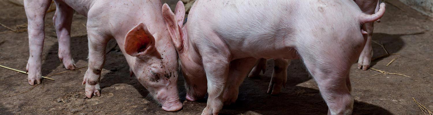
Apart from the initial rise in body temperature, the first sign of disease is usually lameness in one or more pigs in a herd as a result of vesicle formation on the heels of the feet, coronary bands and interdigital spaces. Vesicles are also often present on the snout, lips and tongue, as well as elsewhere in the mouth. Rarely, vesicles occur on the teats of sows suckling piglets. The vesicles are usually flat, blanched areas 5 to 30 mm in diameter, or they may be raised 10 to 20 mm above the surrounding unaffected epithelium. Vesicles on the snout may occasionally resemble bunches of grapes.1, 21 The primary vesicles rupture within a day or two, but secondary vesicles may appear adjacent to those which have already ruptured. This phenomenon has resulted in the clinical signs of the disease being divided into two phases. The first phase covers the time between initial rise in body temperature (40,6 to 42,2 °C) and the appearance of secondary lesions,1 while the second phase lasts one or two days and involves rupture of the secondary vesicles, the temperature returning to normal followed by the resumption of normal behaviour. Complete recovery is usually uneventful.
Apart from vesicular lesions, extensive necrosis of lymphocytes in lymph nodes may occur. It is not known whether the appreciable amounts of virus detectable in lymph nodes is a result of replication at that site or of drainage from other sites of replication.21
Experimental infections of pigs with SMS viruses generally result in a typical vesicular disease.20 The severity of the resulting disease depends upon the virus strain used; some SMS viruses appear to be more pathogenic for pigs than some of the original VESV serotypes.20
Diagnosis and differential diagnosis
The lesions of VES are indistinguishable from those of footand- mouth disease (FMD), swine vesicular disease or vesicular stomatitis. An aetiological diagnosis can only be based on identification of the virus by complement fixation, virus neutralization, or ELISA5 using appropriate antisera, or by specific reverse transcription-polymerase chain reaction assays.11, 13, 16 Suspensions of virus or viral antigens may be detectable directly from vesicular epithelium or from virus cultivated in cell culture. Low levels of virus may be detectable in the blood of acutely infected pigs.21 Since VES is exotic to Africa, diagnosis of this disease in Africa would only be expected in circumstances where direct contact has occurred with pigs or pig products from outside the continent or via a connection with the marine environment.
Control
The rapid eradication of VES from the USA by the slaughterout of affected herds, quarantine of affected premises and enforced heat treatment of swill fed to pigs demonstrates the efficacy of this approach. However, to prevent further recurrence, it is essential to trace the origin of the virus introduction.
References
- BANKOWSKI, R.A., 1965. Vesicular exanthema. Advances in Veterinary Science, 10, 23–64.
- BANKOWSKI, R.A., PERKINS, A.G., STUART, E.E. & KUMMER, M., 1956. Recovery of new immunological types of vesicular exanthema virus. Proceedings of the Sixtieth Annual Meeting of the United States Livestock Sanitary Association, 302–320.
- BARLOUGH, J.E., MATSON, D.O., SKILLING, D.E., BERKE, T., BERRY, E.S., BROWN, R.F. & SMITH, A.W., 1998. Isolation of reptilian calicivirus Crotalus type 1 from feral pinnipeds. Journal of Wildlife Diseases, 34, 451–456.
- BERRY, E.S., SKILLING, D.E., BARLOUGH, J.E., VEDROS, N.A., GAGE, L.J. & SMITH, A.W., 1990. New marine calicivirus serotype infective for swine. American Journal of Veterinary Research, 51, 1184–1187.
- FERRIS, N.P. & OXTOBY, J.M., 1994. An enzyme-linked immunosorbent assay for the detection of marine caliciviruses. Veterinary Microbiology, 42, 229–238.
- GANOVA-RAEVA, L.M., KHUDYAKON, Y.E., FIELDS, H.A. & SMITH, A.W., 2001. Unpublished data.
- GREEN, K.Y, ANDO, T., BALAYAN, M.S., CLARKE, I.N., ESTES, M.K., MATSON, D.O., NAKATA, S., NEILL, J.D., STUDDERT, M.J. & THIEL, H.-J., 2000. Caliciviridae. In: van regenmortel, m.h.v., fauquet, c.m., bishop, d.h.l., calisher, c.h., carsten, e.b., estes, m.k., lemon, s.m., maniloff, j., mayo, m.a., mcgeoch, d.j., pringle, c.r. & wickner, r.b.(eds.). Virus Taxonomy. Seventh Report of the International Committee for the Taxonomy of Viruses. New York, San Diego: Academic Press, pp. 725–735.
- GREEN, K.Y., ANDO, T., BALAYAN, M.S., BERKE, T., CLARKE, I.N., ESTES, M.K., MATSON, D.O., NAKATA, S., NEILL, J.D., STUDDERT, M.J. & THIEL, H.-J., 2000. Taxonomy of the caliciviruses. Journal of Infectious Diseases, 181, Suppl. 2, S322–330.
- LONG, G.G., EVERMANN, J.F. & GORHAM, J.R., 1980. Naturally occurring picornavirus infection of domestic mink. Canadian Journal of Comparative Medicine, 44, 412–417.
- MATSON, D.O., BERKE, T., DINULOS, M.B., POET, E., ZHONG, W.M., DAI, X.M., JIANG, X., GOLDING, B. & SMITH, A.W., 1996. Partial characterization of the genome of nine animal caliciviruses. Archives of Virology, 141, 2443– 2456.
- NEILL, J.D. & SEAL, B.S., 1995. Development of PCR primers for specific amplification of two distinct regions of the genomes of San Miguel sea-lion and vesicular exanthema of swine viruses. Molecular and Cellular Probes, 9, 33–37.
- NEILL, J.D., MEYER, R.F. & SEAL, B.S., 1995. Genetic relatedness of the caliciviruses: San Miguel sea lion and vesicular exanthema of swine viruses constitute a single genotype within the Caliciviridae. Journal of Virology, 69, 4484–4488.
- NUNEZ, J.I., BLANCO, E., HERNANDEZ, T., GOMEZ-TEJEDOR, C., MARTIN, M.J., DOPAZO, J. & SOBRINO, F., 1998. A RT-PCR assay for the differential diagnosis of vesicular viral diseases of swine. Journal of Virological Methods, 72, 227–235.
- PLOWRIGHT, W., 1988. Virus Transmissible Between Wild and Domestic Animals. Zoological Symposium No. 60. London: The Zoological Society of London.
- POET, S.E., SKILLING, D.E., MEGYESI, J.L., GILMARTIN, W.G. & SMITH, A.W., 1996. Detection of a non-cultivatable calicivirus from the white tern (Gygis alba rothschildi). Journal of Wildlife Diseases, 32, 461–467.
- REID, S.M., ANSELL, D.M., FERRIS, N.P., HUTCHINGS, G.H., KNOWLES, N.J. & SMITH, A.W., 1999. Development of a reverse transcription polymerase chain reaction procedure for the detection of marine caliciviruses with potential application for nucleotide sequencing. Journal of Virological Methods, 82, 99–107.
- ROERINK, F., HASHIMOTO, M., TOHYA, Y. & MOCHIZUKI, M., 1999. Organization of the canine calicivirus genome from the RNA polymerase gene to the poly(A) tail. Journal of General Virology, 80, 929–935.
- SEAL, B.S., LUTZE-WALLACE, C., KREUTZ, L.C., SAPP, T., DULAC, G.C. & NEILL, J.D., 1995. Isolation of caliciviruses from skunks that are antigenically and genotypically related to San Miguel sea lion virus. Virus Research, 37, 1–12.
- SMITH, A.W., 1981. Marine reservoirs of caliciviruses. In: steel, j., (ed.). CRC Handbook Series in Zoonoses: Viral Zoonoses. Boca Raton, Florida: CRC Press.
- SMITH, A.W. & BOYT, P.M., 1990. Caliciviruses of ocean origin: A review. Journal of Zoo and Wildlife Medicine, 21, 3–23.
- SMITH, A.W. & MADIN, S.H., 1986. Vesicular exanthema. In: leman, a.d., straw, b., glock, r.d., mengeling, w.l., penney, r.h.c. & scholl, e. (eds.). Diseases of Swine. 6th edn. Ames, Iowa: Iowa State University Press, pp. 358–368.
- SMITH, A.W., AKERS, T.G., MADIN, S.H. & VEDROS, N.A., 1973. San Miguel sea lion virus isolation, preliminary characterization and relationship to vesicular exanthema of swine virus. Nature, 244, 108–110.
- SMITH, A.W., AKERS, T.G. & PRATO, C.M., 1976. Prevalence and distribution of four serotypes of SMSV serum-neutralizing antibodies in wild animal populations. Journal of Wildlife Diseases, 12, 326–334.
- SMITH, A.W., MADIN, S.H., VEDROS, N.A. & BANKOWSKI, R.A., 1977. Host range comparisons of five serotypes of caliciviruses. American Journal of Veterinary Research, 38, 101–105.
- SMITH, A.W. & LATHAM, A.B., 1978. Prevalence of vesicular exanthema of swine antibodies among feral mammals associated with the Southern Californian coastal zones. American Journal of Veterinary Research, 39, 291–296.
- SMITH, A.W., SKILLING, D.E. & RIDGWAY, S., 1983. Calicivirus-induced vesicular disease in cetaceans and probable interspecies transmission. Journal of the American Veterinary Medical Association, 183, 1223–1225.
- SMITH, A.W., RITTER, D.G., RAY, G.C., SKILLING, D.E. & WARTZOK, D., 1983. New calicivirus isolates from faeces of walrus (Odobenus rosmarus). Journal of Wildlife Diseases, 19, 86–89.
- SMITH, A.W., MATTSON, D.E., SKILLING, D.E. & SCHMITZ, J.A., 1983. Isolation and partial characterization of a calicivirus from calves. American Journal of Veterinary Research, 44, 851–855.
- SMITH, A.W., SKILLING, D.E., ENSLEY, P.K., BENIRSCHKE, K. & LESTER, T.L., 1983. Calicivirus isolation and persistence in a pygmy chimpanzee (Pan paniscus). Science, 221, 79–81.
- SMITH, A.W., ANDERSON, M.P., SKILLING, D.E., BARLOUGH, J.E. & ENSLEY, P.K., 1986. First isolation of calicivirus from reptiles and amphibians. American Journal of Veterinary Research, 47, 1718–1721.
- SMITH, A.W., SKILLING, D.E., CHERRY, N., MEAD, J.H. & MATSON, D.O., 1998. Calicivirus emergence from ocean reservoirs: Zoonotic and interspecies movements. Emerging Infectious Diseases, 4, 13–20.
- SMITH, A.W., BERRY, E.S., SKILLING, D.E., BARLOUGH, J.E., POET, S.E., BERKE, T., MEAD, J. & MATSON, D.O., 1998. In vitro isolation and characterization of a calicivirus causing a vesicular disease of the hands and feet. Clinical Infectious Diseases, 26, 434–439.


