- Infectious Diseases of Livestock
- Part 2
- Pseudocowpox
- GENERAL INTRODUCTION: PARAMYXOVIRIDAE AND PNEUMOVIRIDAE
- Rinderpest
- Peste des petits ruminants
- Parainfluenza type 3 infection
- Bovine respiratory syncytial virus infection
- Hendra virus infection
- Paramyxovirus-induced reproductive failure and congenital defects in pigs
- Nipah virus disease
- GENERAL INTRODUCTION: CALICIVIRIDAE AND ASTROVIRIDAE
- Vesicular exanthema
- Enteric caliciviruses of pigs and cattle
- GENERAL INTRODUCTION: RETROVIRIDAE
- Enzootic bovine leukosis
- Jaagsiekte
- Visna-maedi
- Caprine arthritis-encephalitis
- Equine infectious anaemia
- GENERAL INTRODUCTION: PAPILLOMAVIRIDAE
- Papillomavirus infection of ruminants
- Papillomavirus infection of equids
- GENERAL INTRODUCTION: ORTHOMYXOVIRIDAE
- Equine influenza
- Swine influenza
- GENERAL INTRODUCTION: CORONAVIRIDAE
- Porcine transmissible gastroenteritis
- Porcine respiratory coronavirus infection
- Porcine epidemic diarrhoea
- Porcine haemagglutinating encephalomyelitis virus infection
- Porcine deltacoronavirus infection
- Bovine coronavirus infection
- Ovine coronavirus infection
- Equine coronavirus infection
- GENERAL INTRODUCTION: PARVOVIRIDAE
- Porcine parvovirus infection
- Bovine parvovirus infection
- GENERAL INTRODUCTION: ADENOVIRIDAE
- Adenovirus infections
- GENERAL INTRODUCTION: HERPESVIRIDAE
- Equid herpesvirus 1 and equid herpesvirus 4 infections
- Equid gammaherpesvirus 2 and equid gammaherpesvirus 5 infections
- Equine coital exanthema
- Infectious bovine rhinotracheitis/infectious pustular vulvovaginitis and infectious pustular balanoposthitis
- Bovine alphaherpesvirus 2 infections
- Malignant catarrhal fever
- Pseudorabies
- Suid herpesvirus 2 infection
- GENERAL INTRODUCTION: ARTERIVIRIDAE
- Equine viral arteritis
- Porcine reproductive and respiratory syndrome
- GENERAL INTRODUCTION: FLAVIVIRIDAE
- Bovine viral diarrhoea and mucosal disease
- Border disease
- Hog cholera
- Wesselsbron disease
- Louping ill
- West nile virus infection
- GENERAL INTRODUCTION: TOGAVIRIDAE
- Equine encephalitides caused by alphaviruses in the Western Hemisphere
- Old World alphavirus infections in animals
- Getah virus infection
- GENERAL INTRODUCTION: BUNYAVIRIDAE
- Diseases caused by Akabane and related Simbu-group viruses
- Rift Valley fever
- Nairobi sheep disease
- Crimean-Congo haemorrhagic fever
- GENERAL INTRODUCTION: ASFARVIRIDAE
- African swine fever
- GENERAL INTRODUCTION: RHABDOVIRIDAE
- Rabies
- Bovine ephemeral fever
- Vesicular stomatitis and other vesiculovirus infections
- GENERAL INTRODUCTION: REOVIRIDAE
- Bluetongue
- Ibaraki disease in cattle
- Epizootic haemorrhagic disease
- African horse sickness
- Equine encephalosis
- Palyam serogroup orbivirus infections
- Rotavirus infections
- GENERAL INTRODUCTION: POXVIRIDAE
- Lumpy skin disease
- Sheeppox and goatpox
- Orf
- Ulcerative dermatosis
- Bovine papular stomatitis
- Pseudocowpox
- Swinepox
- Cowpox
- Horsepox
- Camelpox
- Buffalopox
- GENERAL INTRODUCTION: PICORNAVIRIDAE
- Teschen, Talfan and reproductive diseases caused by porcine enteroviruses
- Encephalomyocarditis virus infection
- Swine vesicular disease
- Equine picornavirus infection
- Bovine rhinovirus infection
- Foot-and-mouth disease
- GENERAL INTRODUCTION: BORNAVIRIDAE
- Borna disease
- GENERAL INTRODUCTION: CIRCOVIRIDAE AND ANELLOVIRIDAE
- Post-weaning multi-systemic wasting syndrome in swine
- GENERAL INTRODUCTION: PRION DISEASES
- Scrapie
- Bovine spongiform encephalopathy
- Transmissible spongiform encephalopathies related to bovine spongiform encephalopathy in other domestic and captive wild species
Pseudocowpox
This content is distributed under the following licence: Attribution-NonCommercial CC BY-NC  View Creative Commons Licence details here
Pseudocowpox
E MUNZ AND K DUMBELL
Introduction
Pseudocowpox is caused by a parapoxvirus and is characterized by the development of pox lesions on the teats and udders of cows. The virus may also cause cutaneous lesions on the hands of humans (‘milker’s nodule’). The disease has a worldwide distribution. The prevalence may be high — in Britain animals with lesions were detected in 15 out of 16 herds8 and in 13 per cent of 358 cows examined at abattoirs.4 In South Africa pseudocowpox accompanied by poor management practices occasionally causes serious economic losses to dairy farmers as a result of a reduced milk production.5, 6
Aetiology
Pseudocowpox is caused by a parapoxvirus which is very closely related and possibly identical to bovine papular stomatitis virus and less closely related to orf virus. Pseudocowpox virus multiplies in cell cultures derived from bovine and ovine tissues, but not on the chorio-allantoic membranes of embryonated hens’ eggs.
A ‘paravaccinia virus’ has been isolated from the milk of healthy cows,2 and from the semen of two infertile bulls.7
Epidemiology
The virus is usually introduced into susceptible herds by the purchase of infected animals. The infection is then transmitted from animal to animal by contact, milking or biting flies. Milkers, infected parts of milking machines, or teat beakers may transmit the virus from cow to cow unless strict hygiene is applied. Dairy cows in milk are therefore most often infected, the disease seldom occurring in dry cows or heifers. There is no seasonal variation in prevalence.
Because of the short-lived immunity after infection, reinfection may occur during subsequent lactations.
Clinical signs
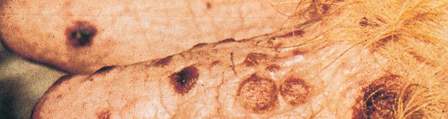
Localized ecthymatous lesions appear on the teats after an incubation period of approximately six days. This is followed within a few days by the formation of papules, vesicles and pustules which become covered with dark, horse-shoe shaped or ring-like scabs (Figure 114.1). The scabs are usually shed within a few weeks but occasionally persist for months. They impart a rough appearance to the teats. Acute lesions may reappear during this period. Secondary bacterial infection and aggravation of the lesions by milking delay healing. Similar lesions may occur on the muzzle and in the mouths of suckling calves.
Personnel handling infected animals may develop pox-like lesions on the hands, forearms or even on the face (milk-er’s nodules). Fever and swelling of lymph nodes, which last two to four weeks, may be observed in humans during the acute stage of the disease.9 The infection in humans takes a mild course unless secondary bacterial infections occur.
Diagnosis and differential diagnosis
A tentative diagnosis based on the appearance of the lesions can quickly be verified by electron microscopic investigation of negatively stained preparations derived from scabs or vesicular material,1 but isolation of the virus in cell culture and its further identification takes several days.3 Serological tests for the detection of antigen or antibodies and allergic tests are not of much value and are no longer used. Pseudocowpox may be confused with teat lesions caused by cowpox, lumpy skin disease, bovine herpes mammilitis, warts or traumatic injuries to the teats and udder. Foot-and-mouth disease and other infections causing skin lesions should also be taken into consideration.4
Control
Topical application of ointments and sprays to prevent bacterial infection and to support the healing process is advisable. Disinfection measures during milking, such as teat dipping in 0,1 per cent potassium permanganate and the use of disposable paper towels for udder washing, are useful in preventing or reducing the spread of the disease. No vaccine is available.

References
- davis, c.m., musil, g. & trochet, j.a., 1969. Electron microscopy for rapid diagnosis of pseudocowpox and milker’s nodule. American Journal of Veterinary Research, 31, 1497–1503.
- dawson, p.s., forbes, d. & stuart, p., 1968. Isolation of a paravaccinia virus from bovine milk. The Veterinary Record, 82, 525–526.
- friedman-kien, a.e., rowe, w.p. & bandfield, w.g., 1963. Milker’s nodules: Isolation of a poxvirus from a human case. Science, 140, 1335–1336.
- gibbs, e.p.j., johnson, r.h. & osborne, a.d., 1970. The differential diagnosis of viral skin infections of the bovine teat. The Veterinary Record, 87, 602–609.
- giesecke, w.h., du preez, j.h. & petzer, i-m., 1989. Practical information on various aspects of udder health in dairy cattle: 39. Udder and teat lesions and related problems (skin diseases of udder and teats). Milk Producer. 26–30 May.
- giesecke, w.h., theodorides, a. & els, h.j., 1971. Pseudocowpox (paravaccinia) in dairy cows. Journal of the South African Veterinary Medical Association, 42, 193–194.
- johnston, w.s. & deas, d.w., 1971. Isolation of a paravaccinia virus from bovine semen. The Veterinary Record, 89, 450.
- nagington, j., tee, g.h. & smith, j.s., 1966. Milker’s nodule virus infections in Dorset and their similarity to orf. The Veterinary Record, 78, 305–308.
- ruzicka, t., schmoeckel, c. & ryckmanns, f., 1983. Ecthyma contagiosum und Melkerknoten. Mu¨nchener Medizinische Wochenschrift, 125, 1103–1104.


