- Infectious Diseases of Livestock
- Part 2
- Teschen, Talfan and reproductive diseases caused by porcine enteroviruses
- GENERAL INTRODUCTION: PARAMYXOVIRIDAE AND PNEUMOVIRIDAE
- Rinderpest
- Peste des petits ruminants
- Parainfluenza type 3 infection
- Bovine respiratory syncytial virus infection
- Hendra virus infection
- Paramyxovirus-induced reproductive failure and congenital defects in pigs
- Nipah virus disease
- GENERAL INTRODUCTION: CALICIVIRIDAE AND ASTROVIRIDAE
- Vesicular exanthema
- Enteric caliciviruses of pigs and cattle
- GENERAL INTRODUCTION: RETROVIRIDAE
- Enzootic bovine leukosis
- Jaagsiekte
- Visna-maedi
- Caprine arthritis-encephalitis
- Equine infectious anaemia
- GENERAL INTRODUCTION: PAPILLOMAVIRIDAE
- Papillomavirus infection of ruminants
- Papillomavirus infection of equids
- GENERAL INTRODUCTION: ORTHOMYXOVIRIDAE
- Equine influenza
- Swine influenza
- GENERAL INTRODUCTION: CORONAVIRIDAE
- Porcine transmissible gastroenteritis
- Porcine respiratory coronavirus infection
- Porcine epidemic diarrhoea
- Porcine haemagglutinating encephalomyelitis virus infection
- Porcine deltacoronavirus infection
- Bovine coronavirus infection
- Ovine coronavirus infection
- Equine coronavirus infection
- GENERAL INTRODUCTION: PARVOVIRIDAE
- Porcine parvovirus infection
- Bovine parvovirus infection
- GENERAL INTRODUCTION: ADENOVIRIDAE
- Adenovirus infections
- GENERAL INTRODUCTION: HERPESVIRIDAE
- Equid herpesvirus 1 and equid herpesvirus 4 infections
- Equid gammaherpesvirus 2 and equid gammaherpesvirus 5 infections
- Equine coital exanthema
- Infectious bovine rhinotracheitis/infectious pustular vulvovaginitis and infectious pustular balanoposthitis
- Bovine alphaherpesvirus 2 infections
- Malignant catarrhal fever
- Pseudorabies
- Suid herpesvirus 2 infection
- GENERAL INTRODUCTION: ARTERIVIRIDAE
- Equine viral arteritis
- Porcine reproductive and respiratory syndrome
- GENERAL INTRODUCTION: FLAVIVIRIDAE
- Bovine viral diarrhoea and mucosal disease
- Border disease
- Hog cholera
- Wesselsbron disease
- Louping ill
- West nile virus infection
- GENERAL INTRODUCTION: TOGAVIRIDAE
- Equine encephalitides caused by alphaviruses in the Western Hemisphere
- Old World alphavirus infections in animals
- Getah virus infection
- GENERAL INTRODUCTION: BUNYAVIRIDAE
- Diseases caused by Akabane and related Simbu-group viruses
- Rift Valley fever
- Nairobi sheep disease
- Crimean-Congo haemorrhagic fever
- GENERAL INTRODUCTION: ASFARVIRIDAE
- African swine fever
- GENERAL INTRODUCTION: RHABDOVIRIDAE
- Rabies
- Bovine ephemeral fever
- Vesicular stomatitis and other vesiculovirus infections
- GENERAL INTRODUCTION: REOVIRIDAE
- Bluetongue
- Ibaraki disease in cattle
- Epizootic haemorrhagic disease
- African horse sickness
- Equine encephalosis
- Palyam serogroup orbivirus infections
- Rotavirus infections
- GENERAL INTRODUCTION: POXVIRIDAE
- Lumpy skin disease
- Sheeppox and goatpox
- Orf
- Ulcerative dermatosis
- Bovine papular stomatitis
- Pseudocowpox
- Swinepox
- Cowpox
- Horsepox
- Camelpox
- Buffalopox
- GENERAL INTRODUCTION: PICORNAVIRIDAE
- Teschen, Talfan and reproductive diseases caused by porcine enteroviruses
- Encephalomyocarditis virus infection
- Swine vesicular disease
- Equine picornavirus infection
- Bovine rhinovirus infection
- Foot-and-mouth disease
- GENERAL INTRODUCTION: BORNAVIRIDAE
- Borna disease
- GENERAL INTRODUCTION: CIRCOVIRIDAE AND ANELLOVIRIDAE
- Post-weaning multi-systemic wasting syndrome in swine
- GENERAL INTRODUCTION: PRION DISEASES
- Scrapie
- Bovine spongiform encephalopathy
- Transmissible spongiform encephalopathies related to bovine spongiform encephalopathy in other domestic and captive wild species
Teschen, Talfan and reproductive diseases caused by porcine enteroviruses
This content is distributed under the following licence: Attribution-NonCommercial CC BY-NC  View Creative Commons Licence details here
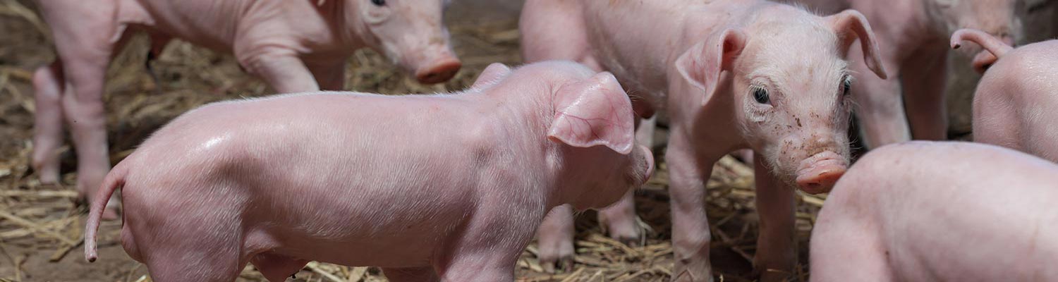
Teschen, Talfan and reproductive diseases caused by porcine enteroviruses
T J L ALEXANDER
Introduction
With the exception of swine vesicular disease (SVD), most infections with porcine enteroviruses, which belong to the family Picornaviridae, cause no apparent disease, but some strains are capable of causing polioencephalomyelitis (Teschen and Talfan), reproductive disorders (stillbirths, foetal mummification, embryonic death and infertility — known collectively by the acronym SMEDI) and, less commonly, pneumonitis or peri- and myocarditis.
Most mammals and birds have their own host-specific array of enteroviruses which are sub-classified into serotypes by serum neutralization and other serological tests.1, 4, 5, 8 Pigs are no exception. They have 11 known serotypes: Teschen/ Talfan disease is caused by serotype 1, and SVD (see Swine vesicular disease) is caused by a distinct porcine enterovirus which is related to the human enterovirus, coxsackie virus 5. With the exception of SVD virus, the porcine enteroviruses are ubiquitous. Although the majority of infections are subclinical, type 1, the Teschen/Talfan virus, may cause polioencephalomyelitis.1, 5, 6 Teschen is the more severe form of polioencephalomyelitis and was first recognized in 1930 in Teschen, which is on the border of the Czeck Republic and Germany. It is endemic in Central Europe, southern Zaire and Madagascar.11 Talfan disease, named after a hill in Wales and known in Denmark as benign enzootic paresis, is a milder disease which has occurred in North America, France, Belgium, Britain, Ireland, Denmark, Greece, Australia and north-east Europe.8, 11 Serotypes 1, 3, 6 and 8 have been reported to be causes of theSMEDIsyndrome,1, 2, 3, 5, 7 which is also caused by porcine parvovirus (Porcine parvovirus infection), encephalomyocarditis virus (Encephalomyocarditis virus infection) and, to some extent, the virus responsible for the porcine reproductive and respiratory syndrome (Porcine reproductive and respiratory syndrome) infections.
Others, for example porcine enterovirus serotype 2, when given experimentally to colostrum-deprived piglets, may produce microscopic lesions of pneumonitis9, 10 but no clinical signs although they may trigger off other pathogenic infectious agents, such as Mycoplasma hyopneumoniae. Serotype 2 has also produced mild polioencephalomyelitis, fibrinous pericarditis and myocarditis in colostrum-deprived piglets.9
Aetiology
The genus Enterovirus is one of five in the family Picornaviridae, the others being Cardiovirus (see Encephalomyocarditis virus infection), Rhinovirus, Hepatovirus, and Aphthovirus.
Enteroviruses (see the introduction, Picornaviridae, and Foot-and-mouth disease) are spherical, 25 to 31 nm in diameter and non-enveloped, with a buoyant density in cesium chloride of 1,34. They contain a core of single-stranded RNA, which is surrounded by a protein capsid but no lipids. They can be grown in porcine epithelial cell cultures, (usually kidney cells) but some can be cultured in cell lines derived from other species.1, 5, 13
Porcine enteroviruses are relatively resistant to heat, pH 2 to 9, commonly used disinfectants and environmental factors.
Epidemiology
Infected pigs shed virus in their faeces, sometimes in large quantities, which contaminates the environment and infects other pigs. The oral–faecal route of infection is probably the most important. Being relatively resistant, the virus can persist for long periods in piggeries and in slurry. Enteroviruses are highly infectious and can be readily carried to other pig farms on boots, clothes, vehicles or other vectors.1, 10
Pathogenesis
Since porcine enteroviruses are so widespread in pig populations, sows tend to have a strong humoral and mucosal-associated immunity. Thus the enteroviruses endemic in a herd are prevented from multiplying in the alimentary tracts of the piglets by secretory IgA derived from their dam’s colostrum. After weaning, however, this rapidly disappears, leaving their alimentary tracts more susceptible to infection. 1, 10 They still have a high level of circulating colostrumderived antibodies, some of which enter the intestinal lumen, reducing the level of multiplication of enteroviruses. The humoral antibodies also prevent spread of these viruses from the alimentary tract into the bloodstream. Low-level multiplication in the intestines in such circumstances stimulates the lymphoid system to produce both mucosal-associated IgA and humoral antibodies which prevent clinical disease and eventually inhibit viral multiplication in the body.5 Individual young pigs thus go through a sequence of subclinical infections with different serotypes during the growing period.1, 10
Natural infection of pigs by enteroviruses is by the oral route.5, 13 The virus multiplies in the intestines and lymph nodes of susceptible pigs and, in the absence of circulating antibody, causes a transient viraemia lasting several days. Virus can be found in most tissues and organs.5, 13 Depending on the strain involved, infection may be silent or may induce the formation of lesions in target organs and clinical signs of disease.
Teschen/Talfan virus invades the central nervous system causing polioencephalomyelitis and paresis, in some animals leading to paralysis of the limbs, particularly the hind limbs.6 Serotypes 1, 3, 6 and 8 may invade the foetuses and cause stillbirths, mummification, embryonic death and infertility (the SMEDI syndrome).3, 7 This is much less common than the SMEDI syndrome caused by porcine parvovirus because enterovirus infections almost always occur well before the gilts reach breeding age so that by the time of mating most gilts are immune.
Clinical signs
In Teschen/Talfan disease the first prodromal signs are mild transient fever, reduced appetite and depression lasting one to three days. It usually goes unnoticed by the farmer. An ascending motor paresis then slowly develops. It may not progress or become severe but manifests as an unsteady gait for several weeks and then resolves itself (Talfan disease), or it may progress to partial or generalized permanent paralysis, affecting particularly the hind limbs (Teschen disease).1, 5, 13 In both cases the pig appears otherwise normal and, provided it can get to the trough and drink, will eat and drink and grow normally. There appears to be no pain.
The mild form (Talfan disease) generally occurs in young pigs several weeks after weaning, presumably at a time when there is no specific secretory IgA in the alimentary tract and before the colostrum-derived antibodies have completely waned.1, 5, 13 It may occur in one or two pigs in a litter, a whole litter or several litters. Mortality is negligible.
The severe form (Teschen disease) tends to occur in a wider spread of age groups, presumably when the virus has recently been introduced into a herd in which there is no herd immunity.1, 5, 13 Affected pigs which become severely paralysed may not be able to get to the feed trough, may develop skin sores and may be attacked by healthy pigs. Some may die from secondary infections.
Gilts that develop SMEDI syndrome generally do not show clinical signs other than reproductive failure. This manifests as the birth of dead piglets and foetuses, mummified foetuses and small litters, failure of gilts thought to be pregnant to farrow, and uneven returns to oestrus.1, 3, 7 The SMEDI syndrome is described in more detail in Porcine parvovirus infection.
Pathology
In pigs with Teschen/Talfan disease there are no gross lesions in the central nervous system (CNS), but in Teschen disease there may be muscle atrophy particularly of the hind limbs and possibly skin lesions of a secondary nature.
Histologically, lesions in the CNS may be seen in the dorsal root ganglia, brain stem and lumbar spinal cord, particularly the ventral horn. These tend to be most pronounced in the grey matter of the spinal cord, dorsal root ganglia and medulla oblongata, and consist of a diffuse infiltration of monocytes, proliferation of microglia, perivascular mononuclear cell cuffing and lymphocytic meningitis, particularly over the cerebellum. Some neuronal degeneration may be observed.1, 6
Electron microscopic studies reveal detachment of ribosomes from the endoplastic reticulum and disappearance of ribosomal clusters in ganglion cells early in the disease process, followed by dilatation of the endoplasmic reticular cisternae, vesiculation and cellular necrosis. Viral crystals may be seen in capillary endothelial cells, in astrocyte processes near capillaries and in neurones.6
Some pigs suffering from an enterovirus viraemia also develop a mild non-specific polyserositis, involving particularly the pericardium. In these cases the pericardial fluid is turbid as a result of the presence of inflammatory cells. Focal myocardial necrosis has been reported.1
Diagnosis
In Teschen/Talfan disease the clinical signs and absence of lesions other than in the CNS are almost pathognomonic. The microscopic lesions in the CNS are not specifically diagnostic. For a specific diagnosis, either rising serum antibody levels in paired serum samples must be demonstrated or virus isolated in tissue culture from the brain tissue and identified. Theoretically, it would be possible to develop specific fluorescent antibody tests (FAT) and polymerase chain reaction (PCR) tests, but clinical disease is so rare in most countries that there has been no incentive to do so.
Similarly, the SMEDI syndrome caused by enteroviruses is so uncommon that no diagnostic tests have been developed. As for the diagnosis of Teschen/Talfan disease, rising serum antibody levels must be demonstrated in the dam, or virus isolated in tissue culture from foetal tissues.
Differential diagnosis
Teschen/Talfan disease has to be distinguished from other infections which cause CNS signs and lesions. These include Aujeszky’s disease (pseudorabies), in which nervous signs occur mainly in suckling piglets, classical swine fever (hog cholera), porcine encephalomyocarditis, rabies, salt poisoning resulting from water deprivation, and bacterial meningitis caused, for example, by Streptococcus suis type 1 or 2.
SMEDI syndrome caused by enteroviruses has to be distinguished from the SMEDI syndrome caused by other infectious agents, including porcine parvovirus, porcine reproductive and respiratory syndrome virus (PRRSV) and encephalomyocarditis virus.
Control
Theoretically, the clinical signs of all these diseases could be prevented by the use of inactivated or attenuated virus vaccines but their prevalence is so low or sporadic that there is no practical call for them. Antibacterial drugs are of no help in treatment. Good husbandry practice (e.g. clean, deep bedding) may be helpful in nursing Teschen/Talfan cases.
References
- derbyshire, j.b., 1999. Enterovirus. In: straw, b.e., d’allaire, s., mengeling, w.l. & taylor, d.j., (eds). Diseases of Swine. Iowa: Iowa State University Press. pp. 145–150.
- de meurichy, w., pensaert, m. & bonte, p., 1976. Het SMEDI-syndrome bij het varken: Rol van de enterovirussen en het parvovirus. Vlaamse Diergeneeskundige Tijdschrift, 45, 241–261.
- dunne, h.w., gobble, j.l., hokanson, j.f., kradel, d.c. & bubash, g.r., 1965. Porcine reproductive failure associated with a newly identified ‘SMEDI’ group of picornaviruses. American Journal of Veterinary Research, 26, 1284–1297.
- dunne, h.w., wang, j.t. & ammerkman, e.h., 1971. Classification of North American porcine enteroviruses: A comparison with European and Japanese strains. Infection and Immunity, 4, 619–631.
- fenner, f.j., gibbs, e.p.j., murphy, f.a., rott, r., studdert, m.j. & white, d.o., 1993. Veterinary Virology. 2nd edn. San Diego: Academic Press.
- holman, j.e., loestner, a. & kasza, l., 1996. Histopathogenesis of porcine poliencephalitis in the germ-free pig. Pathologia Veterinaria, 3, 633–651.
- huang, j., gentry, r.f. & zarkower, a., 1980. Experimental infection of pregnant sows with porcine enteroviruses. American Journal of Veterinary Research, 41, 469–473.
- knowles, n.j., buckley, l.s. & pereira, h.g., 1979. Classification of porcine enteroviruses by antigenic analysis and cytopathic effects in tissue culture. Description of 3 new serotypes. Archives of Virology, 62, 201–208.
- long, j.f., kasza, l. & koestner, a., 1969. Pericarditis and myocarditis in germfree and colostrum-deprived pigs experimentally infected with a porcine polioencephalomyelitis virus. Journal of Infectious Diseases, 120, 245–249.
- meyer, r.c., woods, g.t. & simon, j., 1966. Pneumonitis in an enterovirus infection in swine. Journal of Comparative Pathology, 76, 397–405.
- oden’hal, s., 1983. The Geographical Distribution of Animal Virus Diseases. New York, London: Academic Press, Inc.
- singh, k.v. & bohl, e.h., 1972. The pattern of enteroviral infection in a herd of swine. Canadian Journal of Comparative Medicine, 36, 243–248.
- taylor, d.j., 1995. Pig Diseases. 6th edn. Glasgow: Published by the author.


