- Infectious Diseases of Livestock
- Part 2
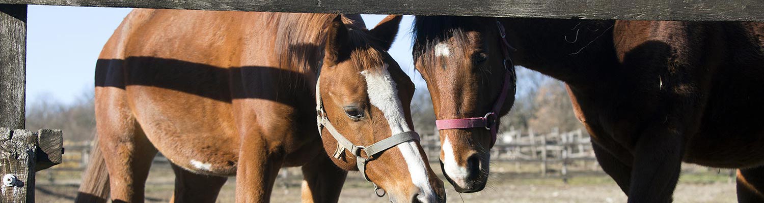
- Hendra virus infection
- GENERAL INTRODUCTION: PARAMYXOVIRIDAE AND PNEUMOVIRIDAE
- Rinderpest
- Peste des petits ruminants
- Parainfluenza type 3 infection
- Bovine respiratory syncytial virus infection
- Hendra virus infection
- Paramyxovirus-induced reproductive failure and congenital defects in pigs
- Nipah virus disease
- GENERAL INTRODUCTION: CALICIVIRIDAE AND ASTROVIRIDAE
- Vesicular exanthema
- Enteric caliciviruses of pigs and cattle
- GENERAL INTRODUCTION: RETROVIRIDAE
- Enzootic bovine leukosis
- Jaagsiekte
- Visna-maedi
- Caprine arthritis-encephalitis
- Equine infectious anaemia
- GENERAL INTRODUCTION: PAPILLOMAVIRIDAE
- Papillomavirus infection of ruminants
- Papillomavirus infection of equids
- GENERAL INTRODUCTION: ORTHOMYXOVIRIDAE
- Equine influenza
- Swine influenza
- GENERAL INTRODUCTION: CORONAVIRIDAE
- Porcine transmissible gastroenteritis
- Porcine respiratory coronavirus infection
- Porcine epidemic diarrhoea
- Porcine haemagglutinating encephalomyelitis virus infection
- Porcine deltacoronavirus infection
- Bovine coronavirus infection
- Ovine coronavirus infection
- Equine coronavirus infection
- GENERAL INTRODUCTION: PARVOVIRIDAE
- Porcine parvovirus infection
- Bovine parvovirus infection
- GENERAL INTRODUCTION: ADENOVIRIDAE
- Adenovirus infections
- GENERAL INTRODUCTION: HERPESVIRIDAE
- Equid herpesvirus 1 and equid herpesvirus 4 infections
- Equid gammaherpesvirus 2 and equid gammaherpesvirus 5 infections
- Equine coital exanthema
- Infectious bovine rhinotracheitis/infectious pustular vulvovaginitis and infectious pustular balanoposthitis
- Bovine alphaherpesvirus 2 infections
- Malignant catarrhal fever
- Pseudorabies
- Suid herpesvirus 2 infection
- GENERAL INTRODUCTION: ARTERIVIRIDAE
- Equine viral arteritis
- Porcine reproductive and respiratory syndrome
- GENERAL INTRODUCTION: FLAVIVIRIDAE
- Bovine viral diarrhoea and mucosal disease
- Border disease
- Hog cholera
- Wesselsbron disease
- Louping ill
- West nile virus infection
- GENERAL INTRODUCTION: TOGAVIRIDAE
- Equine encephalitides caused by alphaviruses in the Western Hemisphere
- Old World alphavirus infections in animals
- Getah virus infection
- GENERAL INTRODUCTION: BUNYAVIRIDAE
- Diseases caused by Akabane and related Simbu-group viruses
- Rift Valley fever
- Nairobi sheep disease
- Crimean-Congo haemorrhagic fever
- GENERAL INTRODUCTION: ASFARVIRIDAE
- African swine fever
- GENERAL INTRODUCTION: RHABDOVIRIDAE
- Rabies
- Bovine ephemeral fever
- Vesicular stomatitis and other vesiculovirus infections
- GENERAL INTRODUCTION: REOVIRIDAE
- Bluetongue
- Ibaraki disease in cattle
- Epizootic haemorrhagic disease
- African horse sickness
- Equine encephalosis
- Palyam serogroup orbivirus infections
- Rotavirus infections
- GENERAL INTRODUCTION: POXVIRIDAE
- Lumpy skin disease
- Sheeppox and goatpox
- Orf
- Ulcerative dermatosis
- Bovine papular stomatitis
- Pseudocowpox
- Swinepox
- Cowpox
- Horsepox
- Camelpox
- Buffalopox
- GENERAL INTRODUCTION: PICORNAVIRIDAE
- Teschen, Talfan and reproductive diseases caused by porcine enteroviruses
- Encephalomyocarditis virus infection
- Swine vesicular disease
- Equine picornavirus infection
- Bovine rhinovirus infection
- Foot-and-mouth disease
- GENERAL INTRODUCTION: BORNAVIRIDAE
- Borna disease
- GENERAL INTRODUCTION: CIRCOVIRIDAE AND ANELLOVIRIDAE
- Post-weaning multi-systemic wasting syndrome in swine
- GENERAL INTRODUCTION: PRION DISEASES
- Scrapie
- Bovine spongiform encephalopathy
- Transmissible spongiform encephalopathies related to bovine spongiform encephalopathy in other domestic and captive wild species
Hendra virus infection
This content is distributed under the following licence: Attribution-NonCommercial CC BY-NC  View Creative Commons Licence details here
Hendra virus infection
Previous authors: M M WILLIAMSON
Current authors:
K HALPIN - Pathology and Pathogenesis Group Leader, BVSc, MVSc, MPH, MANZCVS, PhD, Australian Animal Health Laboratory, 5 Portarlington Road, East Geelong, Victoria, 3219, Australia
JR GIILKERSON - Professor of Veterinary Microbiology, BVSC, BSc (Vet), PhD, Department of Veterinary Biosciences, Faculty of Veterinary and Agricultural Science, University of Melbourne, Building 400, Corner of Flemington Road and Park Drive, Parkville, Victoria, 3010, Australia
Introduction
Hendra virus disease is caused by a virus that is very closely related to Nipah virus (see Nipah virus infection), and the two viruses contribute to the genus name Henipavirus. Hendra virus disease has only occurred in Australia, where fruit bats of the genus Pteropus are the wildlife reservoir host of the virus. Since it emerged in 1994 in the Brisbane suburb of Hendra,28 there have been seven known human cases, four of which were fatal. Horses are the primary spill-over host and are extremely susceptible to infection and capable of amplifying the virus to high titres. All human cases have been infected after high level exposure to infected horse secretions or tissues, putting veterinarians, veterinary nurses and technicians, horse owners and people who work with horses, including trainers at a higher risk.19In response to this, a vaccine has been developed that prevents infection in horses, and thereby prevents infection in humans as well.24
The geographical range of Hendra virus disease has spread along the eastern coast of Australia, reaching as far north as Port Douglas and as far south as Scone in New South Wales (NSW), a distance of more than 2000km7, 40; disease has also been found approximately 300 km inland, in Chinchilla, Queensland. Most outbreaks, which occur on an almost annual basis, have only involved one or two horses and are often seasonal, with more spill-over events occurring in the cooler months. Direct transmission from the wildlife reservoir host to humans has not occurred, despite some humans, particularly wildlife carers, having very close contact with fruit bats.34
While Hendra virus (HeV) is endemic in all four mainland species of Australian fruit bats belonging to the genus Pteropus, black flying foxes (Pteropus alecto) and spectacled flying foxes (Pteropus conspiculatus) are more commonly associated with HeV excretion than other species.6, 32 Virus excretion can occur any time of the year, with a winter peak found particularly in northern NSW and southern Queensland.7 As climatic conditions change and habitat is removed for more human use, the geographical distribution of fruit bats is expected to change, and along with that it is likely that more spill-over events will be reported in new geographical areas.23
Aetiology
Hendra virus is a non-segmented negative-stranded RNA virus in the family Paramyxoviridae, genus Henipavirus. It was discovered in 1994 as the first known henipavirus.27 This genus includes Nipah virus (NiV) and the more recently discovered Mojiang paramyxovirus42 and Cedar virus.20 Cedar virus is not known to cause disease. Mojiang paramyxovirus was implicated in the death of three miners in China in 2012, following potential zoonotic transmission from rats.42
Hendra virus is a large paramyxovirus with a genome consisting of 18, 234 nucleotides. . This increased genome size, compared to other paramyxoviruses, is in part due to the long untranslated regions (UTRs) at the 3′ end of most transcription units, similar to that observed in the filoviruses Marburg and Ebola.37, 38 Like all other viruses in the subfamily Paramyxovirinae, HeV has a genome length which is a multiple of six. Genomes whose lengths deviate from ‘the rule of six’ do not replicate efficiently. It has been proposed that the templates for transcription and replication are nucleoproteins in which each nucleoprotein subunit is associated with six nucleotides of genomic RNA.
The HeV genome consists of six genes that code for six major structural proteins, namely: N nucleocapsid protein (N), phosphoprotein (P), matrix protein (M), fusion protein (F), glycoprotein (G) and large protein (L).
Sequence analysis of five HeV isolates from several outbreaks in horses indicates that the HeV genome is very stable and conserved , with at least 99 per cent nucleotide similarity between isolates when compared to the original isolate from 1994.22
Hendra virus has the morphological and physicochemical properties typical of a paramyxovirus. It is pleomorphic in shape and enveloped with a herring-boned nucleocapsid.15 Virions are 40-600 nm in diameter. Glycoprotein (G) and fusion (F) protein spikes project through a lipid envelope, but unlike NiV, the surface projections of HeV are predominantly double; NiV surface projections are predominantly single.15
Culturing the virus from infected tissues in cell culture is relatively easy. However, HeV is a dangerous human pathogen and requires biosafety level 4 (BSL4) containment (see Diagnosis).
Limited work has been done to assess the stability of the virus. Both HeV and NiV exhibit an extremely broad tolerance to extremes of pH, with viable virus recovered after a 60 minute incubation in solutions ranging from pH 3 to 11 for NiV and pH 4 to 11 for HeV.9 In the same study, henipaviruses survived for more than four days at 22°C in pH-neutral fruit bat urine, but were sensitive to higher temperatures and pH changes. On mango flesh, survival time varied depending on temperature and fruit pH, ranging from two hours to more than two days. Desiccation of viruses substantially reduced survival time to less than two hours. The sensitivity of henipaviruses to pH, temperature and desiccation indicates a need for close contact between hosts for transmission to occur, although under ideal conditions henipaviruses can persist for extended periods facilitating vehicle-borne transmission.9
Epidemiology
Fruit bats of the genus Pteropus are the natural reservoir host of HeV. They are not clinically affected by infection. Pigs, cats, guinea pigs, hamsters, ferrets, and mice have been shown to be susceptible in experimental studies. Dogs can become infected and seroconvert without the development of clinical signs. This has been demonstrated in the field and experimentally.
The first reported HeV disease outbreak occurred in September 1994, when a stable-hand in the Brisbane suburb of Hendra developed influenza-like disease, remaining ill for six weeks before gradually recovering.27, 35 A horse trainer from the same horse stable developed symptoms similar to those of the stable hand but died within six days of developing clinical illness. Thirteen Thoroughbred horses on the property suffering from acute respiratory disease died or were euthanased. Seven other horses developed HeV-specific antibodies and therefore had been exposed to HeV; these horses recovered from the illness but were eventually destroyed due to national regulations. They were not necropsied because of the potential human health risk. A further nine horses from the Hendra stables remained unaffected and were seronegative.27
Approximately 12 months later, in September 1995, a 35-year-old farmer from Mackay, Queensland, was admitted to a hospital with neurological deficits. Serum and cerebrospinal fluid from the farmer were tested for specific neutralizing antibody to HeV and there was a low but significant antibody titre that rose over time until his death 5 weeks later.29 Polymerase chain reaction assay (PCR) confirmed that he was infected with HeV. It became evident in retrospect that the farmer had assisted his wife, a veterinarian, in performing necropsies on two horses that had died on their property one year earlier in August 1994. The property on which the affected horses were in residence is 800 km north of the Brisbane suburb of Hendra, where HeV was first described. The first horse had developed severe respiratory distress and died the same day. The second horse, which was said to have licked the face of the dead horse, died 11 days after the first.33 The farmer is reported to have had close contact with blood and blood-stained foam from the horses’ respiratory tracts, particularly those from the first case. Thus, it is likely that he became infected during these necropsies. He presented with aseptic meningitis and a flu-like syndrome at the time. His wife developed no clinical disease, nor did she have antibodies to HeV.29 Formalin-fixed tissues from the horses necropsied at Mackay were located and indirect immunofluorescence tests and PCR confirmed the presence of HeV viral antigen in tissues from both horses.13
Fruit bats (Pteropodidae), commonly called flying foxes, have a wide distribution in Africa and the Asia-Pacific region, including Australia. They are a known reservoir host of HeV. Antibody to HeV has been detected in all four species of flying foxes that are found on mainland Australia43 although the black flying fox and the spectacled flying fox appear to be the species most likely to contribute to spill-over events in horses.7 The number of seropositive fruit bats varies widely depending on the population and the time of year. In one bat colony, seroprevalence steadily increased from 45 to 69 per cent over a two-year period, supporting a model of endemic infection in the population.4 Absence of disease attributable to HeV infection in fruit bats is supported by experimental observations.10
Dogs can become infected and seroconvert without the development of clinical signs. This has been demonstrated in the field and experimentally.25
Likewise, pigs can become infected experimentally but show no overt signs of disease,17whereas cats infected experimentally develop a fulminating systemic vasculitis with a high mortality rate.26
Experimentally infected Pteropus bats develop subclinical HeV infection with only sporadic low-level viral excretion in urine. Some animals seroconvert and some show evidence of infection by detection of viral genome in excretions (oral and rectal swabs, urine and blood) and viral antigen in tissues.10 The low transmissibility of HeV in fruit bats exposed experimentally is consistent with the low number of field outbreaks of the disease.
Infected horses can shed the virus in naso-pharyngeal secretions before the onset of clinical disease. By the time the disease is apparent, the virus has spread throughout the horse's blood, body fluids and tissues.21 Horses infected with the virus are a transmission risk to humans from 72 hours before the onset of clinical signs up to and after the horse's death, and until the safe disposal of the carcass. The transmission risk increases with disease progression and is highest at the point of the horse's death and during post-mortem contact.
Pathogenesis
Hendra virus enters cells by binding to the receptor Ephrin-B2, which is expressed on neurons, and smooth muscle and endothelial cells of small arteries.3 After receptor binding by the attachment G protein, the F protein, which is cleaved to create two linked polypeptides, F1 and F2, fuses to the host cell membrane, initiating endocytosis.37 Following fusion between the viral envelope and the host cell membrane, the viral ribonucleocapsid is released into the cytoplasm.16 The polymerase complex composed of the polymerase (L) and phosphoprotein (P) initiates transcription of viral mRNAs. As translation of viral mRNA occurs, viral proteins accumulate in the cell, and the polymerase switches from transcription to genome replication.
Newly made genomes are encapsidated by the nucleoprotein (N) and polymerase complexes become associated with packaged nucleocapsids. The glycoproteins are synthesized in the endoplasmic reticulum and mature through the Golgi and are transported to the cell membrane. The cytoplasmic tails of the F and G proteins play a role in the interaction with the matrix (M) protein, which initiates virus maturation and budding.16
The tropism of HeV for endothelial cells results in vasculitis, thrombosis, ischaemia, necrosis and central nervous system parenchymal infection.39, 41
Clinical signs and pathology
Early cases of HeV disease in horses presented with an acute respiratory disease.27 However, as more cases appeared, the spectrum of clinical signs widened to include neurological manifestations and colic-like clinical signs.8 Therefore HeV infected horses can be described as having an acute non-specific illness, showing variable clinical signs that can include fever, tachypnoea, tachycardia, frothy nasal discharge, ataxia, head tilt, circling, seizures, urinary incontinence, altered mentation, recumbency, depression, inappetence, restlessness, and sudden death.2, 12, 21
The incubation period of HeV infection in horses is 3 to 16 days.1, 14, 21 Similarly, the incubation period in humans ranged from a few days to two weeks.30, 35 In humans, mild disease includes fever, headache, drowsiness and influenza-like symptoms.11 Severe disease are often fatal with respiratory and/or neurological signs (e.g. confusion, motor deficits and seizures).30 Relapsing encephalitis is possible after recovery from an acute infection and appears to be due to recrudescence of viral replication in the central nervous system.29, 41 To date, there have only been seven human infections, making the characterization of clinical features less well defined.
All human cases have had high level exposure to infected horse secretions or tissues. Human to human HeV transmission has not been described to date, unlike the closely related NiV where human to human spread has been reported.18
The first fatal human cases of HeV infection died of an acute respiratory illness.35 The second fatal human case suffered from relapsing encephalitis 13 months after exposure,29 with the third and fourth cases succumbing to encephalitis.8, 30 Two of the surviving human cases suffered from a self-limited influenza-like illness at the time of HeV infection.11 The third survivor showed an influenza-like illness that progressed to acute encephalitis and suffered a long and debilitating neurological illness which to this day has not fully resolved.30 This patient had remained seropositive for several years after infection.36
Gross pathology in infected horses includes enlarged and oedematous submandibular, bronchial and/or sternal lymph nodes, dilated pulmonary lymphatics, pulmonary oedema and congestion with gelatinous distension of the subpleural lymphatics, petechial haemorrhages on the pleural surfaces and less frequently, oedema of the mesentery, increased pleural and pericardial fluid and thick foam in the airways.14
Diagnosis
Extreme care must be taken in the handling of samples collected for diagnostic testing of suspected cases of HeV disease as the causative virus is a biosecurity laboratory four (BSL4) agent, in recognition of its status as one of the most dangerous zoonotic agents. Safety precautions for the investigators in the field and in the laboratory are of paramount importance. Certain aspects of the laboratory diagnosis should be conducted only in a BSL4 facility.
Suspicion of HeV infection should be raised when a clinical syndrome consistent with those described above occurs in any area where the natural reservoir host resides. At ante- and post-mortem sampling of specimens staff who are not suitably protected must not come into contact with body fluids and tissues of infected animals. Use of full personal protective equipment including a biohazard face mask is advisable.
Rapid molecular diagnostic tests for HeV are available and should be deployed as quickly as possible once samples are collected. Molecular characterization of isolates is important. Demonstration of HeV antigen by immunohistochemistry in formalin-fixed tissue samples is a rapid and safe diagnostic technique. In areas where HeV has not been found before, confirmation of the diagnosis by virus isolation at a reference laboratory is desirable. Submission of appropriately packed specimens, according to IATA regulations, to an international BSL4 facility is paramount.
Hendra virus has been isolated from lungs, spleen, kidneys, tonsils, meninges and lymph nodes.19, 27 It may also be detected in ante-mortem samples such as blood collected in EDTA, throat and rectal swabs, cerebrospinal fluid and urine.
The virus grows in a range of cell types, including Vero cells and RK13 cells. Cytopathic effects may develop rapidly, in two to three days, but two five-day passages are usually conducted before declaring an isolation attempt unsuccessful. Cytopathic effects are characterized by large syncytia formation, involving fusion of tens to scores of cells. In HeV cultures the nuclei tend to form around the periphery of the syncytial giant cells.15 Confirmation of an isolate can be attempted by real-time PCR of tissue culture supernatant.
Transmission electron microscopic studies of HeV in culture or in lesions in fixed tissue samples showed morphological viral structures characteristic of paramyxovirus infections.
The serum neutralization test (SNT) is the definitive test for detection of antibodies to HeV and is the reference standard for validation of other serological tests.19 This test involves the growth of live virus in cell culture, and its application is recommended only in BSL4 facilities. Care should be taken with sample collection and preparation because toxicity caused by serum samples containing haemolysed erythrocytes or other contaminants may mask low-titred responses.
Enzyme-linked immunosorbent assays (ELISAs) have been developed as screening tests of choice, particularly for surveillance, with a highly accurate one using a soluble G protein as the antigen.5 The assays pose no biosafety concerns, but it should be remembered that where animal populations suspected to be infected are being tested, tissue and blood samples may contain HeV. Sero-surveillance of animals is recommended where there is a need to demonstrate freedom of infection. ELISA positive animals should be confirmed by SNT. A DIVA test is currently being validated and will be used to differentiate vaccinated from naturally infected horses.
Differential diagnosis
The clinical diagnosis of HeV infection in horses presents some difficulties as there are several other causes of fever, sudden death, and respiratory and neurological disease. The following conditions should be considered:
- Neurological disease
- trauma
- lyssavirus infections (Australian bat lyssavirus infection; rabies)
- tetanus
- botulism
- Equid herpesvirus 1 infection
- Equine protozoal myeloencephalitis
- Japanese encephalitis
- West Nile virus infection
- Western equine encephalitis
- Eastern equine encephalitis
- Venezuelan equine encephalitis
- Respiratory disease
- Equid herpesvirus infections
- African horse sickness
- Equine encephalosis
- Bacterial infections
- Inflammatory airway disease
- Equine influenza
- Travel sickness (pleuropneumonia)
- Sudden death
- Toxicities
- Anthrax
- African horse sickness
- Snake bite
- Colic
- Gastrointestinal disease
Control
The control measures in outbreaks of HeV disease are governed by its extreme hazard as a zoonotic agent. It is essential to prevent spread of infection among horses, and to preclude the possibility of infection of humans. Rapid eradication is recommended. This is achieved in Australia by the quarantine of infected premises and the isolation and euthanasia of infected horses, and testing of all horses on the property. It is advisable to keep dogs and cats away from infected animals until quarantine orders are lifted.
Fruit trees and other vegetation that may attract fruit bats should be removed from the proximity of feeding and water troughs and stables. Feeding and water troughs should be covered at night if in an endemic area.
The most powerful weapon in the fight against HeV disease is the vaccine which has been registered for use in Australia since 2012.24 This subunit vaccine, based on the G glycoprotein of HeV, affords protection against virus infection. Horse properties and studs should be managed with strict biosecurity to preclude the introduction of infected animals. Monitoring of vaccination schedules through accurate record-keeping is made easy by an on-line vaccine registry managed by the vaccine manufacturer. Horses must be microchipped, and the microchip is scanned and recorded at all consults. Veterinarians can check records of new patients to ensure vaccine coverage is maintained. Likewise, if a horse becomes ill, the registry can be used to check vaccine status.
Outside the host, HeV will survive in the environment from several hours to several days, depending on the environmental conditions. For disease control purposes, 5 days is presumed to be the maximum survival time under optimal environmental conditions. Appropriate disinfectants include soap and detergents, Virkon®, hypochlorites, iodophors/iodine, biguanidines (e.g. chlorhexidine), and quaternary ammonium compounds.
Currently, there are no approved therapeutics or vaccines to treat or prevent HeV infection in humans. However, a human monoclonal antibody m102.4 is currently being tested in a clinical trial in humans and is showing promising results.31
References
- BALDOCK, F. C., DOUGLAS, I. C., HALPIN, K., FIELD, H., YOUNG, P. L. & BLACK, P. F., 1996. Epidemiological investigations into the 1994 equine morbillivirus outbreaks in Queensland, Australia. Singapore Veterinary Journal, 20, 57-61.
- BALL, M. C., DEWBERRY, T. D., FREEMAN, P. G., KEMSLEY, P. D. & POE, I., 2014. Clinical review of Hendra virus infection in 11 horses in New South Wales, Australia. Aust Vet J, 92, 213-218.
- BONAPARTE, M. I., DIMITROV, A. S., BOSSART, K. N., CRAMERI, G., MUNGALL, B. A., BISHOP, K. A., CHOUDHRY, V., DIMITROV, D. S., WANG, L. F., EATON, B. T. & BRODER, C. C., 2005. Ephrin-B2 ligand is a functional receptor for Hendra virus and Nipah virus. Proc Natl Acad Sci U S A, 102, 10652-10657.
- BREED, A. C., BREED, M. F., MEERS, J. & FIELD, H. E., 2011. Evidence of endemic Hendra virus infection in flying-foxes (Pteropus conspicillatus)--implications for disease risk management. PLoS One, 6, e28816.
- COLLING, A., LUNT, R., BERGFELD, J., MCNABB, L., HALPIN, K., JUZVA, S., NEWBERRY, K., MORRISSY, C., LOOMES, C., WARNER, S., DIALLO, I., KIRKLAND, P., BRODER, C. C., CARLILE, G., LOH, M. H., WAUGH, C., WRIGHT, L., WATSON, J., EAGLES, D., ZUELKE, K., MCCULLOUGH, S. & DANIELS, P., 2018. A network approach for provisional assay recognition of a Hendra virus antibody ELISA: test validation with low sample numbers from infected horses. J Vet Diagn Invest, 30, 362-369.
- EDSON, D., FIELD, H., MCMICHAEL, L., VIDGEN, M., GOLDSPINK, L., BROOS, A., MELVILLE, D., KRISTOFFERSEN, J., DE JONG, C., MCLAUGHLIN, A., DAVIS, R., KUNG, N., JORDAN, D., KIRKLAND, P. & SMITH, C., 2015. Routes of Hendra Virus Excretion in Naturally-Infected Flying-Foxes: Implications for Viral Transmission and Spillover Risk. PLoS One, 10, e0140670.
- FIELD, H., JORDAN, D., EDSON, D., MORRIS, S., MELVILLE, D., PARRY-JONES, K., BROOS, A., DIVLJAN, A., MCMICHAEL, L., DAVIS, R., KUNG, N., KIRKLAND, P. & SMITH, C., 2015. Spatiotemporal Aspects of Hendra Virus Infection in Pteropid Bats (Flying-Foxes) in Eastern Australia. PLoS One, 10, e0144055.
- FIELD, H., SCHAAF, K., KUNG, N., SIMON, C., WALTISBUHL, D., HOBERT, H., MOORE, F., MIDDLETON, D., CROOK, A., SMITH, G., DANIELS, P., GLANVILLE, R. & LOVELL, D., 2010. Hendra virus outbreak with novel clinical features, Australia. Emerg Infect Dis, 16, 338-340.
- FOGARTY, R., HALPIN, K., HYATT, A. D., DASZAK, P. & MUNGALL, B. A., 2088. Henipavirus susceptibility to environmental variables. Virus Reseach, 132, 140-144.
- HALPIN, K., HYATT, A. D., FOGARTY, R., MIDDLETON, D., BINGHAM, J., EPSTEIN, J. H., RAHMAN, S. A., HUGHES, T., SMITH, C., FIELD, H. E., DASZAK, P. & HENIPAVIRUS ECOLOGY RESEARCH, G., 2011. Pteropid bats are confirmed as the reservoir hosts of henipaviruses: a comprehensive experimental study of virus transmission. Am J Trop Med Hyg, 85, 946-951.
- HANNA, J. N., MCBRIDE, W. J., BROOKES, D. L., SHIELD, J., TAYLOR, C. T., SMITH, I. L., CRAIG, S. B. & SMITH, G. A., 2006. Hendra virus infection in a veterinarian. Med J Aust, 185, 562-564.
- HOOPER, P. T., GOULD, A. R., HYATT, A. D., BRAUN, M. A., KATTENBELT, J. A., HENGSTBERGER, S. G. & WESTBURY, H. A., 2000. Identification and molecular characterization of Hendra virus in a horse in Queensland. Aust Vet J, 78, 281-282.
- HOOPER, P. T., GOULD, A. R., RUSSELL, G. M., KATTENBELT, J. A. & MITCHELL, G., 1996. The retrospective diagnosis of a second outbreak of equine morbillivirus infection. Aust Vet J, 74, 244-245.
- HOOPER, P. T. & WILLIAMSON, M. M., 2000. Hendra and Nipah virus infections. Vet Clin North Am Equine Pract, 16, 597-603, xi.
- HYATT, A. D., ZAKI, S. R., GOLDSMITH, C. S., WISE, T. G. & HENGSTBERGER, S. G., 2001. Ultrastructure of Hendra virus and Nipah virus within cultured cells and host animals. Microbes Infect, 3, 297-306.
- LAMB, R. A. & PARKS, G. D., 2007. Paramyxoviridae: The viruses and their replication. Journal, 1, 1449-1496.
- LI, M., EMBURY-HYATT, C. & WEINGARTL, H. M., 2010. Experimental inoculation study indicates swine as a potential host for Hendra virus. Vet Res, 41, 33.
- LUBY, S. P., GURLEY, E. S. & HOSSAIN, M. J., 2009. Transmission of human infection with Nipah virus. Clin Infect Dis, 49, 1743-1748.
- MAHALINGAM, S., HERRERO, L. J., PLAYFORD, E. G., SPANN, K., HERRING, B., ROLPH, M. S., MIDDLETON, D., MCCALL, B., FIELD, H. & WANG, L. F., 2012. Hendra virus: an emerging paramyxovirus in Australia. Lancet Infect Dis, 12, 799-807.
- MARSH, G. A., DE JONG, G., C., BARR, J. A., TACHEDJIAN, M., SMITH, C., MIDDLETON, D., YU, M., TODD, S., FOORD, A. J., HARING, V., PAYNE, J., ROBINSON, R., BROZ, I., CRAMERI, G., FIELD, H. E. & WANG, L. F., 2012. Cedar virus: a novel Henipavirus isolated from Australian bats. PLoS Pathogens, 8(8), e1002836. PLoS Pathogens, 8, e1002836.
- MARSH, G. A., HAINING, J., HANCOCK, T. J., ROBINSON, R., FOORD, A. J., BARR, J. A., RIDDELL, S., HEINE, H. G., WHITE, J. R., CRAMERI, G., FIELD, H. E., WANG, L. F. & MIDDLETON, D., 2011. Experimental infection of horses with Hendra virus/Australia/horse/2008/Redlands. Emerg Infect Dis, 17, 2232-2238.
- MARSH, G. A., TODD, S., FOORD, A., HANSSON, E., DAVIES, K., WRIGHT, L., MORRISSY, C., HALPIN, K., MIDDLETON, D., FIELD, H. E., DANIELS, P. & WANG, L. F., 2010. Genome sequence conservation of Hendra virus isolates during spillover to horses, Australia. Emerg Infect Dis, 16, 1767-1769.
- MCFARLANE, R., BECKER, N. & FIELD, H., 2011. Investigation of the climatic and environmental context of Hendra virus spillover events 1994-2010. PLoS One, 6, e28374.
- MIDDLETON, D., PALLISTER, J., KLEIN, R., FENG, Y. R., HAINING, J., ARKINSTALL, R., FRAZER, L., HUANG, J. A., EDWARDS, N., WAREING, M., ELHAY, M., HASHMI, Z., BINGHAM, J., YAMADA, M., JOHNSON, D., WHITE, J., FOORD, A., HEINE, H. G., MARSH, G. A., BRODER, C. C. & WANG, L. F., 2014. Hendra virus vaccine, a one health approach to protecting horse, human, and environmental health. Emerg Infect Dis, 20, 372-379.
- MIDDLETON, D. J., RIDDELL, S., KLEIN, R., ARKINSTALL, R., HAINING, J., FRAZER, L., MOTTLEY, C., EVANS, R., JOHNSON, D. & PALLISTER, J., 2017. Experimental Hendra virus infection of dogs: virus replication, shedding and potential for transmission. Aust Vet J, 95, 10-18.
- MIDDLETON, D. J., RIDDELL, S., KLEIN, R., ARKINSTALL, R., HAINING, J., FRAZER, L., MOTTLEY, C., EVANS, R., JOHNSON, D. & PALLISTER, J., 2017. Experimental Hendra virus infection of dogs: virus replication, shedding and potential for transmission. Australian Veterinary Journal, 95, 10-18.
- MURRAY, P. K., ROGER, R., SELVEY, L., SELLECK, P., HYATT, A., GOULD, A., GLEESON, L., HOOPER, P. & WESTBURY, H., 1995. A novel morbillivirus pneumonia of horses and its transmission to humans. Emerging Infectious Diseases, 1, 31-33.
- MURRAY, P. K., ROGER, R., SELVEY, L., SELLECK, P., HYATT, A., GOULD, A., GLEESON, L., HOOPER, P. & WESTBURY, H., 1995. A novel morbillivirus pneumonia of horses and its transmission to humans. Emerging Infectious Diseases, 1, 31-33.
- O'SULLIVAN, J. D., ALLWORTH, A. M., PATERSON, D. L., SNOW, T. M., BOOTS, R., GLEESON, L. J., GOULD, A. R., HYATT, A. D. & BRADFIELD, J., 1997. Fatal encephalitis due to novel paramyxovirus transmitted from horses. Lancet, 349, 93-95.
- PLAYFORD, E. G., MCCALL, B., SMITH, G., SLINKO, V., ALLEN, G., SMITH, I., MOORE, F., TAYLOR, C., KUNG, Y. H. & FIELD, H., 2010. Human Hendra virus encephalitis associated with equine outbreak, Australia, 2008. Emerg Infect Dis, 16, 219-223.
- PLAYFORD, E. G., MUNRO, T., MAHLER, S. M., ELLIOTT, S., GEROMETTA, M., HOGER, K. L., JONES, M. L., GRIFFIN, P., LYNCH, K. D., CARROLL, H., EL SAADI, D., GILMOUR, M. E., HUGHES, B., HUGHES, K., HUANG, E., DE BAKKER, C., KLEIN, R., SCHER, M. G., SMITH, I. L., WANG, L. F., LAMBERT, S. B., DIMITROV, D. S., GRAY, P. P. & BRODER, C. C., 2020. Safety, tolerability, pharmacokinetics, and immunogenicity of a human monoclonal antibody targeting the G glycoprotein of henipaviruses in healthy adults: a first-in-human, randomised, controlled, phase 1 study. Lancet Infect Dis, 20, 445-454.
- PLOWRIGHT, R. K., FIELD, H. E., SMITH, C., DIVLJAN, A., PALMER, C., TABOR, G., DASZAK, P. & FOLEY, J. E., 2008. Reproduction and nutritional stress are risk factors for Hendra virus infection in little red flying foxes (Pteropus scapulatus). Proc Biol Sci, 275, 861-869.
- ROGERS, R. J., DOUGLAS, I. C., BALDOCK, F. C., GLANVILLE, R. J., SEPPANEN, K. T., GLEESON, L. J., SELLECK, P. N. & DUNN, K. J., 1996. Investigation of a second focus of equine morbillivirus infection in coastal Queensland. Aust Vet J, 74, 243-244.
- SELVEY, L. A., TAYLOR, R., ARKLAY, A. & GERRARD, J., 1996. Screening of bat carers for antibodies to equine morbillivirus. . Communicable Diseases Intelligence 20, 477-478.
- SELVEY, L. A., WELLS, R. M., MCCORMACK, J. G., ANSFORD, A. J., MURRAY, K., ROGERS, R. J., LAVERCOMBE, P. S., SELLECK, P. & SHERIDAN, J. W., 1995. Infections of humans and horses by a newly described morbillivirus. Medical Journal of Australia, 162, 642-645.
- TAYLOR, C., PLAYFORD, E. G., MCBRIDE, W. J., MCMAHON, J. & WARRILOW, D., 2012. No evidence of prolonged Hendra virus shedding by 2 patients, Australia. Emerg Infect Dis, 18, 2025-2027.
- WANG, L., HARCOURT, B. H., YU, M., TAMIN, A., ROTA, P. A., BELLINI, W. J. & EATON, B. T., 2001. Molecular biology of Hendra and Nipah viruses. Microbes Infect, 3, 279-287.
- WANG, L. F., YU, M., HANSSON, E., PRITCHARD, L. I., SHIELL, B., MICHALSKI, W. P. & EATON, B. T., 2000. The exceptionally large genome of Hendra virus: support for creation of a new genus within the family Paramyxoviridae. J Virol, 74, 9972-9979.
- WEINGARTL, H. M., BERHANE, Y. & CZUB, M., 2009. Animal models of henipavirus infection: a review. Vet J, 181, 211-220.
- WILLIAMSON, K. M., WHEELER, S., KERR, J., BENNETT, J., FREEMAN, P. G., KOHLHAGEN, J., PEEL, A. J., EBY, P., MERRITT, T., HOUSEN, T., DALTON, C. & DURRHEIM, D. N., 2020. Hendra in the Hunter Valley. One Health,
- WONG, K. T., ROBERTSON, T., ONG, B. B., CHONG, J. W., YAIW, K. C., WANG, L. F., ANSFORD, A. J. & TANNENBERG, A., 2009. Human Hendra virus infection causes acute and relapsing encephalitis. Neuropathol Appl Neurobiol, 35, 296-305.
- WU, Z., YANG, L., YANG, F., REN, X., JIANG, J., DONG, J., SUN, L., ZHU, Y., ZHOU, H. & JIN, Q., 2014. Novel Henipa-like virus, Mojiang Paramyxovirus, in rats, China, 2012. Emerging Infectious Diseases, 20, 1064-1066.
- YOUNG, P. L., HALPIN, K., SELLECK, P. W., FIELD, H., GRAVEL, J. L., KELLY, M. A. & MACKENZIE, J. S., 1996. Serologic evidence for the presence in Pteropus bats of a paramyxovirus related to equine morbillivirus. Emerg Infect Dis, 2, 239-240.


